. 2012 Jun;23(6):609-22.
doi: 10.1089/hum.2011.114. Epub 2012 Feb 21.

Negative regulation-resistant p53 variant enhances oncolytic adenoviral gene therapy

Affiliations

Negative regulation-resistant p53 variant enhances oncolytic adenoviral gene therapy

Taeyoung Koo et al. Hum Gene Ther. 2012 Jun.

Abstract

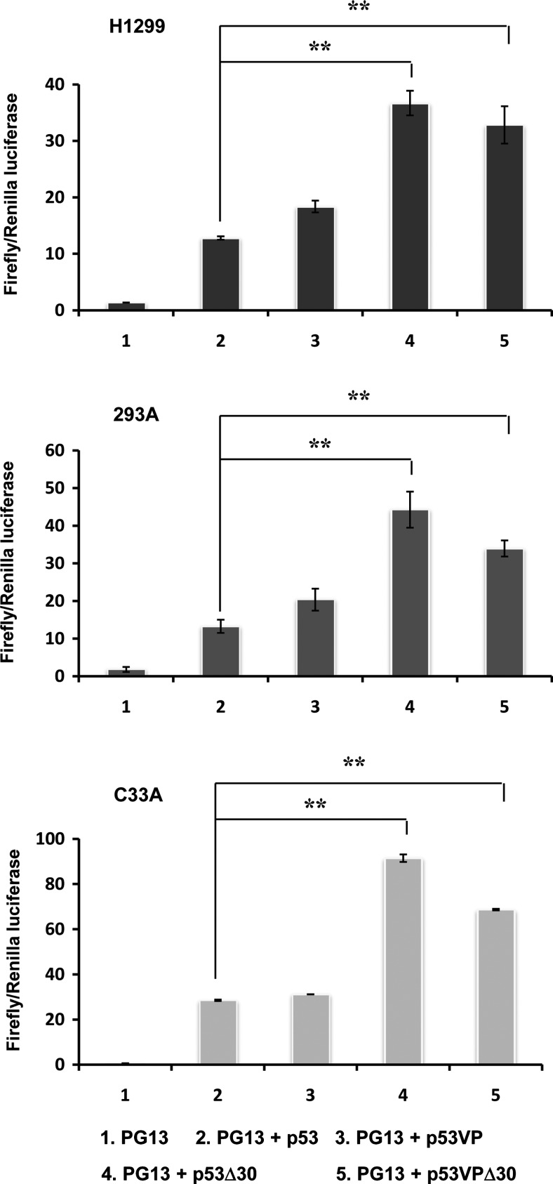
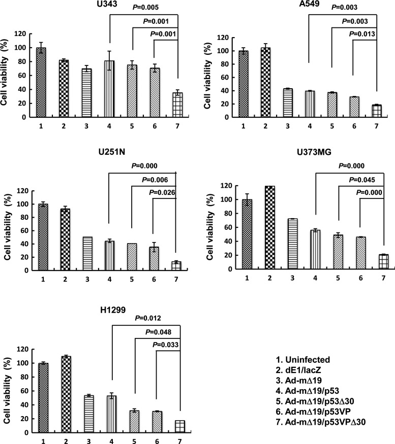
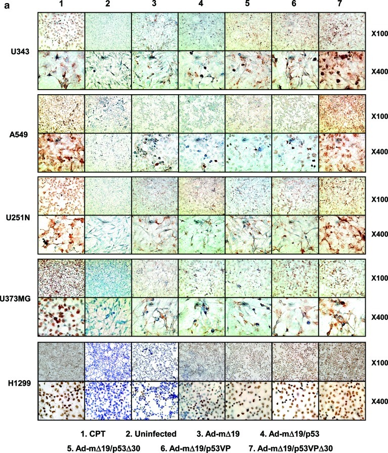
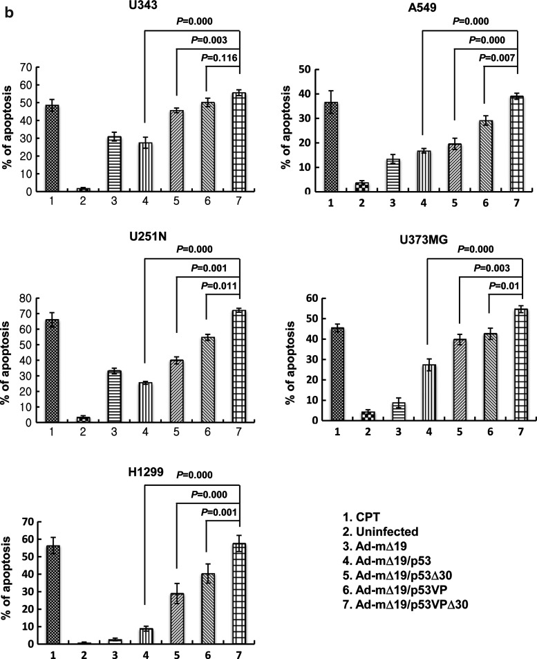
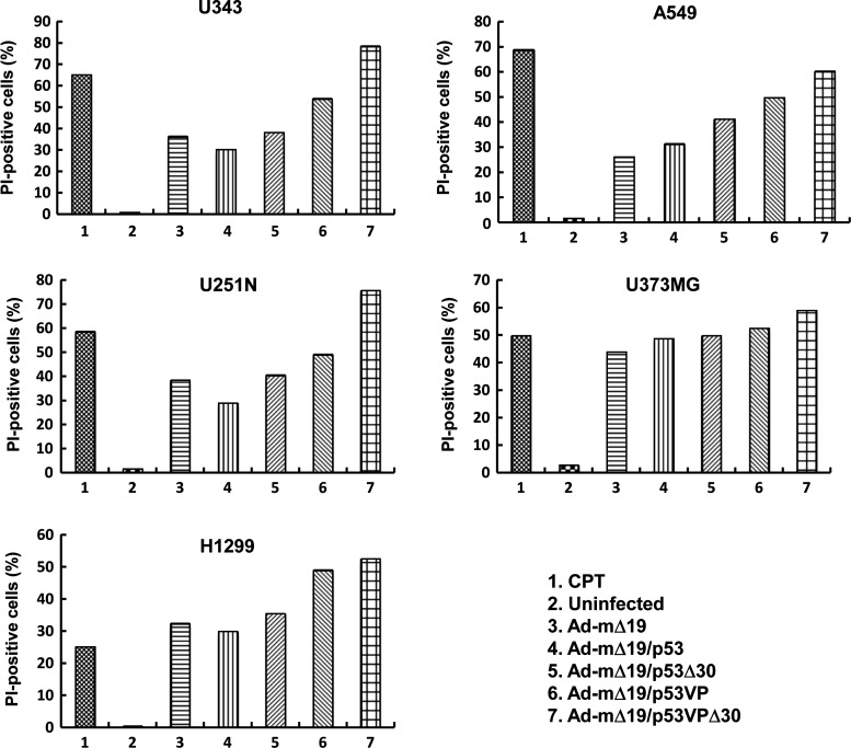
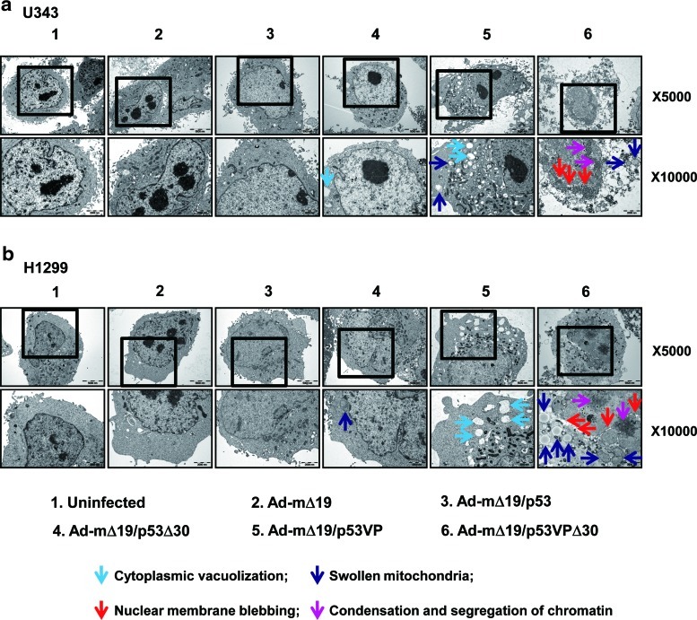
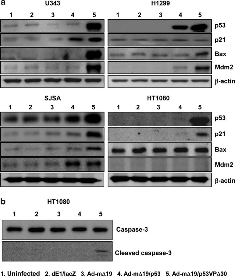
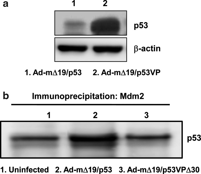
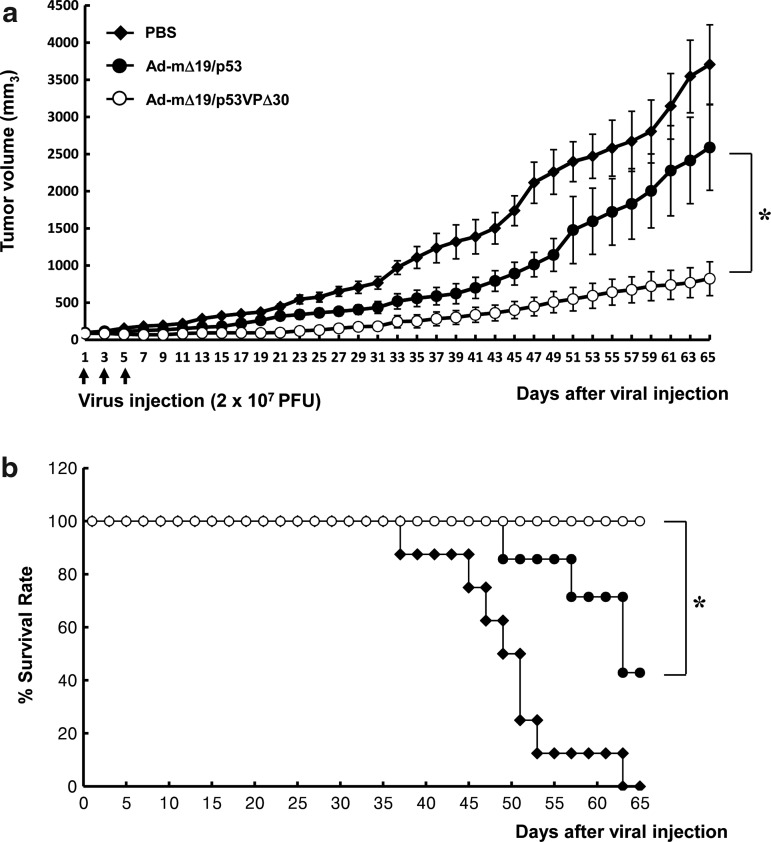
Intact p53 function is essential for responsiveness to cancer therapy. However, p53 activity is attenuated by the proto-oncoprotein Mdm2, the adenovirus protein E1B 55kD, and the p53 C-terminal domain. To confer resistance to Mdm2, E1B 55kD, and C-terminal negative regulation, we generated a p53 variant (p53VPΔ30) by deleting the N-terminal and C-terminal regions of wild-type p53 and inserting the transcriptional activation domain of herpes simplex virus VP16 protein. The oncolytic adenovirus vector Ad-mΔ19 expressing p53VPΔ30 (Ad-mΔ19/p53VPΔ30) showed greater cytotoxicity than Ad-mΔ19 expressing wild-type p53 or other p53 variants in human cancer cell lines. We found that Ad-mΔ19/p53VPΔ30 induced apoptosis through accumulation of p53VPΔ30, regardless of endogenous p53 and Mdm2 status. Moreover, Ad-mΔ19/p53VPΔ30 showed a greater antitumor effect and increased survival rates of mice with U343 brain cancer xenografts that expressed wild-type p53 and high Mdm2 levels. To our knowledge, this is the first study reporting a p53 variant modified at the N terminus and C terminus that shows resistance to degradation by Mdm2 and E1B 55kD, as well as negative regulation by the p53 C terminus, without decreased trans-activation activity. Taken together, these results indicate that Ad-mΔ19/p53VPΔ30 shows potential for improving p53-mediated cancer gene therapy.


Figures

FIG. 1.
FIG. 1.
Transcriptional activation by wild-type p53, p53Δ30, p53VP, or p53VPΔ30. H1299, 293, and C33A cells were cotransfected with the luciferase reporter PG13-Luc (PG13) and pCEP4 expressing the p53 variants. Luciferase activity was determined 48 hr after transfection. Data are expressed as the mean (±SEM) firefly-to-Renilla ratio, and are representative of three independent experiments. **p<0.01.
FIG. 2.
FIG. 2.
Oncolytic effects of Ad-mΔ19 expressing p53 variants. Monolayers of cancer cells were infected with Ad-mΔ19, Ad-mΔ19/p53, Ad-mΔ19/p53Δ30, Ad-mΔ19/p53VP, or Ad-mΔ19/p53VPΔ30 (A549, U251N, and U373MG, 5 MOI; U343 and H1299, 10 MOI). A replication-incompetent Ad, dE1/lacZ, served as a negative control. Cell viability was determined by the 3-(4,5-dimethylthiazol-2-yl)-2,5-diphenyltetrazolium bromide (MTT) assay. The percentage of viable cells was normalized to that of uninfected cells, and results are expressed as means±SEM (n=3) and are representative of three independent experiments.
FIG. 3.
FIG. 3.
Enhanced apoptosis in cancer cells as assessed by the terminal deoxynucleotidyltransferase dUTP nick end-labeling (TUNEL) assay. (a) Forty-eight hours after camptothecin treatment (CTP, 1 μM) or infection with Ad-mΔ19, Ad-mΔ19/p53, Ad-mΔ19/p53Δ30, Ad-mΔ19/p53VP, or Ad-mΔ19/p53VPΔ30 (MOI, 3), apoptotic cells were labeled with 3,3-diaminobenzidine using terminal deoxynucleotidyltransferase and counterstained with methyl green. Brown staining indicates positive staining for DNA strand breakage. Representative fields of three independent experiments are shown (original magnifications, ×100 and ×400). (b) The percentage of apoptotic cells induced by each treatment was calculated as the number of brown-stained cells per 2000 cells. The percentage of apoptotic cells is expressed as means±SEM (n=3), and data are representative of three independent experiments. Color images available online at www.liebertonline.com/hum
FIG. 3.
FIG. 3.
Enhanced apoptosis in cancer cells as assessed by the terminal deoxynucleotidyltransferase dUTP nick end-labeling (TUNEL) assay. (a) Forty-eight hours after camptothecin treatment (CTP, 1 μM) or infection with Ad-mΔ19, Ad-mΔ19/p53, Ad-mΔ19/p53Δ30, Ad-mΔ19/p53VP, or Ad-mΔ19/p53VPΔ30 (MOI, 3), apoptotic cells were labeled with 3,3-diaminobenzidine using terminal deoxynucleotidyltransferase and counterstained with methyl green. Brown staining indicates positive staining for DNA strand breakage. Representative fields of three independent experiments are shown (original magnifications, ×100 and ×400). (b) The percentage of apoptotic cells induced by each treatment was calculated as the number of brown-stained cells per 2000 cells. The percentage of apoptotic cells is expressed as means±SEM (n=3), and data are representative of three independent experiments. Color images available online at www.liebertonline.com/hum
FIG. 4.
FIG. 4.
Induction of apoptosis. Cells were infected with Ad-mΔ19, Ad-mΔ19/p53, Ad-mΔ19/p53Δ30, Ad-mΔ19/p53VP, or Ad-mΔ19/p53VPΔ30 (MOI, 5) or treated with camptothecin (CPT, 1 μM). Cells collected 48 hr postinfection were stained with propidium iodide (PI) and analyzed by fluorescence-activated cell sorting. Results are expressed as the percentage of PI-positive cells, and are representative of five independent experiments.
FIG. 5.
FIG. 5.
Detection of apoptosis by transmission electron microscopy (TEM). (a) U343 cells and (b) H1299 cells were infected with each vector (MOI, 1). Thirty-six hours postinfection, the cells were harvested and analyzed by TEM. Uninfected cells showed normal cellular morphology, whereas cells infected with Ad-mΔ19/p53VPΔ30 exhibited cytoplasmic changes (cytoplasmic vacuolization, light blue arrow; swollen mitochondria, dark blue arrow) and nuclear changes (nuclear membrane blebbing, red arrow; chromatin condensation and segregation, pink arrow). Original magnifications: ×5000 and ×10,000. Color images available online at www.liebertonline.com/hum
FIG. 6.
FIG. 6.
Activation of the apoptotic signal transduction pathway. Cells were infected with dE1/lacZ, Ad-mΔ19, Ad-mΔ19/p53, or Ad-mΔ19/p53VPΔ30 (MOI, 1). Twenty-four hours after infection, cell lysates were analyzed by Western blot with antibodies against p53 and downstream targets p21, Bax, Mdm2, caspase-3, and cleaved caspase-3; β-actin was used as an internal control.
FIG. 7.
FIG. 7.
Resistance of oncolytic Ad expressing p53VPΔ30 to E1B 55kD- and Mdm2-mediated negative regulation. (a) U343 cells were infected with Ad-mΔ19/p53 or Ad-mΔ19/p53VP (MOI, 1). Twenty-four hours postinfection, cell lysate was analyzed by Western blot with antibodies against p53; β-actin was used as an internal control. (b) SJSA cells were infected with Ad-mΔ19/p53 or Ad-mΔ19/p53VPΔ30 (MOI, 1). Twenty-four hours after infection, the cell lysate was first immunoprecipitated with anti-Mdm2 monoclonal antibody, and analyzed by Western blot with anti-p53 monoclonal antibody.
FIG. 8.
FIG. 8.
Tumor growth suppression and survival benefit by Ad-mΔ19/p53VPΔ30 in U343 human cancer xenografts established in male athymic nude mice. Subcutaneous tumors derived from U343 cells were treated with Ad-mΔ19/p53 or Ad-mΔ19/p53VPΔ30 (2×10⁷ PFU); negative controls were treated with PBS. (a) Tumor volume was measured every 2 days after treatment. The arrow indicates when treatments were administered. Results are expressed as means±SEM (each group, n=6). (b) Survival curve. The percentage of surviving mice was determined by monitoring tumor growth-related events (tumor size >2000 mm3) over a period of 65 days. *p<0.05.

Similar articles

Cited by

References

    1. Barak Y. Juven T. Haffner R. Oren M. mdm2 expression is induced by wild type p53 activity. EMBO J. 1993;12:461–468. - PMC - PubMed
    1. Chene P. Inhibiting the p53-MDM2 interaction: An important target for cancer therapy. Nat. Rev. Cancer. 2003;3:102–109. - PubMed
    1. Choi K.J. Kim J.H. Lee Y.S., et al. Concurrent delivery of GM-CSF and B7-1 using an oncolytic adenovirus elicits potent antitumor effect. Gene Ther. 2006;13:1010–1020. - PubMed
    1. de Belle I. Huang R.P. Fan Y., et al. p53 and Egr-1 additively suppress transformed growth in HT1080 cells but Egr-1 counteracts p53-dependent apoptosis. Oncogene. 1999;18:3633–3642. - PubMed
    1. Donehower L.A. Bradley A. The tumor suppressor p53. Biochim. Biophys. Acta. 1993;1155:181–205. - PubMed

Publication types

Substances