

Review
doi: 10.1016/j.neuroimage.2012.01.021.
Epub 2012 Jan 10.
FreeSurfer
Affiliations
- PMID: 22248573
- PMCID: PMC3685476
- DOI: 10.1016/j.neuroimage.2012.01.021
Item in Clipboard
Review
FreeSurfer
Neuroimage.
.
Abstract
FreeSurfer is a suite of tools for the analysis of neuroimaging data that provides an array of algorithms to quantify the functional, connectional and structural properties of the human brain. It has evolved from a package primarily aimed at generating surface representations of the cerebral cortex into one that automatically creates models of most macroscopically visible structures in the human brain given any reasonable T1-weighted input image. It is freely available, runs on a wide variety of hardware and software platforms, and is open source.
Copyright © 2012 Elsevier Inc. All rights reserved.
Figures
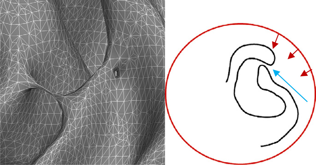
Left: examples of two geometrically different topological defects that are topologically equivalent: a handle that bridges a sulcus, and a hole in the bank of a gyrus. Right: a graphical example of the difficulty of using surface deformation techniques to model the cortical surface. Typically we want a smooth surface, but much of the cortical surface is buried deep inside folds forcing surfaces to pass through regions (indicated by the blue arrow) where the evolving surface has to bunch up to get enough surface area inside the fold to model the surface. Another problem is finding energy terms that will draw the surface into the deep fissure, and away from the narrow opening, which also means pulling it away from the true cortical surface to traverse the sulcal opening and arrive at the boundary on the other side.
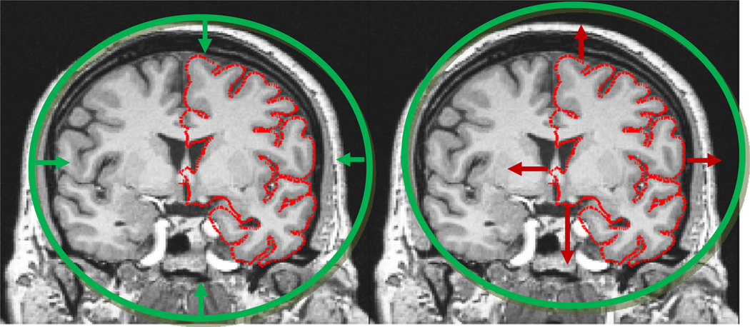
Left: the approach taken with deformable models. A surface of known topology but incorrect geometry (a sphere) is driven by an energy functional towards the desired pial surface shown in red. The difficulty stems from finding terms that will generate a smooth surface but will allow it to pass through an intermediate representation that can push enough surface area into e.g. the sylvian fissure. Right: in contrast, deforming the topologically incorrect surface model outwards to the surface of the sphere is a relatively simple computational problem.
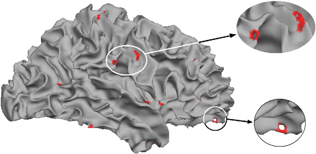
Example of the typical spatial distribution of topological defects.
Example of a topological defect (left), an inaccurate correction (center) and an accurate correction (right) (thanks to Florent Ségonne).
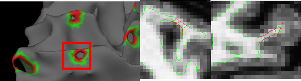
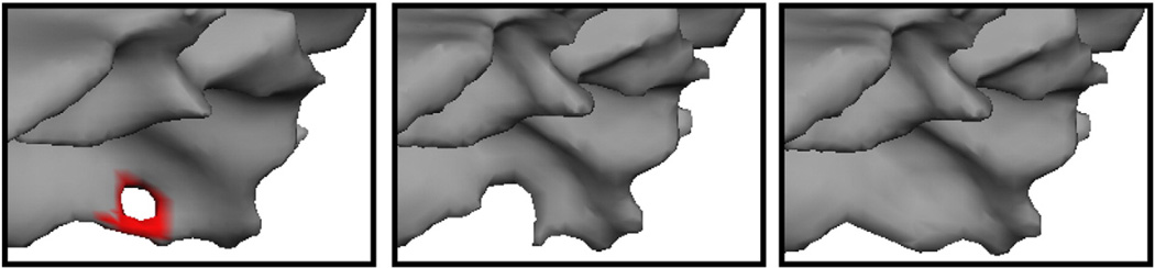
Example of a surface defect (left), and it's representation in two orthogonal slices (center and right). The red portion in the left-hand image represents the region found to contain the defect. The red portion of the surface in the center and right images represent the surface that has been added to fill the hole.
References
-
- Becker C, Fischl, Greve, DeLuca, LaViolette, O'Keefe, Fischman, Rentz, Sperling, Johnson . Amyloid deposition related to cortical thinning. Australia: 14th Annual Meeting of the Organization for Human Brain Mapping Sydney; 2008.
-
- Collins D, Evans A. Animal: validation and applications of non-linear registration-based segmentation. Int. J. Pattern Recognit. Artif. Intell. 1997;11:1271–1294.
-
- Dale A. Cognitive Science, vol. Ph.D. San Diego: University of California at San Diego; 1994. Source localization and spatial discriminant analysis of event-related potentials: linear approaches.
-
- Dale AM, Fischl B, Sereno MI. Cortical surface-based analysis I: segmentation and surface reconstruction. Neuroimage. 1999;9:179–194. - PubMed
Publication types
MeSH terms
Grants and funding
- U24 RR021382/RR/NCRR NIH HHS/United States
- R01EB006758/EB/NIBIB NIH HHS/United States
- R01 NS052585-01/NS/NINDS NIH HHS/United States
- P41 RR014075/RR/NCRR NIH HHS/United States
- R01 NS052585/NS/NINDS NIH HHS/United States
- AG022381/AG/NIA NIH HHS/United States
- 1S10RR023401/RR/NCRR NIH HHS/United States
- R01 RR016594/RR/NCRR NIH HHS/United States
- S10 RR023043/RR/NCRR NIH HHS/United States
- S10 RR023401/RR/NCRR NIH HHS/United States
- R01 AG022381/AG/NIA NIH HHS/United States
- R01 NS070963/NS/NINDS NIH HHS/United States
- P41-RR14075/RR/NCRR NIH HHS/United States
- S10 RR019307/RR/NCRR NIH HHS/United States
- U54 EB005149/EB/NIBIB NIH HHS/United States
- 1S10RR019307/RR/NCRR NIH HHS/United States
- 1S10RR023043/RR/NCRR NIH HHS/United States
- 1R21NS072652-01/NS/NINDS NIH HHS/United States
- RC1 AT005728/AT/NCCIH NIH HHS/United States
- 1R01NS070963/NS/NINDS NIH HHS/United States
- R21 NS072652/NS/NINDS NIH HHS/United States
- RC1AT005728-01/AT/NCCIH NIH HHS/United States
- P41 RR006009/RR/NCRR NIH HHS/United States
- R01 EB006758/EB/NIBIB NIH HHS/United States
LinkOut - more resources
Full Text Sources
Other Literature Sources
Medical
