. 2012 Feb;163(2):628-35.
doi: 10.1016/j.virusres.2011.12.021. Epub 2012 Jan 11.

Coronavirus infection of rat dorsal root ganglia: ultrastructural characterization of viral replication, transfer, and the early response of satellite cells

Affiliations

Coronavirus infection of rat dorsal root ganglia: ultrastructural characterization of viral replication, transfer, and the early response of satellite cells

Yan-Chao Li et al. Virus Res. 2012 Feb.

Abstract

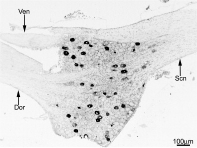
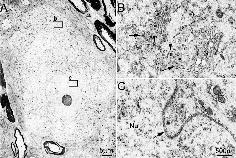
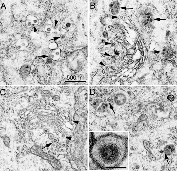
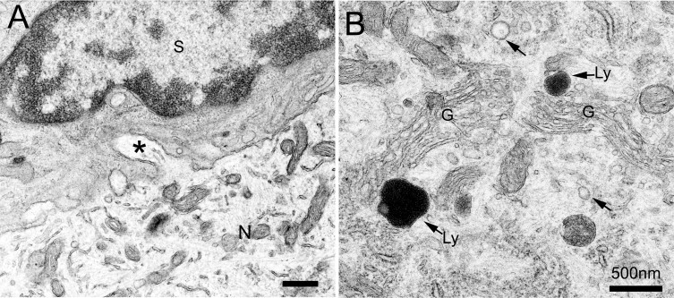
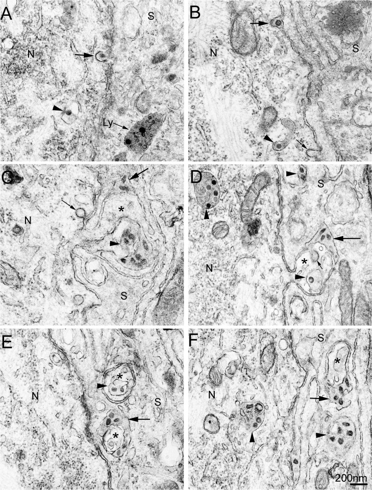
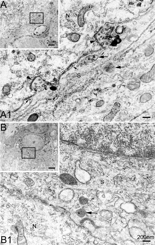
Swine hemagglutinating encephalomyelitis virus (HEV) has been shown to have a capability to gain access to the cell bodies of sensory neurons after peripheral inoculation, resulting in ganglionic infection. It is not clearly understood how this virus is replicated within and released from the sensory neurons, and it remains to know how satellite cells response to the HEV invasion. By ultrastructurally examining HEV-infected rat dorsal root ganglia, we found that HEV in the cell bodies of infected neurons budded from endoplasmic reticulum-Golgi intermediate compartments, and were assembled either individually within small vesicles or in groups within large vesicles. The progeny virions were released from the sensory neurons mainly by smooth-surfaced vesicle-mediated secretory pathway, which occurred predominantly at the perikaryal projections and infoldings of sensory neurons. Released HEV particles were subsequently taken up by the adjacent satellite cells. Almost all virus particles in the cytoplasm of satellite cells were contained in groups within vesicles and lysosome-like structures, suggesting that these glial cells may restrict the local diffusion of HEV. These observations give some insights into the pathogenesis of coronavirus infection and are thought to help understand the interactions between sensory neurons and their satellite cells.


Figures

Fig. 1
Fig. 1
HEV antigen in the DRG. Scale bar: 100 μm. A representative light micrograph shows the immunohistochemical distribution of HEV antigen in a longitudinally cut DRG. Dor: dorsal root; Ven: ventral root; Scn: sciatic nerve.
Fig. 2
Fig. 2
Morphogenesis of HEV in the DRG neuron. Scale bars: A, 5 μm; B and C, 500 nm. Panel A shows an HEV-infected DRG neuron. The details in insets b and c are given in panels B and C, respectively, showing that morphogenesis of HEV occurs exclusively in the perikaryal cytoplasm. In panel B, viral budding profiles (arrows) can be observed in ER and in the lateral rims of Golgi complexes (G). Besides small vesicles enclosing single virions, large vesicles (arrowheads) containing more than one virion are also found in the Golgi areas. No virus-related structures are seen within the nucleus (C), but nuclear envelope invaginations are often encountered (arrow).
Fig. 3
Fig. 3
The replication and assembly of HEV in the DRG neurons. Scale bar: 500 nm. Panel A shows the budding profiles of HEV at different stages in the ER areas. Arrowheads indicate electron-dense crescent segments attached to the external membrane of ER cisternae, while arrows indicate larger crescent segments that have bulged into the lumen of ER cisternae. Panel B shows groups of virions enclosed within large smooth-surfaced vesicle (arrows) in the Golgi areas. The arrowheads indicate virus particles present in the dilated rims of the trans-Golgi network. Panel C shows progeny virions enclosed individually within small vesicles in a trans-Golgi network. The arrow indicates a coated vesicle containing a single virion. Similar coating structures are also present on a small empty vesicle as well as part of the trans-Golgi networks (arrowheads). Panel D shows that virus-containing vesicles are attached to each other (arrows). The inset in panel D is a high-power electron micrograph, showing a virus enclosed within a vesicle. A layer of surface projections can be seen surrounding the viral envelope beneath the vesicle membrane.
Fig. 4
Fig. 4
Electron microphotographs from the vehicle controls. Scale bars: 500 nm. A: No virus particles are seen in either the neurons (N) or the surrounding SCs (S). The intercellular space between neuronal cell bodies and their SCs are generally narrow with a distance of about 20 nm. The neuronal-SC boundary is complicated by the presence of numerous projections, which arose from either the neuron or the adjacent SCs. The neuronal projections occur more frequently. In single sections, they are sometimes seen continuous with the neuronal cell surface (asterisk). B: In the neurons, lysosomes (labeled arrows) and vesicles (arrows) of varying size are frequently located near the Golgi apparatuses (G). No virus particles are found within the vesicles or the lysosome-like structures.
Fig. 5
Fig. 5
HEV transfer between DRG neurons and their SCs. Scale bar: 200 nm. Neurons (N) and SCs (S) are marked in all images. Arrowheads indicate virus-containing vesicular structures in the cytoplasm of neurons or SCs. Large arrows indicate extracellular virus particles between the neuronal cell bodies and their SCs. Small arrows indicate empty coated vesicles or invaginations. Top row panels show coated vesicle-mediated HEV transfer. Panel A shows a coated vesicle containing a single virion fusing with the plasma membrane of an infected neuron. A lysosome-like structure (arrow labeled by Ly), which contains several virus-like particles, is found in the cytoplasm of the adjacent SCs. Panel B shows a virion located extracellularly between an infected neuron and its SC, where the invaginated plasma membrane of the infected neuron is covered with a layer of coating decorations (larger arrow). Panels in middle and bottom rows show HEV transfer by use of large smooth-surface vesicles, which is found to occur in neuronal perikaryal specializations. In Panels C and D, the neuronal projections are seen arising from the neuronal cell body and appear as microvillus-like structures (asterisks). Panels E and F show cross-sectioned neuronal projections (asterisks), which are embedded within the cytoplasm of SCs without continuity with the neuronal cell bodies.
Fig. 6
Fig. 6
HEV transfer and the reactions of SCs. Scale bars: A and B, 2 μm, A1 and B1, 200 nm. Neurons (N) and SCs (S) are marked in all images. Panel A shows the boundary of an infected neuron and its SC. Details of the inset are shown in panel A1 under higher magnification. Numerous virions are distributed extracellularly near infolded cell surface of the infected neuron, where subsurface cisternae are seen closely attached to the invaginated plasma membrane on the cytoplasmic side (arrowheads). A few virions (small arrows) are also noted in the narrow extracellular space along smooth parts of the neuronal cell surface. In the cytoplasm of the SC, there are many vesicles (large arrows) containing groups of virus particles. Panel B shows a SC adjacent to an infected neuron. Details of the inset are shown in panel B1 under higher magnification. HEV particles present in the SC are located within lysosome-like structures (arrow), but no single virions or viral budding profiles are found.

References

    1. Alemañ N., Quiroga M.I., López-Peña M., Vázquez S., Guerrero F.H., Nieto J.M. Induction and inhibition of apoptosis by pseudorabies virus in the trigeminal ganglion during acute infection of swine. J. Virol. 2001;75:469–479. - PMC - PubMed
    1. Andries K., Pensaert M.B. Immunofluorescence studies on the pathogenesis of hemagglutinating encephalomyelitis virus infection in pigs after oronasal inoculation. Am. J. Vet. Res. 1980;41:1372–1378. - PubMed
    1. Bai W.Z., Li Y.C., Hirano N., Tohyama K., Hashikawa T. Transneuronal infection and associated immune response in the central nervous system induced by hemagglutinating encephalomyelitis virus following rat hindpaw inoculation. Neurosci. Res. 2008;61(Suppl. 1):S135.
    1. Chasey D., Alexander D.J. Morphogenesis of avian infectious bronchitis virus in primary chick kidney cells. Arch. Virol. 1976;52:101–111. - PMC - PubMed
    1. Clarke J.K., McFerran J.B. An electron microscopic study of haemagglutinating encephalomyelitis virus of pigs. J. Gen. Virol. 1971;13:339–344. - PubMed

LinkOut - more resources