. 2012 Apr;142(4):978-88.
doi: 10.1053/j.gastro.2011.12.055. Epub 2012 Jan 13.

HCV infection induces a unique hepatic innate immune response associated with robust production of type III interferons

Affiliations

HCV infection induces a unique hepatic innate immune response associated with robust production of type III interferons

Emmanuel Thomas et al. Gastroenterology. 2012 Apr.

Abstract

Background & aims: Polymorphisms in the IL28B gene have been associated with clearance of hepatitis C virus (HCV), indicating a role for type III interferons (IFNs) in HCV infection. Little is known about the function of type III IFNs in intrinsic antiviral innate immunity.

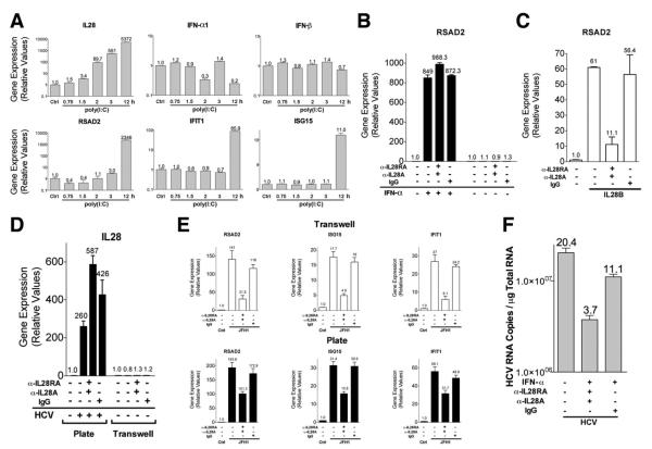
Methods: We used in vivo and in vitro models to characterize the role of the type III IFNs in HCV infection and analyzed gene expression in liver biopsy samples from HCV-infected chimpanzees and patients. Messenger RNA and protein expression were studied in HCV-infected hepatoma cell lines and primary human hepatocytes.

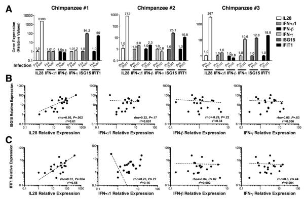
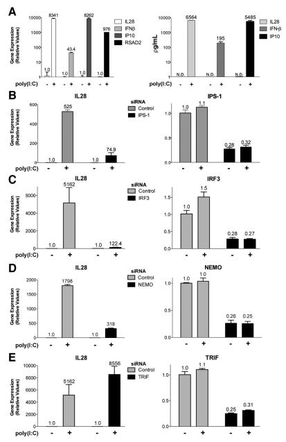
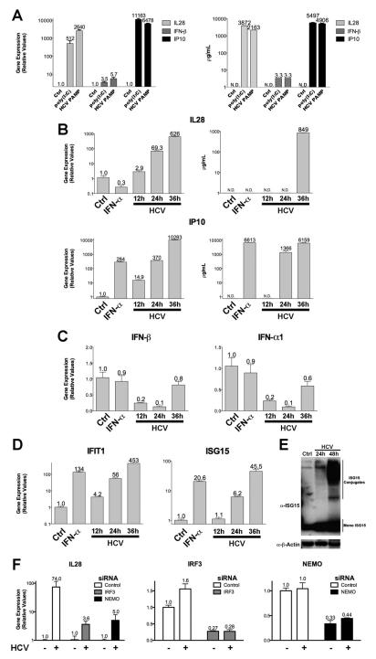
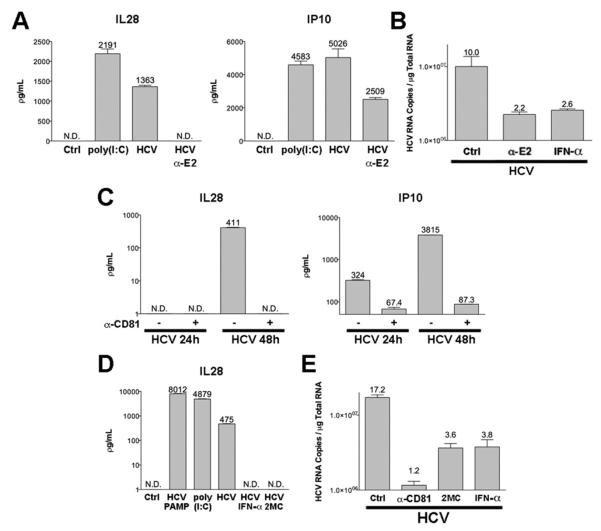
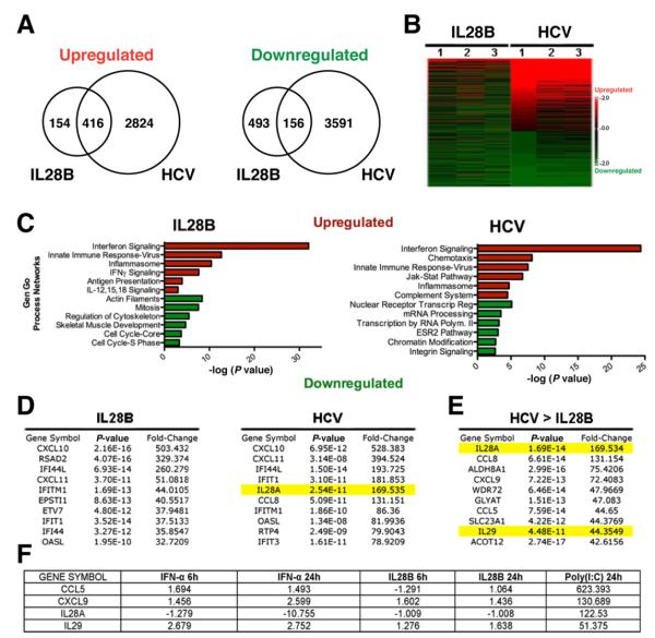
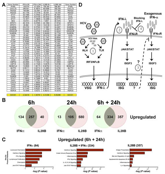
Results: HCV infection of primary human hepatocytes induced production of chemokines and type III IFNs, including interleukin (IL)-28, and led to expression of IFN-stimulated genes (ISGs). Chimpanzees infected with HCV showed rapid induction of hepatic type III IFN, associated with up-regulation of ISGs and minimal induction of type I IFNs. In liver biopsy specimens from HCV-infected patients, hepatic expression of IL-28 correlated with levels of ISGs but not of type I IFNs. HCV infection produced extensive changes with gene expression in addition to ISGs in primary human hepatocytes. The induction of type III IFNs is regulated by IFN regulatory factor 3 and nuclear factor κB. Type III IFNs up-regulate ISGs with a different kinetic profile than type 1 IFNs and induce a distinct set of genes, which might account for their functional differences.

Conclusions: HCV infection results predominantly in induction of type III IFNs in livers of humans and chimpanzees; the level of induction correlates with hepatic levels of ISGs. These findings might account for the association among IL-28, level of ISGs, and recovery from HCV infection and provide a therapeutic strategy for patients who do not respond to IFN therapy.


Figures

Figure 1
Figure 1
Induction of IL-28 and ISGs in liver biopsy specimens of HCV-infected chimpanzees and humans. (A) qPCR analysis of IL-28, IFN-α1, IFN-β, IFN-γ, ISG15, and IFIT1 expression levels in chimpanzees before and after infection. The postinfection biopsies were performed shortly after infection (<4 weeks; see Materials and Methods for specific information on each animal). Correlation of (B) ISG15 and (C) IFIT1 mRNA levels with IL-28, IFN-α1, IFN-β, and IFN-γ expression, as determined by qPCR, in livers of patients (n = 19) with chronic hepatitis C genotype 1 infection. Dashed lines represent log-log regression. P value represents significance of the Spearman correlation coefficient (ρ). r2 = goodness of fit of the regression line.
Figure 2
Figure 2
Preferential induction of IL-28 by viral mimetics in hepatocytes is mediated by the IRF3 and nuclear factor κB signaling pathways. (A) Analysis of IL-28, IFN-β, IP10, and RSAD2 mRNA levels by qPCR and protein production of IL-28, IFN-β, and IP10 by ELISA in HepG2 cells following transfection with poly(I:C) (6 μg/mL for 24 hours). qPCR analysis of IL-28 mRNA levels in HepG2 cells first treated with siRNAs for 3 days targeting (B) IPS-1, (C) IRF3, (D) NEMO, and (E) TRIF and then transfected with or without poly(I:C) (6 μg/mL for 24 hours). Quantification of siRNA-mediated specific gene suppression is shown in the right panel. Results are representative of more than three experiments. Nontargeting siRNA represents control. N.D., not detected.
Figure 3
Figure 3
Robust induction of IL-28 and ISGs by viral mimetics and following HCV infection of PHHs. (A) Analysis of IL-28, IFN-β, and IP10 mRNA levels by qPCR and ELISA in PHHs following transfection with poly(I:C) (6 μg/mL for 24 hours) or HCV PAMP RNA (6 μg/mL for 24 hours). PHHs infected with JFH1 virus (MOI of 5) for 12, 24, and 36 hours or treated with IFN-α (100 U/mL) for 12 hours. (B) Analysis of IL-28 and IP10 mRNA levels by qPCR and protein production by ELISA. qPCR analysis of (C) IFN-β and IFN-α1 as well as (D) IFIT1 and ISG15 mRNA expression. (E) Western blot analysis of ISG15 and β-actin on PHHs infected with JFH1 virus (MOI of 5) for 24 and 48 hours. (F) qPCR analysis of IL-28 mRNA levels in PHHs first treated with siRNAs for 3 days targeting IRF3 and NEMO and then infected with or without JFH1 (MOI of 5) for 36 hours. Quantification of siRNA-mediated specific gene suppression is shown in the right panels. Results are representative of more than three experiments. Nontargeting siRNA represents control. N.D., not detected.
Figure 4
Figure 4
Blocking of IL-28 induction following HCV infection in PHHs by anti-E2 and CD81 antibodies and antiviral molecules. (A) Quantification of IL-28 and IP10 protein production by ELISA in PHHs with or without pretreatment with anti-E2 antibodies (10 μg/mL) for 1 hour and subsequently infected with JFH1 (MOI of 5) for 52 hours or following transfection with poly(I:C) (6 μg/mL for 24 hours). (B) qPCR analysis of intracellular HCV RNA 52 hours after infection with JFH1 (MOI of 5) in PHHs alone or pretreated with anti-E2 antibodies (10 μg/mL) for 1 hour or treated with IFN-α (100 U/mL) for 48 hours. (C) Quantitation of IL-28 and IP10 protein production by ELISA in PHHs with or without pretreatment with anti-CD81 antibodies (10 μg/mL) for 1 hour and subsequently infected with JFH1 (MOI of 5) for 24 and 48 hours. (D) Quantitation of IL-28 protein production by ELISA in PHHs transfected with HCV-PAMP RNA (6 μg/mL for 24 hours) or poly(I:C) (6 μg/mL for 24 hours) or infected with JFH1 (MOI of 5) for 52 hours alone or treated with IFN-α (100 U/mL) or 2-methylcytidine (20 μmol/L) for 48 hours. (E) qPCR analysis of intracellular HCV RNA 52 hours after infection with JFH1 (MOI of 5) in PHH alone or pretreated with CD81 antibodies (10 μg/mL) for 1 hour or treated with 2-methylcytidine (20 μmol/L) or IFN-α (100 U/mL) for 48 hours. Numbers above bars represent HCV RNA copies χ 106. Results are representative of two experiments. N.D., not detected.
Figure 5
Figure 5
Changes in gene expression following HCV infection are mediated by IFN-dependent and independent pathways. Microarray analysis of PHHs treated with IL28B (25 ng/mL for 24 hours) and JFH1 (MOI of 5 for 36 hours). (A) Venn diagrams displaying the number or probe sets up-regulated and down-regulated (>2.0-fold change) following treatment. (B) Heat maps generated using microarray data from 3 replicates. (C) GeneGo networks that are significantly up-regulated or down-regulated by IL28B and JFH1. (D) Top 10 genes ranked by fold induction following treatment with IL28B and JFH1. (E) Top 10 genes ranked by fold induction following JFH1 infection whose expression levels were not significantly up-regulated by IL28B. (F) Fold induction of genes up-regulated by JFH1 that are also induced by poly(I:C) (6 μg/mL) but minimally induced by IL28B (25 ng/mL) or IFN-α (10U/mL), by microarray analysis, for the indicated durations of treatment. Data from 3 replicates.
Figure 6
Figure 6
Autocrine and paracrine IL-28 production contributes to ISG induction following poly(I:C) treatment and HCV infection. (A) qPCR analysis of IL-28, IFN-α1, IFN-β, RSAD2, IFIT1, and ISG15 mRNAs in HepG2 cells following transfection of poly(I:C) (6 μg/mL) for 0.75, 1.5, 2, 3, and 12 hours. In the following experiments, antibodies against IL28A (15 μg/mL) and IL28RA (20 μg/mL) or equal concentrations of immunoglobulin G were used. (B) qPCR analysis of RSAD2 mRNA level in PHHs treated with IFN-α (100 U/mL) for 12 hours with or without pretreatment with antibodies. (C) qPCR analysis of RSAD2 mRNA levels in PHHs treated with IL28B (25 ng/mL) for 24 hours with or without pretreatment with antibodies. (D) PHHs were plated on bottom wells and infected with JFH1 virus (MOI of 5). The virus was then removed after 4 hours and PHHs plated on Transwell membranes were then placed into the wells for coculturing. The cocultures were treated with antibodies. qPCR analysis of IL-28 mRNA levels of cells in the bottom wells (left) and Transwells (right) was determined 48 hours later. (E) qPCR analysis of RSAD2, ISG15, and IFIT1 expression levels in PHHs, plated on Transwell membranes (top panels), exposed to media from cells infected with JFH1 (MOI of 5, bottom panels), and treated with antibodies. (F) qPCR analysis of intracellular HCV RNA levels 52 hours after infection with JFH1 (MOI of 5) in PHH alone or treated with IFN-α (100 U/mL) and antibodies for 48 hours. Numbers above bars represent HCV RNA copies χ 10. Results are representative of 2 experiments.
Figure 7
Figure 7
Differential effects of type III IFN and IFN-α on gene expression and model of HCV infection and subsequent innate immune responses in hepatocytes. (A) Top 42 probe sets, ranked by fold induction, following treatment with IL28B (25 ng/mL) and IFN-α (10 U/mL) for the indicated duration. Data from 3 replicates. (B) Venn diagrams displaying the numbers of probe sets up-regulated (>2.0-fold change) following the indicated treatment time points. For the 6h + 24h Venn diagrams, probe sets up-regulated (>2.0-fold change) at either 6- and 24-hour time points were combined for each treatment and then compared. (C) GeneGo networks that are significantly up-regulated by IL28B and/or IFN-α using the 6h + 24h probe set lists. (D) This proposed model highlights the role of type III IFNs in intrinsic antiviral innate immunity to HCV in the liver. (1) HCV infects hepatocytes, resulting in the detection of viral RNA by the RIG-I–like helicases and/or Toll-like receptors and leading to activation of IRF3 and nuclear factor (NF) κB. They translocate to the nucleus binding to the promoters of virus-stimulated genes (VSGs), resulting in the production of type III IFNs. (2) Type III IFNs are secreted and bind to its cognate receptor, leading to the up-regulation of ISGs. (3) In a subset of chronically infected patients, type III IFNs continually induce genes that block IFN-α from further up-regulating ISGs, leading to nonresponse to exogenous IFN. (4) Blocking type III IFN up-regulation of genes may render nonresponders sensitive to the antiviral activity of exogenous type I IFN by allowing it to up-regulate antiviral ISGs.

Similar articles

Cited by

References

    1. Kawai T, Akira S. The roles of TLRs, RLRs and NLRs in pathogen recognition. Int Immunol. 2009;21:317–337. - PMC - PubMed
    1. Iwasaki A, Medzhitov R. Regulation of adaptive immunity by the innate immune system. Science. 2010;327:291–295. - PMC - PubMed
    1. Takeuchi O, Akira S. Innate immunity to virus infection. Immunol Rev. 2009;227:75–86. - PMC - PubMed
    1. Beutler BA. TLRs and innate immunity. Blood. 2009;113:1399–1407. - PMC - PubMed
    1. Onomoto K, Onoguchi K, Takahasi K, et al. Type I interferon production induced by RIG-I-like receptors. J Interferon Cytokine Res. 2010;30:875–881. - PubMed

Publication types

MeSH terms