. 2012 Mar 1;363(1):258-65.
doi: 10.1016/j.ydbio.2011.12.047. Epub 2012 Jan 10.

Sgt1 acts via an LKB1/AMPK pathway to establish cortical polarity in larval neuroblasts

Affiliations

Sgt1 acts via an LKB1/AMPK pathway to establish cortical polarity in larval neuroblasts

Ryan O Andersen et al. Dev Biol. .

Abstract

Drosophila neuroblasts are a model system for studying stem cell self-renewal and the establishment of cortical polarity. Larval neuroblasts generate a large apical self-renewing neuroblast, and a small basal cell that differentiates. We performed a genetic screen to identify regulators of neuroblast self-renewal, and identified a mutation in sgt1 (suppressor-of-G2-allele-of-skp1) that had fewer neuroblasts. We found that sgt1 neuroblasts have two polarity phenotypes: failure to establish apical cortical polarity at prophase, and lack of cortical Scribble localization throughout the cell cycle. Apical cortical polarity was partially restored at metaphase by a microtubule-induced cortical polarity pathway. Double mutants lacking Sgt1 and Pins (a microtubule-induced polarity pathway component) resulted in neuroblasts without detectable cortical polarity and formation of "neuroblast tumors." Mutants in hsp83 (encoding the predicted Sgt1-binding protein Hsp90), LKB1, or AMPKα all show similar prophase apical cortical polarity defects (but no Scribble phenotype), and activated AMPKα rescued the sgt1 mutant phenotype. We propose that an Sgt1/Hsp90-LKB1-AMPK pathway acts redundantly with a microtubule-induced polarity pathway to generate neuroblast cortical polarity, and the absence of neuroblast cortical polarity can produce neuroblast tumors.


Figures

Figure 1
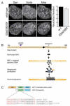
Figure 1. Identification of a mutant in sgt1 that is required for larval brain development
(A) Wild type and l(3)s2383 / Df(3R)Exel6147 (l(3)s2383 / Df) third instar larval brains stained for the neuroblast markers Miranda (Mira) and Deadpan (Dpn) plus Scribble (Scrib) which decorates the cortex of all cells in the brain. Right: histogram of the average neuroblast number per brain lobe (bar, standard deviation) in each genotype. Note that the mutant brains have fewer neuroblasts and Scrib protein is cytoplasmic. (B) Schematic of the “sequence capture” and deep sequencing strategy used to identify the lesion in the l(3)s2383 mutant chromosome. Although the chromosome was generated in a P element mutagenesis, we found a 15 nucleotide deletion unrelated to the P element insertion that showed the mutant phenotype when transheterozygous to Df(3R)Exel6147. (C) The l(3)s2383 mutation resulted in an in-frame five amino acid deletion in the Drosophila Sgt1 protein, within the N-terminal CS domain (green); the C-terminal Sgt1-specific SGS domain is shown (white).
Figure 2
Figure 2. sgt1 mutant neuroblasts have defects in establishing apical cortical polarity
Wild type and sgt1s2383 / sgt1s2383 (sgt1) mutant larval brain neuroblasts stained for the indicated cortical proteins plus α-tubulin (not shown) and phospho-histone H3 (PH3; not shown). Prophase neuroblasts were identified as PH3+ without a bipolar spindle; metaphase neuroblasts were identified as PH3+ with a bipolar spindle. Scale bar, 5 μm. (A) Wild type prophase neuroblasts show apical enrichment of the Par complex (Baz, aPKC) and Pins complex (Pins) and Insc; quantification in (A’). (B) sgt1 prophase neuroblasts show a nearly complete loss of apical localized Par complex (Baz, aPKC), Pins complex (Pins) and Insc; quantification in (B’). (C) Wild type metaphase neuroblasts show apical enrichment of the Par complex (Baz, aPKC) and Pins complex (Pins) and Insc; quantification in (C’). (D) sgt1 metaphase neuroblasts have recovered substantial apical protein localization compared to their prophase phenotype, although it is not fully back to wild type levels; quantification in (D’). (A’-D’) Quantification of the phenotypes shown in A-D. Dark green, normal asymmetric; light green, expanded asymmetric; red, cytoplasmic. Number of neuroblasts scored shown in each bar.
Figure 3
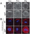
Figure 3. sgt1 mutant neuroblasts have defects in Dlg/Scrib cortical localization
(A, B) Wild type prophase or metaphase larval neuroblasts; Scrib is uniform cortical, and Dlg is uniform cortical with apical enrichment at metaphase. (C, D) sgt1s2383 / sgt1s2383 (sgt1) metaphase larval neuroblasts; Scrib is cytoplasmic, and Dlg is mostly cytoplasmic except for persistent apical enrichment at metaphase. Scale bar, 5 μm.
Figure 4
Figure 4. Sgt1 and microtubules act in redundant pathways to generate apical protein localization in metaphase neuroblasts
Wild type and sgt1s2383 / sgt1s2383 (sgt1) metaphase larval neuroblasts stained for the indicated Pins complex proteins and the mitotic marker mitotic marker phospho-histone H3 (not shown in A; blue in B). Scale bar, 5 μm. (A) Wild type and sgt1 mutant neuroblasts show apical localization of Insc and Pins proteins, and apical enrichment of Dlg protein. Quantification of apical asymmetry: WT (Pins, 100%, n=50; Insc, 100%, n=40; Dlg, 94%, n=34), sgt1 (Pins, 83%, n=35; Insc, 82%, n=23; Dlg, 83%, n=29) (B) Wild type and sgt1 mutants treated with 10 μM Colcemid to depolymerize microtubules. Wild type neuroblasts show normal apical localization of the Pins complex (Pins, 100%, n=20; Insc, 100%, n=26; Dlg, 100%, n=20), whereas sgt1 mutants lack detectable cortical localization of Pins complex proteins (Pins, 6%, n=31; Insc, 14%, n=22; Dlg, 7%, n=15).
Figure 5
Figure 5. sgt1 pins double mutants larval brains have ectopic, apolar neuroblasts
Wild type, sgt1s2383 / sgt1s2383 (sgt1) single mutants, pinsp62 / pinsp62 (pins) single mutants, or sgt1 pins double mutant larval brains stained for the indicated cortical polarity proteins and the mitotic marker phospho-histone H3 (not shown). Scale bar, 5 μm (A-J) and 25 μm (K-N). (A) Wild type metaphase neuroblasts showing normal cortical polarity; apical up. Quantification of normal asymmetric localization for each protein: aPKC, 100%, n=30; Insc, 100%, n=16; Dlg, 83%, n=29; Mira, 100%, n=20; Numb, 100%, n=20. (B) sgt1 pins double mutant metaphase neuroblasts lack all cortical polarity. Quantification of asymmetric localization for each protein: aPKC, 6%, n=64; Insc, 5%, n=39; Dlg, 0%, n=45; Mira, 10%, n=79; Numb, 7%, n=41. (C-F) Third instar larval brains stained for the neuroblast marker Miranda (Mira); the number of central brain neuroblasts in each genotype is given below each panel (average +/- standard deviation). (C) Wild type showing normal neuroblast numbers. (D,E) sgt1 and pins single mutant brains showing reduced neuroblast numbers. (F) sgt1 pins double mutant brain showing a large increase in neuroblast numbers.(Ahmed et al., 2003)
Figure 6
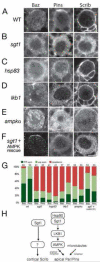
Figure 6. sgt1, hsp83, LKB1, and AMPKαhave similar neuroblast polarity phenotypes
Prophase larval brain neuroblasts stained for the Par complex protein Bazooka (Baz), the Pins complex protein Pins, and the cortical protein Scribble (Scrib). Quantification in (G). Scale bar, 5 μm. (A) Wild type. Baz and Pins form apical cortical crescents; Scrib is uniform cortical. (B) sgt1s2383 / Df(3R)6147 (sgt1). Baz and Pins are delocalized; Scrib is cytoplasmic. (C) hsp8313F3/hsp83582 (hsp83). Baz and Pins are delocalized; Scrib is uniform cortical. (D) lkb14A4-2 / lkb14A4-2 (lkb1). Baz and Pins are delocalized; Scrib is uniform cortical. (E) ampk1 / ampk1 (ampkα). Baz and Pins are delocalized; Scrib is uniform cortical. (F) sgt1s2383 / Df(3R)6147; tub-gal4 UAS-ampkαTD(sgt1 + AMPK rescue). Baz and Pins form apical cortical crescents; Scrib is cytoplasmic. (G) Quantification of the Baz and Pins phenotypes for the genotypes listed in A-F. Dark green, normal asymmetric; light green, expanded asymmetric; red, cytoplasmic. Number of neuroblasts scored shown in each bar. (H) Model for Sgt1 regulation of neuroblast cortical polarity. Sgt1/Hsp90 activate LKB1 which activates AMPK to promote apical Par/Pins complex localization by an unknown mechanism; spindle microtubules provide a redundant pathway generating Par/Pins apical localization at metaphase. Sgt1 acts by an Hsp90/LKB1/AMPK-independent mechanism to promote Scribble (Scrib) uniform cortical localization.

Similar articles

Cited by

References

    1. Ahmed A, Chandra S, Magarinos M, Vaessin H. Echinoid mutants exhibit neurogenic phenotypes and show synergistic interactions with the Notch signaling pathway. Development. 2003;130:6295–6304. - PubMed
    1. Atwood Chabu, C., Penkert Doe, Prehoda. Cdc42 acts downstream of Bazooka to regulate neuroblast polarity through Par-6-aPKC. J Cell Sci. 2007 - PMC - PubMed
    1. Bansal P, Mishra A, High A, Abdulle R, Kitagawa K. sgt1 dimerization is negatively regulated by protein kinase CK2-mediated phosphorylation at S361. J Biol Chem. 2009 - PMC - PubMed
    1. Barros CS, Phelps CB, Brand AH. Drosophila nonmuscle myosin II promotes the asymmetric segregation of cell fate determinants by cortical exclusion rather than active transport. Dev Cell. 2003;5:829–840. - PubMed
    1. Bland ML, Lee RJ, Magallanes JM, Foskett JK, Birnbaum MJ. AMPK Supports Growth in Drosophila by Regulating Muscle Activity and Nutrient Uptake in the Gut. Dev Biol. 2010 - PMC - PubMed

Publication types

MeSH terms

LinkOut - more resources