. 2012 Apr;97(1):130-6.
doi: 10.1016/j.exer.2012.01.003. Epub 2012 Jan 9.

SFRP1 promoter methylation and expression in human trabecular meshwork cells

Affiliations

SFRP1 promoter methylation and expression in human trabecular meshwork cells

Weiming Mao et al. Exp Eye Res. 2012 Apr.

Abstract

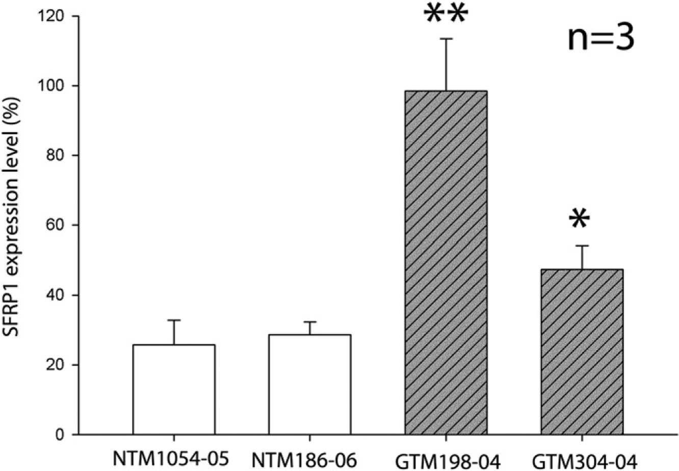
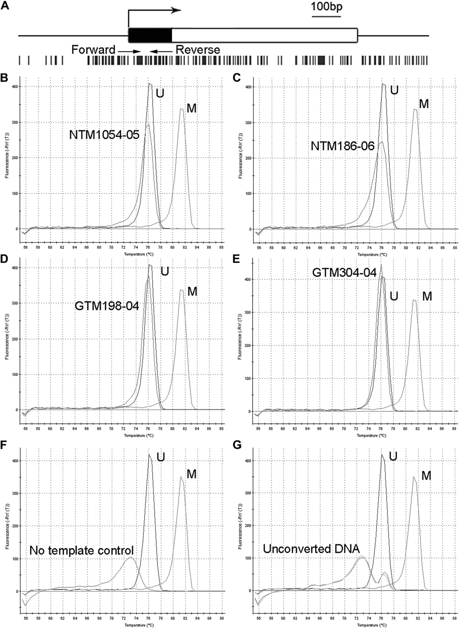
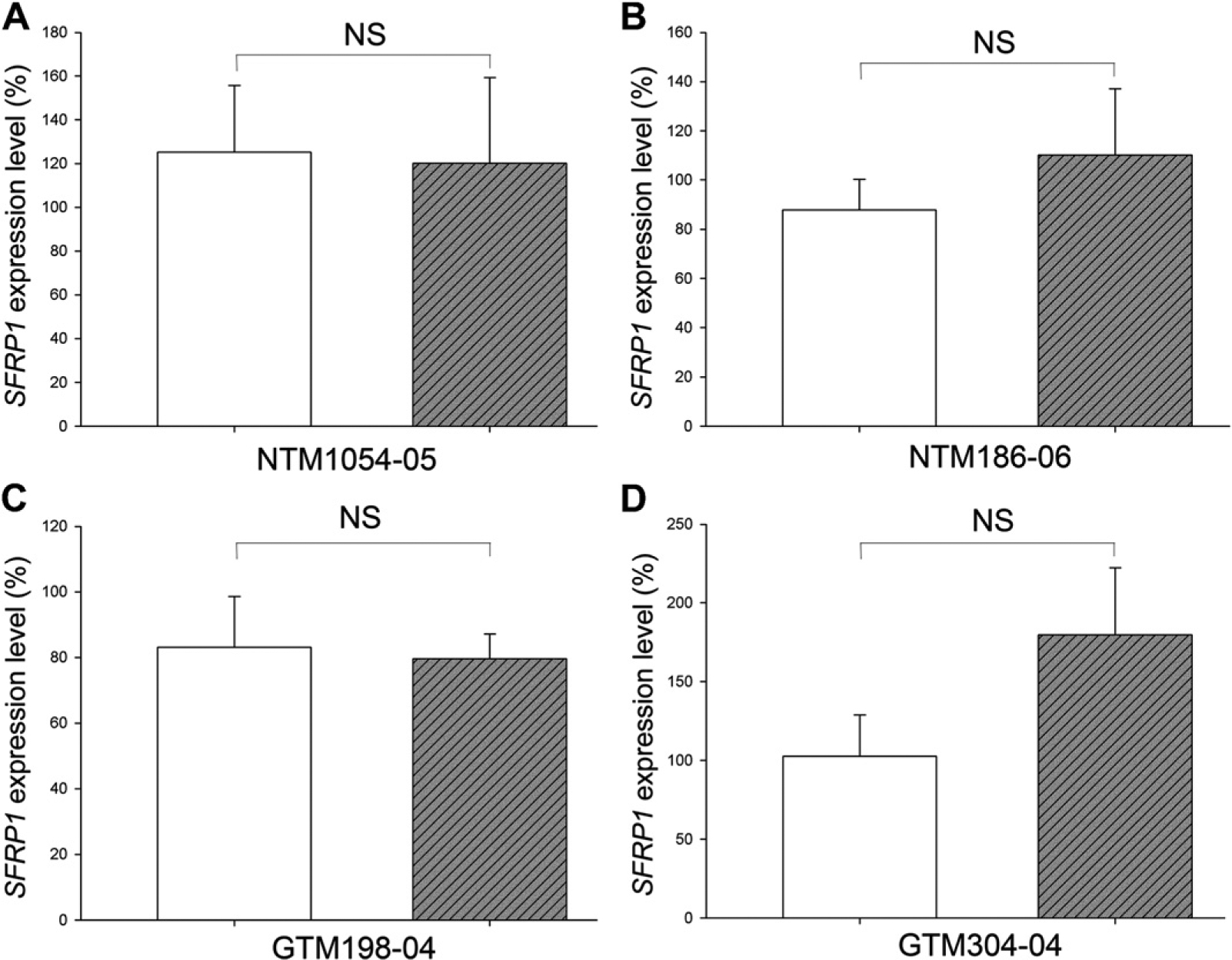
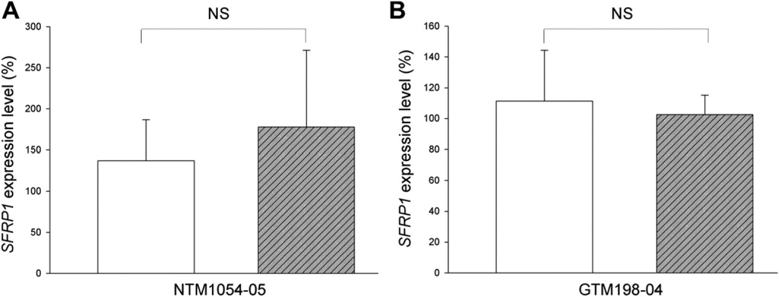
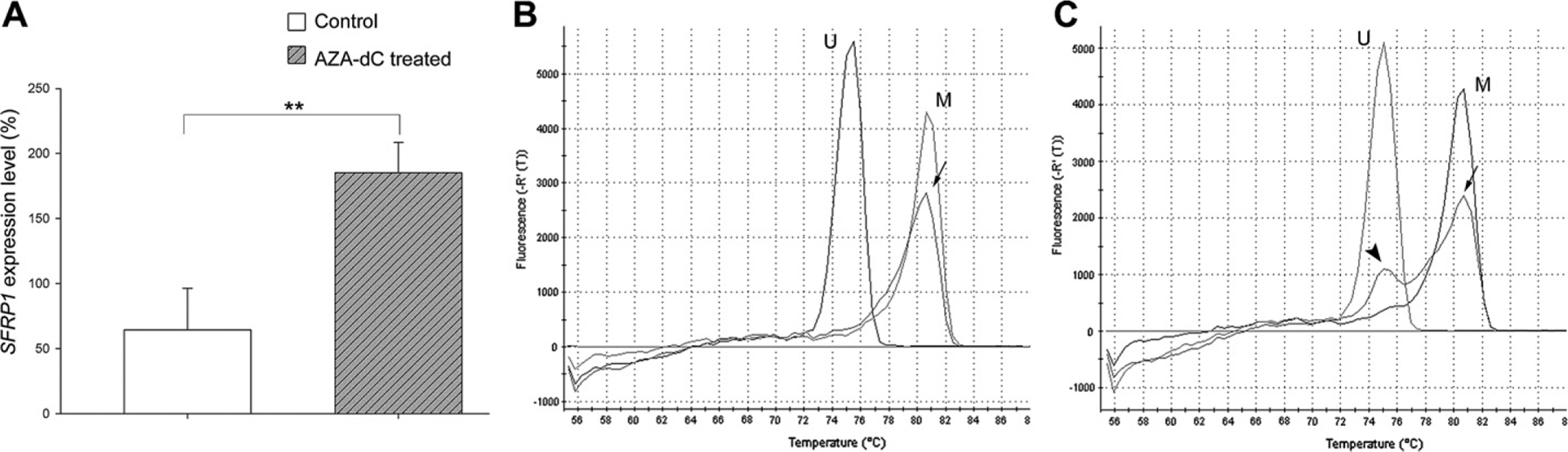
Glaucoma is a leading cause of blindness worldwide. In primary open angle glaucoma (POAG) patients, impaired trabecular meshwork (TM) function results in elevated intraocular pressure (IOP), which is the primary risk factor of developing optic neuropathy. Our previous studies showed that Wnt signaling pathway components are expressed in the human TM (HTM), and the Wnt inhibitor, secreted frizzled-related protein 1 (SFRP1) is elevated in the glaucomatous TM (GTM). Elevated SFRP1 increased IOP in mice eyes and in perfusion cultured anterior segments of the human eye. However, the cause of elevated SFRP1 in the GTM remains unknown. Promoter methylation plays a key role in regulating SFRP1 expression in certain cancer cells. In light of this, we studied whether promoter methylation is also involved in SFRP1 differential expression in the TM. Two normal TM (NTM) and two GTM cell strains were cultured for an additional 7 days after they were confluent. RNA and genomic DNA (gDNA) were isolated simultaneously to compare SFRP1 expression levels by quantitative PCR (qPCR) and to determine SFRP1 promoter methylation status by bisulfite conversion and methylation-sensitive high resolution melting analysis (MS-HRM). To study whether DNA methylation inhibitors affect SFRP1 expression in TM cells, the four TM cell strains were treated with or without 2 μM 5-aza-2'-deoxycytidine (AZA-dC) for 4 days. RNA was isolated to compare SFRP1 expression by qPCR. In addition, a human cancer cell line, NCI-H460, was used as a positive control. We found that the two GTM cell strains had significantly higher expression levels of SFRP1 than the two NTM cell strains. However, the SFRP1 promoter of all four TM cell strains was unmethylated. In addition, AZA-dC treatment did not affect SFRP1 expression in any of the TM cell strains (n = 3, p > 0.05). In contrast, the hypermethylated SFRP1 promoter of NCI-H460 cells was partially demethylated by the same treatment. AZA-dC treatment also elevated SFRP1 expression by approximately two fold in NCI-H460 cells (n = 3, p < 0.01). Our data suggest that the differential expression of SFRP1 in HTM cells is not due to differential promoter methylation.


Figures

Fig. 1.
Fig. 1.
Differential expression of SFRP1 in HTM cells. Four different HTM cell strains were cultured in 35 mm dishes for an additional 7 days after they were confluent. The expression level of SFRP1 of each cell strain was studied by qPCR. The expression level of SFRP1 was first normalized to GAPDH, and then to a GTM198–04 sample, whose SFRP1 expression level was set at 100%. All experiments were performed in biological triplicates (n = 3). Bars = means ± standard deviation (SD). *: p < 0.05 in comparison with either NTM cell strain. **: p < 0.01 in comparison with either NTM cell strain.
Fig. 2.
Fig. 2.
The methylation status of the SFRP1 promoter of HTM cells. The CpG islands in the SFRP1 promoter region are shown in (A). Black bar: the 5′ untranslated region. White bar: the coding region. Large arrow: transcriptional start site. Vertical black bars: CpG sites. Small arrows: the primers for MS-HRM. (B–E): MS-HRM analysis of the SFRP1 promoter of HTM cells. gDNA from the same cells as described in Fig. 1 was used for MS-HRM after bisulfite conversion. The first order derivative of fluorescence intensity was plotted against temperature. Each peak represents the Tm of the PCR product. The difference in the Tm between completely unmethylated control DNA (U) and methylated control DNA (M) was approximately 5 °C. The Tm of all four HTM cell strains was almost the same as that of the unmethylated control DNA. Negative control studies included PCR reactions without DNA template (No template control) (F), or with unmethylated or methylated DNA that were not subjected to bisulfite conversion (Unconverted DNA) (G). All experiments were performed in biological triplicates, and representative data are shown.
Fig. 3.
Fig. 3.
AZA-dC treatment did not affect SFRP1 expression in confluent HTM cells. Confluent HTM cells cultured for additional 7 days were subjected to treatment with (hatched bars) or without (open bars) 2 μM AZA-dC for 4 days. The expression levels of SFRP1 were studied by qPCR. The expression level of SFRP1 was first normalized to GAPDH, and then to a control sample, whose SFRP1 expression level was set at 100%. All experiments were performed in biological triplicates (n = 3). Bars = means ± standard deviation (SD). NS: not significant (p > 0.05).
Fig. 4.
Fig. 4.
AZA-dC treatment did not affect SFRP1 expression in non-confluent HTM cells. Eighty percent confluent HTM cells were subjected to treatment with (hatched bars) or without (open bars) 2 μM AZA-dC for 4 days. The expression levels of SFRP1 were studied by qPCR. The expression level of SFRP1 was first normalized to GAPDH, and then to a control sample, whose SFRP1 expression level was set at 100%. All experiments were performed in biological triplicates (n = 3). Bars = means ± SD. NS: not significant (p > 0.05).
Fig. 5.
Fig. 5.
AZA-dC treatment induced SFRP1 expression and demethylation of the SFRP1 promoter in NCI-H460 cells. NCI-H460 cells were treated with or without 2 μM AZA-dC for 4 days. (A) The expression level of SFRP1 of each cell strain was studied by qPCR. The expression level of SFRP1 was first normalized to GAPDH, and then to a control sample, whose SFRP1 expression level was set at 100%. The experiment was performed in biological triplicates (n = 3). Bars = means ± SD. **: p < 0.01. (B–C) MS-HRM studies showed that NCI-H460 cells without AZA-dC treatment had methylated SFRP1 promoters (B, arrow). AZA-dC treatment caused demethylation of the SFRP1 promoter in some cells, which resulted in two peaks on the melting curve (C). The major peak (arrow) represented the methylated promoters while the minor peak (arrowhead) represented the unmethylated promoters. U: unmethylated DNA control. M: methylated DNA control.

Similar articles

Cited by

References

    1. The AGIS Investigators, 2000. The Advanced Glaucoma Intervention Study (AGIS): 7. The relationship between control of intraocular pressure and visual field deterioration. Am. J. Ophthalmol 130, 429–440. - PubMed
    1. Baylin SB, Ohm JE, 2006. Epigenetic gene silencing in cancer – a mechanism for early oncogenic pathway addiction? Nat. Rev. Cancer 6, 107–116. - PubMed
    1. Bovolenta P, Esteve P, Ruiz JM, Cisneros E, Lopez-Rios J, 2008. Beyond Wnt inhibition: new functions of secreted frizzled-related proteins in development and disease. J. Cell. Sci 121, 737–746. - PubMed
    1. Finch PW, He X, Kelley MJ, Uren A, Schaudies RP, Popescu NC, Rudikoff S, Aaronson SA, Varmus HE, Rubin JS, 1997. Purification and molecular cloning of a secreted, frizzled-related antagonist of Wnt action. Proc. Natl. Acad. Sci. U S A 94, 6770–6775. - PMC - PubMed
    1. Fleenor DL, Shepard AR, Hellberg PE, Jacobson N, Pang IH, Clark AF, 2006. TGFbeta2-induced changes in human trabecular meshwork: implications for intraocular pressure. Invest. Ophthalmol. Vis. Sci 47, 226–234. - PubMed

MeSH terms