. 2011 Dec;2(12):1279-90.
doi: 10.18632/oncotarget.411.

Selenium is a modulator of circadian clock that protects mice from the toxicity of a chemotherapeutic drug via upregulation of the core clock protein, BMAL1

Affiliations

Selenium is a modulator of circadian clock that protects mice from the toxicity of a chemotherapeutic drug via upregulation of the core clock protein, BMAL1

Yan Hu et al. Oncotarget. 2011 Dec.

Abstract

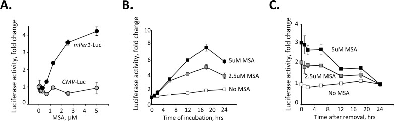
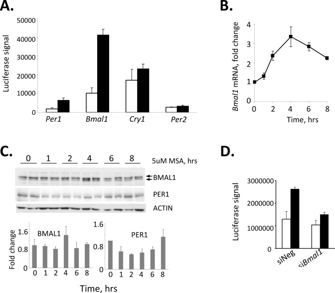
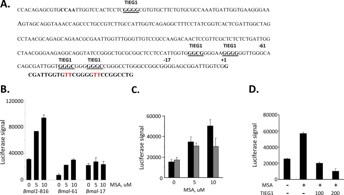
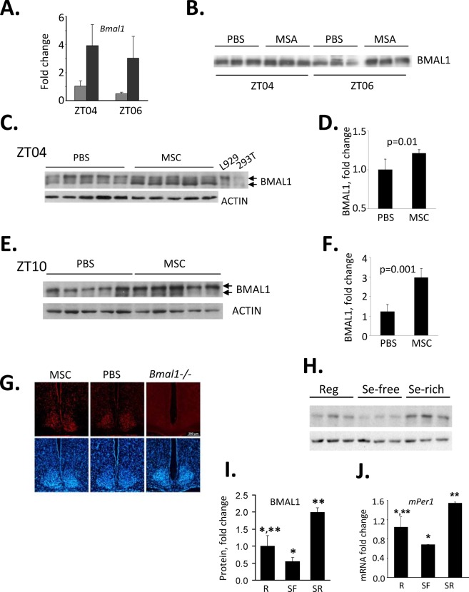
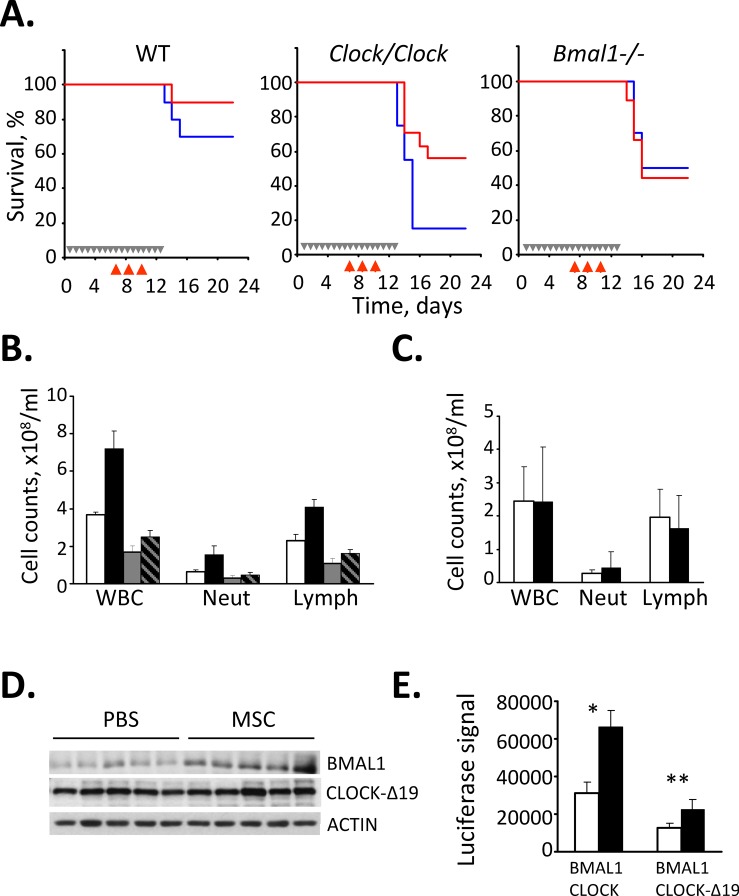
Selenium compounds are known as cancer preventive agents and are also able to ameliorate the toxicity associated with anti-cancer radiation and chemotherapy in mouse models. Sensitivity to the toxicity of chemotherapy is also modulated by the circadian clock, molecular time-keeping system that underlie daily fluctuations in multiple physiological and biochemical processes. Here we show that these two mechanisms are interconnected. By screening a library of small molecules in a cell-based reporter system, we identified L-methyl-selenocysteine as a positive regulator of the core clock protein, BMAL1. L-methyl-selenocysteine up-regulates BMAL1 at the transcriptional level both in cultured cells and in mice. We also show that in tissue culture selenium exerts its action by interfering with TIEG1-mediated repression of Bmal1 promoter. Selenium treatment fails to protect BMAL1-deficient mice from toxicity induced by the chemotherapeutic agent cyclophosphamide but does protect Clock mutant mice deficient in circadian rhythm control but having normal BMAL1. These findings define selenium as circadian modulator and indicate that the tissue protective effect of selenium results, at least in part, from up-regulation of BMAL1 expression and subsequent enhancement of CLOCK/BMAL1-mediated transcription.


Figures

Figure 1
Figure 1. Selenium specifically activates circadian promoter in a time and dose-dependent manner
(A). Selenium activates Per1 promoter in a dose-dependent manner. L929 cells carrying luciferase reporter genes under the control of either mPer1 or CMV promoters were treated with the indicated concentrations of MSA for 18 hrs. Luciferase activity was measured in cell lysates and normalized for β-Gal expression. The experiment was performed in triplicate; the values presented are mean fold-change relative to that in untreated cells ± standard error. (B). Selenium activates Per1 promoter in a time-dependent manner. L929-Per1-Luc cells were treated with different concentrations of MSA (0, 2, 5μM). Cells were collected at various time points over a period of 36 hrs and luciferase activity was measured. (C) L929-Per1-Luc cells were pretreated with different concentrations of MSA (0, 2, 5μM) for 18 hrs. MSA was then washed out and cells were collected at various time points over a period of 24 hr for measurement of luciferase activity. Values are mean from triplicates with error bars indicating standard deviation.
Figure 2
Figure 2. Selenium-dependent up-regulation of the Per1 promoter is mediated through BMAL1
(A) Treatment with selenium results in acute induction of the Bmal1 promoter. 293T cells were transfected with indicated reporters; 24hrs after transfection cells were treated with 5uM MSA for 6 hrs and luciferase activity was measured in cell lysates. Values represent mean from triplicates with error bars indicating standard deviation. (B) Treatment with selenium results in an increase of endogenous Bmal1 mRNA. L929 cells were treated with 5uM of MSA for indicated times. The relative abundance of Bmal1 mRNA was determined by real-time RT-PCR using the comparative delta Ct method. The final measurements were normalized based on Gapdh mRNA expression and are shown as the fold-change. (C) Selenium increases the abundance of BMAL1 protein. L929 cells were treated with 5uM of MSA for indicated times. The abundance of BMAL1 and PER1 in whole cell extracts was analyzed by Western blot (top panel). Two individual samples for each time point are shown. Lower panel presents quantitative analysis of BMAL1 and PER1 expression showing an increase in BMAL1 in 4hrs after MSA treatment followed by an increase in PER1. (D) Suppression of BMAL1 by specific siRNA abrogates selenium-mediated increase in Per1 activation. L929 cells containing the Per1-Luc construct were transfected with siRNA against BMAL1 or control non-targeting (-) siRNA. 72hrs post-transfection, cells were treated with 5μM MSA and luciferase activity was measured in cell lysates. The values represent mean ± standard deviation.
Figure 3
Figure 3. Selenium mediates Bmal1 upregulation through TIEG1 repressor binding sites
(A) Nucleotide sequence of the putative core promoter of the mBmal1 gene. The transcription start site of ORF is designated +1. Potential TIEG1 binding sites are underlined. The Bmal1 promoter fragments used (Bmal1-61 and Bmal1-17) are indicated by bold number above the truncation sites (-17 and -61). (B). Selenium up-regulates Bmal1 promoters that contain the most proximal TIEG1 binding-sites. 293T cells were transfected with one of three Bmal1 promoter fragmented reporter genes (816bp, 61bp, or 17bp upstream from +1bp transcription start site). 18hour post transfection cells were treated with or without indicated concentrations of selenium (MSA) and harvested 9 hours later to be analyzed for luciferase expression. (C). Selenium-mediated up-regulation of Bmal1 gene is compromised in a promoter lacking the two most proximal TIEG1 sites. Bmal1-816 was used as a template to generate a promoter lacking the two proximal TIEG1-sites, which were previously determined to be responsible for Bmal1 gene repression [32]. The primer used in the site-directed mutagenesis is shown below the two TIEG1 proximal sites, and the specific mutated base pairs are shown in red. Cells were transfected with either wild-type Bmal1-816 reporter gene (black bars) or TIEG1 binding site mutant Bmal1-816 (gray bars) and treated without or with MSA at 5 and 10uM.
Figure 4
Figure 4. Selenium up-regulates Bmal1 expression in mouse liver
(A) Selenium administration results in acute induction of Bmal1 transcript in mouse liver. C57BL/6J mice received singe i.p. injection of MSC (10mg/kg) or PBS at ZT03 (zeitgeber time, ZT00 corresponds to the time of lights on). Livers were collected 1 and 3 hrs later (ZT04 and ZT06 respectively), and relative abundance of Bmal1 mRNA was measured by real-time RT-PCR or PBS at ZT03 (zeitgeber time, ZT00 corresponds to the time of lights on). Data present mean values for three mice ± standard deviation. (B) Selenium-induced activation of the Bmal1 promoter results in increase in BMAL1 protein in liver. Western blot analysis of total lysates of liver obtained from the same animals as in (A). (C-F) Selenium increases BMAL1 protein in liver when administered by oral gavage. Western blots (C,E) and their quantitative analysis (D,F). Twenty animals received either PBS or MSC (7.5 mg/kg, oral gavage) daily for two weeks. Tissue samples were collected on day 15 at either ZT04 (C,D) or ZT10 (E,F) and probed with anti-BMAL1 antibody. Each lane represents tissue lysate obtained from an individual animal. Actin was used to control for loading. ImageQuant software was used for Western blots quantitation. Cell lysates from L929 and 293T cells were used as positive controls for endogenous and ectopically expressed BMAL1 respectively. (G) Selenium has no effect on BMAL1 expression in the SCN. Animals were fed daily with either PBS or MSC (7.5 mg/kg, oral gavage) for two weeks. Animals were then sacrificed for preparation of SCN tissue sections. The 12 μm sections were stained with anti-BMAL1 antibody followed by staining with a fluorescently-labeled secondary antibody (red). DAPI (blue) was used to stain the nuclei of all cells. SCN sections obtained from untreated Bmal1−/− mice are shown as controls for specificity of the antibody. Consistent with previous reports [52], no daily variations in BMAL1 immuno-reactivity were detected. Representative sections of the brains collected at ZT02 are shown. (H) The abundance of BMAL1 protein in mouse livers correlates with the amount of selenium in their diets. Animals were fed regular (Reg), selenium-free (SF) or selenium-enriched (SR) diets for 6 weeks. Tissue samples were collected at ZT10 and used for Western blotting with anti-BMAL1 antibody. Each lane represents tissue lysate obtained from an individual animal. (I) Quantitation of the Western blot (G) using ImageQuant software. Expression of BMAL1 was normalized based on ACTIN levels. Values represent mean ± standard error, n=3/group. *p=0.06; **p=0.007 (Student’s t-test). (J) Selenium-induced changes in the level of BMAL1 correlate with increased level of its direct transcriptional target Per1 in the liver. Same animals as in G were used. Relative mRNA abundance was determined by real-time RT-PCR using the comparative delta Ct method. The final measurements were normalized by Gapdh mRNA expression. Values represent mean ± standard deviation, n=3/group. *p=0.04; **p=0.01 (Student’s t-test).
Figure 5
Figure 5. Selenium rescues cyclophosphamide-sensitive Clock mutant mice from drug-induced toxicity
(A) Kaplan-Meyer survival curves. Wild type (WT), Clock mutant and Bmal1−/− mice were given either PBS (blue) or MSC (red, 7.5 mg/kg) daily for 2 weeks and injected with CY, 3x150mg/kg. Grey arrows in the Kaplan-Meyer plot indicate days of PBS or MSC administration; red arrows indicate days of CY injection. Treatment with MSC dramatically increased the 25-day survival rate of CY-treated Clock/Clock mice but had no effect on survival of Bmal1 knockout animals. The experiment was performed three times with similar results. (B) Selenium treatment decreases CY-induced neutropenia and leukocytopenia. Total white blood cells (WBC), neutrophils (Neut) and lymphocyte (Lymph) counts in peripheral blood of wild type and Clock mutant mice treated with PBS or MSC and CY as described for Fig. 5A. MSC-induced increase in survival of CY-treated Clock mutant mice correlates with significant increase in the number of circulating lymphocytes (Student’s t-test p=0.015, n=5/group). (C) Selenium has no effect on the number of circulating lymphocytes in CY-treated Bmal1−/− mice. Mice were treated with PBS or MSC and CY as described in Fig. 5A. The cell count data are mean values for 8 mice. Please note that absolute values cannot be directly compared between the Clock/Clock (Fig. 4A) and Bmal1−/− mice since the experiments were not performed simultaneously. (D) Selenium administration increases BMAL1 protein abundance in the livers of Clock/Clock mice. Western blot analysis of liver extracts of five individual Clock/Clock mice fed with either PBS or MSC with anti-BMAL1 and anti-CLOCK antibodies. No effect on CLOCK-Δ19 expression level was detected. (E) Selenium increases CLOCK-Δ19/BMAL1-mediated activation of a responsive promoter. HEK293T cells were transfected with Per1-Luciferase reporter, HA-BMAL1 and either full-length wild type CLOCK or deletion mutant CLOCK (CLOCK-Δ19) and treated with indicated concentrations of MSA. Luciferase activity of cell lysates was normalized with β-Gal. Bars represent mean values ± standard error. Asterisk indicate statistically significant increase in promoter activation; *p=0.0011, **p=0.0012 (Student’s t-test).

Similar articles

Cited by

References

    1. Rayman MP. The importance of selenium to human health. Lancet. 2000;356:233–241. - PubMed
    1. Rayman MP. Selenium in cancer prevention: a review of the evidence and mechanism of action. Proc Nutr Soc. 2005;64:527–542. - PubMed
    1. Zeng H, Combs GF., Jr Selenium as an anticancer nutrient: roles in cell proliferation and tumor cell invasion. J Nutr Biochem. 2008;19:1–7. - PubMed
    1. Bhattacharya A, Toth K, Durrani FA, Cao S, Slocum HK, Chintala S, Rustum YM. Hypoxia-specific drug tirapazamine does not abrogate hypoxic tumor cells in combination therapy with irinotecan and methylselenocysteine in well-differentiated human head and neck squamous cell carcinoma a253 xenografts. Neoplasia. 2008;10:857–865. - PMC - PubMed
    1. Cao S, Durrani FA, Rustum YM. Selective modulation of the therapeutic efficacy of anticancer drugs by selenium containing compounds against human tumor xenografts. Clin Cancer Res. 2004;10:2561–2569. - PubMed

Publication types

MeSH terms

LinkOut - more resources