. 2012 Mar 2;287(10):7821-33.
doi: 10.1074/jbc.M111.327254. Epub 2012 Jan 16.

The Drosophila juvenile hormone receptor candidates methoprene-tolerant (MET) and germ cell-expressed (GCE) utilize a conserved LIXXL motif to bind the FTZ-F1 nuclear receptor

Affiliations

The Drosophila juvenile hormone receptor candidates methoprene-tolerant (MET) and germ cell-expressed (GCE) utilize a conserved LIXXL motif to bind the FTZ-F1 nuclear receptor

Travis J Bernardo et al. J Biol Chem. .

Abstract

Juvenile hormone (JH) has been implicated in many developmental processes in holometabolous insects, but its mechanism of signaling remains controversial. We previously found that in Drosophila Schneider 2 cells, the nuclear receptor FTZ-F1 is required for activation of the E75A gene by JH. Here, we utilized insect two-hybrid assays to show that FTZ-F1 interacts with two JH receptor candidates, the bHLH-PAS paralogs MET and GCE, in a JH-dependent manner. These interactions are severely reduced when helix 12 of the FTZ-F1 activation function 2 (AF2) is removed, implicating AF2 as an interacting site. Through homology modeling, we found that MET and GCE possess a C-terminal α-helix featuring a conserved motif LIXXL that represents a novel nuclear receptor (NR) box. Docking simulations supported by two-hybrid experiments revealed that FTZ-F1·MET and FTZ-F1·GCE heterodimer formation involves a typical NR box-AF2 interaction but does not require the canonical charge clamp residues of FTZ-F1 and relies primarily on hydrophobic contacts, including a unique interaction with helix 4. Moreover, we identified paralog-specific features, including a secondary interaction site found only in MET. Our findings suggest that a novel NR box enables MET and GCE to interact JH-dependently with the AF2 of FTZ-F1.


Figures

FIGURE 1.
FIGURE 1.
Isoform-specific expression of ftz-f1 in the S2 cell line. A, structural organization of the ftz-f1 gene. Black bars, exons; arrows, promoters for the α and β isoforms. Each isoform possesses a unique coding sequence at the 5′ end. Rectangles above the exons indicate probes specific for the α (white) and β (gray) isoforms as well as a common probe (striped). Alternative polyadenylation sites are indicated by vertical arrows. Shown at the top is the size of ftz-f1 in kilobases. B, total RNA was isolated from S2 cells cultured in the presence of 1 × 10⁻⁶ m ecdysone for the time period indicated above each lane and from prepupae (PP). RNA samples were analyzed by Northern blot hybridization with radioactive probes for α-specific, β-specific, or common exons. Transcript sizes are indicated on the right. rp49 expression was used as a loading control.
FIGURE 2.
FIGURE 2.
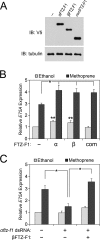
Both FTZ-F1 isoforms mediate JH-dependent activation of E75A. A, expression of ectopic FTZ-F1 proteins used in B and C was confirmed by Western blot hybridization (IB) using anti-V5 antibody, with tubulin as a control for loading. B, S2 cells were transfected with empty plasmid or expression plasmid encoding αFTZ-F1, βFTZ-F1, or common domains of FTZ-F1 as indicated (x axis) and then treated with ethanol solvent (light bars) or 1 × 10⁻⁶ m methoprene (dark bars) for 1 h. Total RNA was extracted, and E75A expression was measured as -fold abundance against rp49 by quantitative RT-PCR (y axis). Bars, mean ± S.D. from three independent experiments. *, significant (p < 0.05) JH-dependent increase in E75A expression; **, significant JH-independent E75A activation. C, S2 cells were incubated for 48 h with double-stranded RNA targeting the 5′ region of the αftz-f1 transcript (αftz-f1 dsRNA), transfected with empty plasmid or βftz-f1-encoding plasmid as indicated on the x axis, and treated with ethanol (light bars) or 1 × 10⁻⁶ m methoprene (dark bars) for 1 h. Expression of E75A relative to rp49 was measured as in B. Bars, mean ± S.D. from three independent experiments. *, significant (p < 0.05) change in E75A expression.
FIGURE 3.
FIGURE 3.
JH-dependent interaction of MET and GCE with FTZ-F1 requires helix 12. A and B, S2 cells were transfected with 4× UAS-TATA-Luc reporter construct, along with expression vectors for GAL4 and p65 fused to FTZ-F1, MET, or GCE as indicated (y axis). After transfection, cells were treated with ethanol (light gray) or 5.0 × 10⁻⁶ m methoprene (dark gray) for 24 h. Luciferase activity was normalized to β-galactosidase activity from a constitutive reporter (x axis). Interactions of MET (A) and GCE (B) were measured with αFTZ-F1, βFTZ-F1, and C-terminally truncated βFTZ-F1ΔH12. Data are shown as the mean ± S.D. from at least three independent experiments. *, significant (p < 0.05) difference in JH-dependent reporter activity between βFTZ-F1 and βFTZ-F1ΔH12. C, equal expression of fusion proteins was confirmed by Western blot (IB) using anti-FLAG or anti-HA antibody as indicated, with anti-tubulin antibodies used as a loading control. *, a nonspecific band.
FIGURE 4.
FIGURE 4.
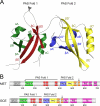
Structure of MET and GCE. A, three-dimensional structure of the two PAS folds from MET comprising residues 134–506, as predicted by Phyre2. α-Helices and β-strands are colored according to the annotated motifs PAS A (green), PAS B (blue), and PAC (yellow) and sequence blocks I–III (red). Each PAS fold contains five β-strands (βA--βE and βA′–βE′) and three or four α-helices (αA–αC and αA′–αC′). The fourth α-helix present only in PAS Fold 2 is denoted by an asterisk. A similar model was observed for GCE (not shown). B, schematic illustration of MET and GCE protein structures. Shown are the bHLH domain; the PAS A, PAS B, and PAC motifs; sequence blocks I–V; and the acidic (Ac), proline/serine (P/S), and glutamine (QR) regions described in this paper. An arrowhead indicates the location of an LXXLL sequence in MET; the vertical arrows indicate the novel LIXXL NR boxes in MET and GCE identified in this report. Numbers at the bottom of each schematic designate residue position.
FIGURE 5.
FIGURE 5.
Critical MET, GCE residues required for interaction with FTZ-F1. A, alignment of sequences from αIV, αV, and the QR of MET and GCE homologs from D. melanogaster, D. grimshawi, A. aegypti, T. castaneum, and N. vitripennis as indicated. Start and end position are shown for each sequence. Identical residues are highlighted in black; similar residues are highlighted in gray. Residues targeted for point mutation, including a conserved leucine (Leu-554 in MET, Leu-418 in GCE) in the putative NR box and conserved glutamine/glutamatic acid residues in the QR are indicated with an asterisk. B and C, S2 cells were transfected with 4× UAS-TATA-Luc reporter construct, along with GAL4 and p65 fusion proteins as indicated (y axis). After transfection, cells were treated with ethanol (light gray) or 5.0 × 10⁻⁶ m methoprene (dark gray) for 24 h. Luciferase activity was normalized to constitutive β-galactosidase activity (x axis). Interaction with βFTZ-F1 was measured for MET (B) and GCE (C) using the wild type or mutant proteins indicated. Data are shown as the mean ± S.D. and are the result of at least three independent experiments. *, significant difference (p < 0.05). Western blots (IB) at the bottom of each panel show equivalent expression of wild type and mutant proteins.
FIGURE 6.
FIGURE 6.
Complex formation between the MET, GCE NR box, and the FTZ-F1 LBD. Shown are lowest energy structures from refined docking of MET and GCE NR boxes to the FTZ-F1 LBD (PDB entry 2XHS). AF2 helices from FTZ-F1 (H3, H3′, H4, and H12) are shown in green. NR box peptides for MET (LYLIENLQK, residues 552–560) and GCE (LRLIQNLQK, residues 416–424) are shown in orange and red, respectively. In the bottom illustration of each panel, the model is rotated by 60º. A, superposition of MET and GCE peptide backbones with the empirically derived FTZ NR box (STLRALLTN, residues 107–115), shown in blue (20). B and C, molecular details of the FTZ-F1·MET (B) and FTZ-F1·GCE (C) interactions. The core LIXXL residues of MET and GCE and their interacting residues in FTZ-F1 are shown. Side chains are colored according to atom type: oxygen (bright red), nitrogen (blue), sulfur (yellow), and carbon (gray for FTZ-F1, orange for MET, red for GCE).
FIGURE 7.
FIGURE 7.
Hydrophobic residues in the FTZ-F1 AF2 are essential to interaction with MET and GCE. S2 cells were transfected with 4× UAS-TATA-Luc reporter and the indicated GAL4 and p65 fusion constructs (y axis). After transfection, cells were treated with ethanol (light gray) or 5.0 × 10⁻⁶ m methoprene (dark gray) for 24 h. Luciferase activity was normalized to constitutive β-galactosidase activity (x axis). Interaction of MET (A) and GCE (B) was measured with wild type and mutant βFTZ-F1 proteins bearing amino acid substitutions (V621D, R625A, V635D, M639D, L642D, L792, M796D, or E795A) or a truncation (ΔH12) in the LBD. Data are shown as mean ± S.D. from three independent experiments. *, significant (p < 0.05) difference in JH-dependent interaction compared with wild type βFTZ-F1. Western blots below each panel show equivalent expression of wild type and mutant proteins.

Similar articles

Cited by

References

    1. Dubrovsky E. B. (2005) Hormonal cross-talk in insect development. Trends Endocrinol. Metab. 16, 6–11 - PubMed
    1. Tan A., Tanaka H., Tamura T., Shiotsuki T. (2005) Precocious metamorphosis in transgenic silkworms overexpressing juvenile hormone esterase. Proc. Natl. Acad. Sci. U.S.A. 102, 11751–11756 - PMC - PubMed
    1. Konopova B., Jindra M. (2007) Juvenile hormone resistance gene methoprene-tolerant controls entry into metamorphosis in the beetle Tribolium castaneum. Proc. Natl. Acad. Sci. U.S.A. 104, 10488–10493 - PMC - PubMed
    1. Parthasarathy R., Tan A., Palli S. R. (2008) bHLH-PAS family transcription factor methoprene-tolerant plays a key role in JH action in preventing the premature development of adult structures during larval-pupal metamorphosis. Mech. Dev. 125, 601–616 - PMC - PubMed
    1. Charles J. P., Iwema T., Epa V. C., Takaki K., Rynes J., Jindra M. (2011) Ligand-binding properties of a juvenile hormone receptor, methoprene-tolerant. Proc. Natl. Acad. Sci. U.S.A. 108, 21128–21133 - PMC - PubMed

Publication types

MeSH terms

Substances

LinkOut - more resources