Review
. 2012 Mar;90(3):260-70.
doi: 10.1038/icb.2011.112. Epub 2012 Jan 17.

The interplay between endoplasmic reticulum stress and inflammation

Affiliations
Review

The interplay between endoplasmic reticulum stress and inflammation

Sumaira Z Hasnain et al. Immunol Cell Biol. 2012 Mar.

Abstract

Endoplasmic reticulum (ER) stress may be both a trigger and consequence of chronic inflammation. Chronic inflammation is often associated with diseases that arise because of primary misfolding mutations and ER stress. Similarly, ER stress and activation of the unfolded protein response (UPR) is a feature of many chronic inflammatory and autoimmune diseases. In this review, we describe how protein misfolding and the UPR trigger inflammation, how environmental ER stressors affect antigen presenting cells and immune effector cells, and present evidence that inflammatory factors exacerbate protein misfolding and ER stress. Examples from both animal models of disease and human diseases are used to illustrate the complex interactions between ER stress and inflammation, and opportunities for therapeutic targeting are discussed. Finally, recommendations are made for future research with respect to the interaction of ER stress and inflammation.


Figures

Figure 1
Figure 1
ER stress, UPR signalling and NF‐κB activation. The three branches of the UPR, activating transcription factor 6 (ATF6), protein kinase RNA‐like ER kinase (PERK) and inositol requiring enzyme 1 (IRE1), are activated when the chaperone GRP78 that is usually bound to these factors is recruited to misfolded proteins accumulating in the ER. ATF6 activation requires its migration to the trans Golgi network (TGN) where it's cleaved by site 1/2 proteases (S1P/S2P) leading to interaction with AKT and NF‐κB activation. Autophosphorylation of PERK results in the phosphorylation of eIF2α, which can inhibit protein translation and lead to decreased IκBα production and thereby, induce NF‐κB transcription. Phosphorylated IRE1 binds the adaptor protein TNF‐receptor activating factor 2 (TRAF2), which can activate the JNK/AKT pathway, and phosphorylate NF‐κB protein IκB kinase (IKK) leading to cleavage of Ikβα and activation of NF‐κB. UPR‐independent Ca2+ and ROS release and GRP78 that leaks into the cytosol have also been proposed to activate NF‐κB to induce inflammation. In contrast, during mild ER stress IRE1‐ and PERK‐dependent production of Ccaat‐enhancer binding proteins (C‐EBP) and A20 can inhibit activation of NF‐κB in response to inflammatory stimuli, including microbial proteins and inflammatory cytokines. A full colour version of this figure is available at the Immunology and Cell Biology journal online.
Figure 2
Figure 2
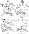
ER stress and its possible effects on antigen presentation. (a) Intracellular self and pathogen proteins are degraded by the proteasome to form peptides that are transported to the ER and coupled to the major histocompatibility complex (MHC) Class I molecules, before being transported to the cell surface where they are recognised by immune cells, for example, dendritic cells (DC). Professional APCs can also present antigens on MHC Class II. (b) During ER‐associated protein degradation (ERAD), misfolded proteins accumulated within the ER are translocated to the proteasome for degradation into peptides. Misfolding can thereby result in greater presentation on MHC Class I at the cell surface resulting in increased likelihood of activation of autoreactive T cells. (c) Misfolded proteins unable to be cleared by ERAD activate PERK and IRE1 and their downstream factors eIF2α and JNK, respectively, which can activate autophagy; a process by which misfolded protein aggregates from the ER are engulfed and degraded by lysosomal proteins. Autophagy in APCs can result in reduced protein processing by the proteasome from the ER, hence reduced peptide loading and reduced MHC Class I presentation at the cell surface, although there is some evidence for derivation of Class I peptides from autophagolysosomes. Autophagy could also result in increased presentation of self‐proteins on MHC Class II. (d) During the UPR, GRP78 dissociates from PERK, allowing the phosphorylation of eIF2α, which can inhibit protein translation, potentially resulting in reduced synthesis of MHC molecules and an overall reduction in protein degradation and peptide loading onto MHC, and therefore reduced antigen presentation. A full colour version of this figure is available at the Immunology and Cell Biology journal online.
Figure 3
Figure 3
Demonstration of the importance of inflammation on the phenotype of misfolding disease. Production of goblet cell mucins stained with Alcian blue and PAS in mice with a Muc2 mucin misfolding mutation (Winnie) and in Winnie mice treated with the anti‐inflammatory drug 6‐thioguanine (6‐TG). Goblet cells in Winnie mice have small blue‐staining thecae (a small reservoir of granules of Muc2 for secretion) and large accumulations of pink‐staining misfolded protein. After treatment with 6‐TG, mucin production is restored (large blue thecae of stored mucin granules), although a small amount of misfolded protein is still seen. A full colour version of this figure is available at the Immunology and Cell Biology journal online.
Figure 4
Figure 4
Schematic illustration of the interactions between protein misfolding, the UPR and inflammation. Protein misfolding can be caused by a primary defect, such as a genetic mutation, or can occur as a secondary event as a consequence of microbial ER stressors (for example, bacterial toxins), host ER stressors (for example, hypoxia and increased ATP levels) and/or of inflammation (for example, ROS, cytokines). Misfolding triggers the UPR and NF‐κB activation (Figure 1) activating a network of signalling and transcriptional events that result in increased leukocyte recruitment and T‐cell activation leading to increased inflammation. Inflammation once it develops exacerbates ER stress potentially leading to unresolved cycles of inflammation in susceptible individuals. A full colour version of this figure is available at the Immunology and Cell Biology journal online.

References

    1. Hotamisligil GS. Endoplasmic reticulum stress and the inflammatory basis of metabolic disease. Cell 2010; 140: 900–917. - PMC - PubMed
    1. Matus S, Glimcher LH, Hetz C. Protein folding stress in neurodegenerative diseases: a glimpse into the ER. Curr Opin Cell Biol 2011; 23: 239–252. - PubMed
    1. Mhaille AN, McQuaid S, Windebank A, Cunnea P, McMahon J, Samali A et al Increased expression of endoplasmic reticulum stress‐related signaling pathway molecules in multiple sclerosis lesions. J Neuropathol Exp Neurol 2008; 67: 200–211. - PubMed
    1. Colbert RA, DeLay ML, Klenk EI, Layh‐Schmitt G. From HLA‐B27 to spondyloarthritis: a journey through the ER. Immunol Rev 2010; 233: 181–202. - PMC - PubMed
    1. Hybiske K, Fu Z, Schwarzer C, Tseng J, Do J, Huang N et al Effects of cystic fibrosis transmembrane conductance regulator and DeltaF508CFTR on inflammatory response, ER stress, and Ca2+ of airway epithelia. Am J Physiol Lung Cell Mol Physiol 2007; 293: L1250–L1260. - PubMed

MeSH terms

Substances