

Mitotic centromere-associated kinesin (MCAK): a potential cancer drug target
- PMID: 22249213
- PMCID: PMC3282097
- DOI: 10.18632/oncotarget.416
Mitotic centromere-associated kinesin (MCAK): a potential cancer drug target
Abstract
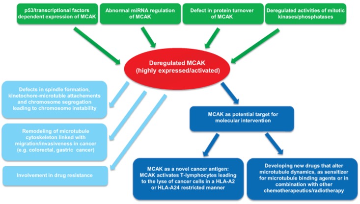
The inability to faithfully segregate chromosomes in mitosis results in chromosome instability, a hallmark of solid tumors. Disruption of microtubule dynamics contributes highly to mitotic chromosome instability. The kinesin-13 family is critical in the regulation of microtubule dynamics and the best characterized member of the family, the mitotic centromere-associated kinesin (MCAK), has recently been attracting enormous attention. MCAK regulates microtubule dynamics as a potent depolymerizer of microtubules by removing tubulin subunits from the polymer end. This depolymerizing activity plays pivotal roles in spindle formation, in correcting erroneous attachments of microtubule-kinetochore and in chromosome movement. Thus, the accurate regulation of MCAK is important for ensuring the faithful segregation of chromosomes in mitosis and for safeguarding chromosome stability. In this review we summarize recent data concerning the regulation of MCAK by mitotic kinases, Aurora A/B, Polo-like kinase 1 and cyclin-dependent kinase 1. We propose a molecular model of the regulation of MCAK by these mitotic kinases and relevant phosphatases throughout mitosis. An ever-increasing quantity of data indicates that MCAK is aberrantly regulated in cancer cells. This deregulation is linked to increased malignance, invasiveness, metastasis and drug resistance, most probably due to increased chromosomal instability and remodeling of the microtubule cytoskeleton in cancer cells. Most interestingly, recent observations suggest that MCAK could be a novel molecular target for cancer therapy, as a new cancer antigen or as a mitotic regulator. This collection of new data indicates that MCAK could be a new star in the cancer research sky due to its critical roles in the control of genome stability and the cytoskeleton. Further investigations are required to dissect the fine details of the regulation of MCAK throughout mitosis and its involvements in oncogenesis.
Conflict of interest statement
The authors declare no conflict of interest.
Figures

Similar articles
-
Molecular insight into the regulation and function of MCAK.Crit Rev Biochem Mol Biol. 2015 Jul-Aug;51(4):228-45. doi: 10.1080/10409238.2016.1178705. Epub 2016 May 5. Crit Rev Biochem Mol Biol. 2015. PMID: 27146484 Review.
-
PLK1 phosphorylates mitotic centromere-associated kinesin and promotes its depolymerase activity.J Biol Chem. 2011 Jan 28;286(4):3033-46. doi: 10.1074/jbc.M110.165340. Epub 2010 Nov 15. J Biol Chem. 2011. PMID: 21078677 Free PMC article.
-
Spatiotemporal dynamics of Aurora B-PLK1-MCAK signaling axis orchestrates kinetochore bi-orientation and faithful chromosome segregation.Sci Rep. 2015 Jul 24;5:12204. doi: 10.1038/srep12204. Sci Rep. 2015. PMID: 26206521 Free PMC article.
-
Aurora B phosphorylates centromeric MCAK and regulates its localization and microtubule depolymerization activity.Curr Biol. 2004 Feb 17;14(4):273-86. doi: 10.1016/j.cub.2004.01.055. Curr Biol. 2004. PMID: 14972678
-
Mitosis: MCAK under the aura of Aurora B.Curr Biol. 2004 May 4;14(9):R346-8. doi: 10.1016/j.cub.2004.04.022. Curr Biol. 2004. PMID: 15120087 Review.
Cited by
-
Functional analysis of phosphorylation of the mitotic centromere-associated kinesin by Aurora B kinase in human tumor cells.Cell Cycle. 2015;14(23):3755-67. doi: 10.1080/15384101.2015.1068481. Epub 2015 Jul 6. Cell Cycle. 2015. PMID: 26148251 Free PMC article.
-
Regulating PLK1 dynamics by Cullin3/KLHL22-mediated ubiquitylation.Cell Cycle. 2013 Aug 15;12(16):2528-9. doi: 10.4161/cc.25839. Epub 2013 Jul 29. Cell Cycle. 2013. PMID: 23907128 Free PMC article. No abstract available.
-
Bioinformatics analysis of SRSF1-controlled gene networks in colorectal cancer.Oncol Lett. 2017 Nov;14(5):5393-5399. doi: 10.3892/ol.2017.6900. Epub 2017 Sep 6. Oncol Lett. 2017. PMID: 29113173 Free PMC article.
-
Spatial regulation of MCAK promotes cell polarization and focal adhesion turnover to drive robust cell migration.Mol Biol Cell. 2021 Apr 1;32(7):590-604. doi: 10.1091/mbc.E20-05-0301. Epub 2021 Feb 10. Mol Biol Cell. 2021. PMID: 33566676 Free PMC article.
-
Oncogenic role of kinesin proteins and targeting kinesin therapy.Cancer Sci. 2013 Jun;104(6):651-6. doi: 10.1111/cas.12138. Epub 2013 Apr 4. Cancer Sci. 2013. PMID: 23438337 Free PMC article. Review.
References
Publication types
MeSH terms
Substances
Grants and funding
LinkOut - more resources
Full Text Sources
Other Literature Sources
Research Materials
