. 2012 Jan 23;196(2):247-59.
doi: 10.1083/jcb.201109113. Epub 2012 Jan 16.

A molecular switch on an arrestin-like protein relays glucose signaling to transporter endocytosis

Affiliations

A molecular switch on an arrestin-like protein relays glucose signaling to transporter endocytosis

Michel Becuwe et al. J Cell Biol. .

Abstract

Endocytosis regulates the plasma membrane protein landscape in response to environmental cues. In yeast, the endocytosis of transporters depends on their ubiquitylation by the Nedd4-like ubiquitin ligase Rsp5, but how extracellular signals trigger this ubiquitylation is unknown. Various carbon source transporters are known to be ubiquitylated and endocytosed when glucose-starved cells are exposed to glucose. We show that this required the conserved arrestin-related protein Rod1/Art4, which was activated in response to glucose addition. Indeed, Rod1 was a direct target of the glucose signaling pathway composed of the AMPK homologue Snf1 and the PP1 phosphatase Glc7/Reg1. Glucose promoted Rod1 dephosphorylation and its subsequent release from a phospho-dependent interaction with 14-3-3 proteins. Consequently, this allowed Rod1 ubiquitylation by Rsp5, which was a prerequisite for transporter endocytosis. This paper therefore demonstrates that the arrestin-related protein Rod1 relays glucose signaling to transporter endocytosis and provides the first molecular insights into the nutrient-induced activation of an arrestin-related protein through a switch in post-translational modifications.


Figures

Figure 1.
Figure 1.
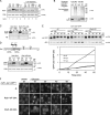
The arrestin-related protein Rod1 is involved in the glucose-induced endocytosis of the lactate transporter, Jen1. (A) WT and rod1Δ cells were grown in lactate medium, and 14C-labeled lactate transport activity was followed over time after glucose addition. A representative experiment of more than three repetitions is shown. The values represent the mean ± SEM of triplicate measurements. (B) Cells in which Jen1 is tagged with GFP at the chromosomal locus were grown in lactate medium, and Jen1-GFP subcellular localization was followed after glucose addition for the indicated times in WT and rod1Δ strains. Bar, 5 µm. (C) Total protein extracts from a similar experiment as in B were prepared at the indicated times after glucose addition and immunoblotted with the indicated antibodies. (D, top) A plasmid-encoded, galactose-inducible Jen1-GFP-His6 construct was expressed in end3Δ (control) and end3Δ rod1Δ cells. Total protein extracts were prepared at several time points after glucose addition and immunoblotted with anti-GFP or anti-PGK (loading control) antibodies. Arrowheads indicate ubiquitylated Jen1 species. (Bottom) Jen1-GFP-His6 was purified under denaturing conditions by Ni-based chromatography 10 min after glucose addition. The eluates were loaded twice and incubated with anti-GFP or anti-ubiquitin (P4D1) antibodies. (E) A rod1Δ strain expressing Jen1-GFP tagged at the chromosomal locus was transformed with an empty plasmid (Ø) or plasmid-encoded Rod1-3HA or Rod1-PYm-3HA, and the cells were imaged after 4 h of growth on lactate medium, and 30 min after the addition of glucose. Bar, 5 µm.
Figure 2.
Figure 2.
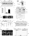
Dynamic modifications of Rod1 by phosphorylation and ubiquitylation are regulated by glucose availability. (A) A WT strain expressing a plasmid-encoded Rod1-3HA was grown overnight in glucose medium (exponential phase), then 4 h in lactate medium, before glucose was added for the indicated times. Total protein extracts were either treated or untreated with calf intestinal phosphatase (CIP) and immunoblotted with anti-HA and anti-PGK antibodies. (B) A WT strain expressing plasmid-encoded Rod1-3HA and 6xHis-tagged ubiquitin was grown for 4 h in lactate medium before glucose was added for 10 min. 6xHis-tagged ubiquitin conjugates were purified under denaturing conditions by Ni-based chromatography. The lysates (Lys.) and eluates (El.) were immunoblotted with an anti-ubiquitin antibody, and with an anti-HA antibody to detect Rod1. (C) Strains expressing a plasmid-encoded Rod1-3HA or a variant mutated on both PPxY motifs (Rod1-PYm-3HA) were grown as in B. Total protein extracts were immunoblotted with an anti-HA antibody. A nonspecific, cross-reactive band was used as internal loading control. P/Ub refers to the molecular weight of phosphorylated or ubiquitylated Rod1, respectively. (D, top) Representation of Rod1 primary sequence. The numbers below show the amino acid boundaries of the predicted arrestin-related domains (“Arrestin_N”/PF00339 and “Arrestin_C”/PF02752) as determined by Pfam. The position of the Rod1 lysines that were mutated to raise the Rod1-KR-3HA construct (below) is displayed above the scheme. (Bottom) A strain expressing a plasmid-encoded Rod1-3HA or a mutant form in which the four lysines depicted in the scheme above are mutated to arginine (Rod1-KR-3HA) were grown as in A. Total protein extracts were immunoblotted with anti-HA and anti-PGK antibodies. P/Ub refers to the molecular weight of phosphorylated or ubiquitylated Rod1, respectively. (E) A rod1Δ strain in which Jen1 is tagged with GFP at the chromosomal locus was transformed with an empty plasmid (ø), or plasmids encoding Rod1-3HA or Rod1-KR-3HA, and grown for 3 h in lactate medium before glucose was added for the indicated times. Total protein extracts were immunoblotted with an anti-GFP antibody. Band intensities were quantified using ImageJ and the GFP*/Jen1-GFP ratio is plotted over time. (F) Jen1-GFP subcellular localization was followed in the same strains as in E grown in similar conditions. Bar, 5 µm.
Figure 3.
Figure 3.
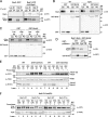
The Snf1/PP1 glucose-signaling pathway regulates Rod1 activation and Jen1 endocytosis. (A, left) WT and snf1Δ cells expressing plasmid-encoded Rod1-3HA or Rod1-PYm-3HA were grown in glucose overnight and then 4 h in lactate medium. Crude extracts were prepared, treated with phosphatase where indicated, and immunoblotted with the indicated antibodies. The asterisk indicates ubiquitylated Rod1 as determined by the absence of this band in cells expressing Rod1-PYm (lane 9). (Right) snf1Δ cells expressing plasmid-encoded Rod1-3HA and 6xHis-tagged ubiquitin were grown overnight in glucose-containing medium (exponential phase). 6xHis-tagged ubiquitin conjugates were purified under denaturing conditions by Ni-based chromatography. The lysates (Lys.) and eluates (El.) were immunoblotted with an anti-ubiquitin antibody, and with an anti-HA antibody to detect Rod1. (B) The snf1Δ mutant is unable to grow on alternate sugars such as galactose or nonfermentable carbon sources, but the additional deletion of MIG1, encoding a Snf1-regulated transcriptional repressor, allows growth on galactose (Vallier and Carlson, 1994). Transport activity of 14C-labeled lactate was therefore assayed in the snf1Δ mig1Δ and snf1Δ mig1Δ rod1Δ strains transformed with a galactose-inducible Jen1-GFP-His6 construct after 2 h of growth in galactose medium. A representative experiment of three repetitions is shown. The values represent the mean ± SEM of triplicate measurements. (C) Crude extracts were prepared from the strains described in B. Crude extracts were prepared at the indicated times and immunoblotted with anti-GFP and anti-PGK antibodies. GFP* indicates the size of a proteolytic fragment indicative of Jen1-GFP degradation in the vacuole. Band intensities were quantified using ImageJ and the Jen1-GFP/GFP* ratio is plotted over time. (D) Jen1-GFP localization was followed in strains as in B after 2 h of induction. (E) A yeast two-hybrid screen using full-length Rod1 as the bait led to the identification of the C-terminal region of Reg1, a PP1 regulatory subunit. Rsp5 was used as a positive control. Average β-galactosidase activity values (Miller units) for each combination are presented. (F) WT and reg1Δ strains expressing plasmid-encoded Rod1-3HA were grown as indicated and total protein extracts were treated with phosphatase when indicated, and immunoblotted with the indicated antibodies. (G) WT and reg1Δ strains in which Jen1 is tagged with GFP at the chromosomal locus were grown for 4 h in lactate medium, and Jen1-GFP subcellular localization was followed at the indicated time after glucose addition by fluorescence microscopy. Bar, 5 µm. (H) The same strains as in G were grown in similar conditions. Samples were collected at the indicated times after glucose treatments and total protein extracts were immunoblotted with anti-GFP antibodies. GFP* indicates the size of a proteolytic fragment indicative of Jen1-GFP degradation in the vacuole.
Figure 4.
Figure 4.
Rod1 ubiquitylation is regulated by a phospho-dependent binding to 14-3-3 proteins. (A) WT cells expressing a plasmid-encoded Rod1-GFP construct were transformed with an empty plasmid or a plasmid encoding 3HA-Rps5. Immunoprecipitation of 3HA-Rsp5 was performed from cells grown as indicated. The input (Lys) and immunoprecipitated (IP) fractions were immunoblotted with the indicated antibodies. (B) WT and reg1Δ cells expressing Rod1-3HA tagged at the chromosomal locus were transformed with a plasmid encoding GST-Bmh2 or GST alone as a control. GST pull-downs were performed from cells harvested after overnight growth in glucose medium (exponential phase). The input (Lys) and pull-down (PD) fractions were immunoblotted with the indicated antibodies. Asterisk indicates a nonspecific band that sometimes cross-reacted with the anti-GST antibody. (C) WT cells in which Rod1 is tagged with GFP at the chromosomal locus were transformed with a plasmid encoding GST-Bmh2 or GST alone. GST pull-downs were performed from cells grown as indicated, and the input (Lys) and pull-down (PD) fractions were immunoblotted with the indicated antibodies. (D) WT or snf1Δ strains in which Rod1 is tagged with GFP at the chromosomal locus were transformed with a plasmid encoding GST-Bmh2 or GST alone and grown overnight (exponential phase) in glucose medium. GST pull-downs were performed, and input (Lys) and pull-down (PD) fractions were immunoblotted with the indicated antibodies. Asterisk indicates a nonspecific band that sometimes cross-reacted with the anti-GST antibody. (E) WT or bmh1Δ bmh2Δ strains expressing a plasmid-encoded Rod1-GFP were grown as indicated. Total protein extracts were treated with phosphatase when indicated to better visualize ubiquitylation, and immunoblotted with the indicated antibodies. (F) bmh1Δ bmh2Δ strains expressing a plasmid-encoded Rod1-GFP (WT or PYm) were grown as indicated and treated as in E.
Figure 5.
Figure 5.
Model for the regulation of transporter endocytosis by intracellular signaling through arrestin-related protein activation. When yeast cells are grown in lactate medium, Snf1, the yeast AMPK homologue, is active and phosphorylates Rod1 to inactivate it (red arrows). Glucose addition triggers Jen1 endocytosis, which depends on Rod1 activation through its PP1-mediated dephosphorylation and subsequent Rsp5-mediated ubiquitylation, which are coordinated by 14-3-3 proteins (green arrows). The subcellular compartment at which Rod1 acts on Jen1 endocytosis may be the plasma membrane, or internal compartments (dashed lines). See the Discussion for details. Noteworthy, the Snf1/PP1 pathway also controls the transcriptional reprogramming of cells in response to glucose fluctuation, including the expression of the JEN1 gene (Lodi et al., 2002), illustrating a robust physiological regulation in which transcriptional and post-translational events are coordinated.

Similar articles

Cited by

References

    1. Alvarez C.E. 2008. On the origins of arrestin and rhodopsin. BMC Evol. Biol. 8:222 10.1186/1471-2148-8-222 - DOI - PMC - PubMed
    1. Andoh T., Hirata Y., Kikuchi A. 2002. PY motifs of Rod1 are required for binding to Rsp5 and for drug resistance. FEBS Lett. 525:131–134 10.1016/S0014-5793(02)03104-6 - DOI - PubMed
    1. Argenzio E., Bange T., Oldrini B., Bianchi F., Peesari R., Mari S., Di Fiore P.P., Mann M., Polo S. 2011. Proteomic snapshot of the EGF-induced ubiquitin network. Mol. Syst. Biol. 7:462 10.1038/msb.2010.118 - DOI - PMC - PubMed
    1. Bhalla V., Daidié D., Li H., Pao A.C., LaGrange L.P., Wang J., Vandewalle A., Stockand J.D., Staub O., Pearce D. 2005. Serum- and glucocorticoid-regulated kinase 1 regulates ubiquitin ligase neural precursor cell-expressed, developmentally down-regulated protein 4-2 by inducing interaction with 14-3-3. Mol. Endocrinol. 19:3073–3084 10.1210/me.2005-0193 - DOI - PubMed
    1. Bhandari D., Trejo J., Benovic J.L., Marchese A. 2007. Arrestin-2 interacts with the ubiquitin-protein isopeptide ligase atrophin-interacting protein 4 and mediates endosomal sorting of the chemokine receptor CXCR4. J. Biol. Chem. 282:36971–36979 10.1074/jbc.M705085200 - DOI - PubMed

Publication types

MeSH terms

Substances