

Modic type I change may predict rapid progressive, deforming disc degeneration: a prospective 1-year follow-up study
- PMID: 22249308
- PMCID: PMC3366121
- DOI: 10.1007/s00586-012-2147-9
Modic type I change may predict rapid progressive, deforming disc degeneration: a prospective 1-year follow-up study
Abstract
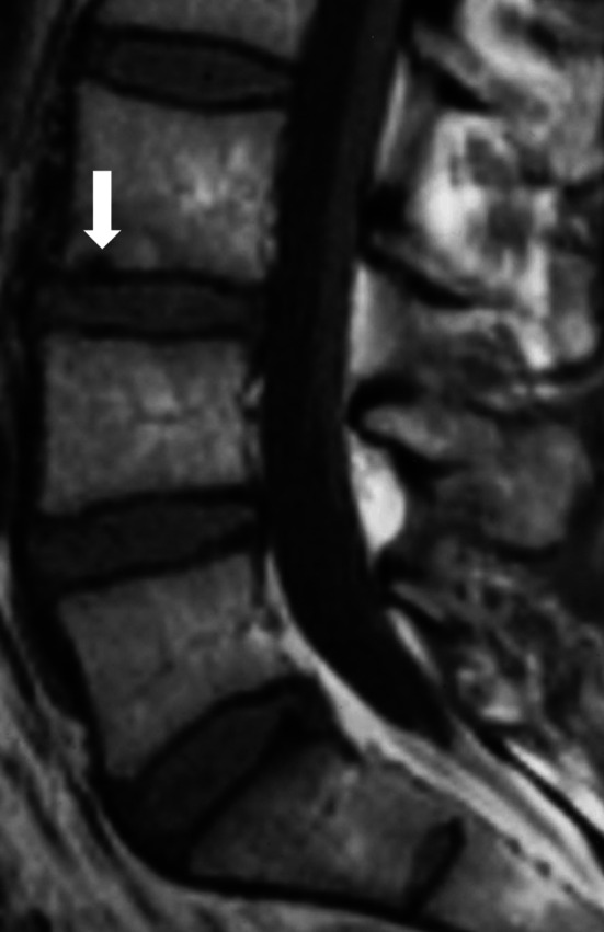
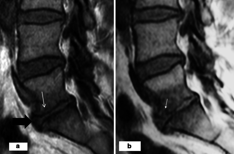
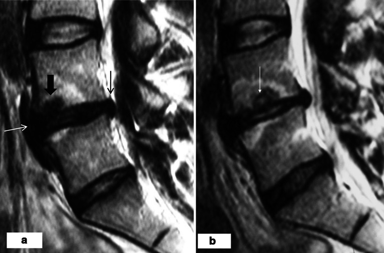
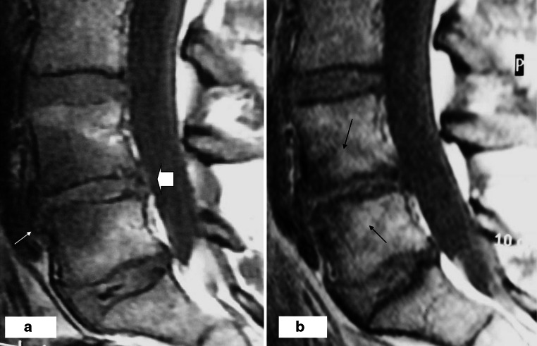
Introduction: This prospective magnetic resonance imaging (MRI) study in chronic low-back pain (CLBP) patients evaluated the natural course of degenerative lumbar spine changes in relation to Modic 1 type changes (M1) within 1 year.
Materials and methods: From 3,811 consecutive CLBP patients referred to lumbar spine MRI 54 patients with a large M1 were selected using strict exclusion criteria to exclude specific back disorders. Follow-up MRI was obtained within 11-18 months.
Results: At baseline M1 was associated with an adjacent endplate lesion in 96% of the cases. In follow-up, an unstable M1 was associated both with an increase of endplate lesions, decrease of disc height and change in disc signal intensity, most found at L4/5 or L5/S1. In disc spaces without M1, progression of degenerative changes was rare.
Conclusion: Endplate deformation, decreasing disc height and change of disc signal intensity appear essential features of accelerated degenerative process associated with M1.
Figures

Similar articles
-
Chronic low back pain in relation to Modic changes, bony endplate lesions, and disc degeneration in a prospective MRI study.Eur Spine J. 2016 Sep;25(9):2873-81. doi: 10.1007/s00586-016-4715-x. Epub 2016 Aug 1. Eur Spine J. 2016. PMID: 27480265
-
[The study on the relationship between modic change and disc height together with lumbar hyperosteogeny].Zhonghua Wai Ke Za Zhi. 2013 Jul;51(7):610-4. Zhonghua Wai Ke Za Zhi. 2013. PMID: 24256586 Chinese.
-
Analysis of Correlation Between Vertebral Endplate Change and Lumbar Disc Degeneration.Med Sci Monit. 2017 Oct 15;23:4932-4938. doi: 10.12659/msm.904315. Med Sci Monit. 2017. PMID: 29032381 Free PMC article.
-
From Modic 1 vertebral-endplate subchondral bone signal changes detected by MRI to the concept of 'active discopathy'.Ann Rheum Dis. 2015 Aug;74(8):1488-94. doi: 10.1136/annrheumdis-2015-207317. Epub 2015 May 14. Ann Rheum Dis. 2015. PMID: 25977562 Review.
-
A definition and clinical grading of Modic changes.J Orthop Res. 2022 Feb;40(2):301-307. doi: 10.1002/jor.25240. Epub 2021 Dec 27. J Orthop Res. 2022. PMID: 34910328 Review.
Cited by
-
Magnetic Resonance Imaging Characteristics Associated with Treatment Success from Basivertebral Nerve Ablation: An Aggregated Cohort Study of Multicenter Prospective Clinical Trials Data.Pain Med. 2022 Jul 20;23(Suppl 2):S34-S49. doi: 10.1093/pm/pnac093. Pain Med. 2022. PMID: 35856328 Free PMC article.
-
Correlations between Modic change and degeneration in 3-joint complex of the lower lumbar spine: A retrospective study.Medicine (Baltimore). 2018 Sep;97(38):e12496. doi: 10.1097/MD.0000000000012496. Medicine (Baltimore). 2018. PMID: 30235755 Free PMC article.
-
Modic type 2 changes are fibroinflammatory changes with complement system involvement adjacent to degenerated vertebral endplates.JOR Spine. 2022 Dec 23;6(1):e1237. doi: 10.1002/jsp2.1237. eCollection 2023 Mar. JOR Spine. 2022. PMID: 36994463 Free PMC article.
-
Chronic low back pain in relation to Modic changes, bony endplate lesions, and disc degeneration in a prospective MRI study.Eur Spine J. 2016 Sep;25(9):2873-81. doi: 10.1007/s00586-016-4715-x. Epub 2016 Aug 1. Eur Spine J. 2016. PMID: 27480265
-
Relationship between Endplate Defects, Modic Change, Disc Degeneration, and Facet Joint Degeneration in Patients with Low Back Pain.Biomed Res Int. 2019 Jul 11;2019:9369853. doi: 10.1155/2019/9369853. eCollection 2019. Biomed Res Int. 2019. PMID: 31380443 Free PMC article.
References
-
- Weishaupt D, Zanetti M, Hodler J, Min K, Fuchs B, Pfirrmann CW, Boos N. Painful lumbar disk derangement: relevance of endplate abnormalities at MR imaging. Radiology. 2001;218:420–427. - PubMed
-
- Kuisma M, Karppinen J, Niinimäki J, Ojala R, Haapea M, Heliövaara M, Korpelainen R, Taimela S, Natri A, Tervonen O. Modic changes in endplates of lumbar vertebral bodies. Prevalence and association with low back and sciatic pain among middle-aged male workers. Spine. 2007;32:1116–1122. doi: 10.1097/01.brs.0000261561.12944.ff. - DOI - PubMed
-
- Weishaupt D, Zanetti M, Hodler J, Boos N. MR imaging of the lumbar spine: prevalence of intervertebral disk extrusion and sequestration, nerve root compression, end plate abnormalities, and osteoarthritis of the facet joints in asymptomatic volunteers. Radiology. 1998;209:661–666. - PubMed
-
- Modic MT, Steinberg PM, Ross JS, Masaryk TJ, Carter JR. Degenerative disk disease: assessment of changes in vertebral body marrow with MR imaging. Radiology. 1988;166:193–199. - PubMed
Publication types
MeSH terms
LinkOut - more resources
Full Text Sources
Other Literature Sources
Medical
