. 2012 Feb;71(2):148-61.
doi: 10.1097/NEN.0b013e318244477d.

Cerebellar ataxia in patients with mitochondrial DNA disease: a molecular clinicopathological study

Affiliations

Cerebellar ataxia in patients with mitochondrial DNA disease: a molecular clinicopathological study

Nichola Zoe Lax et al. J Neuropathol Exp Neurol. 2012 Feb.

Abstract

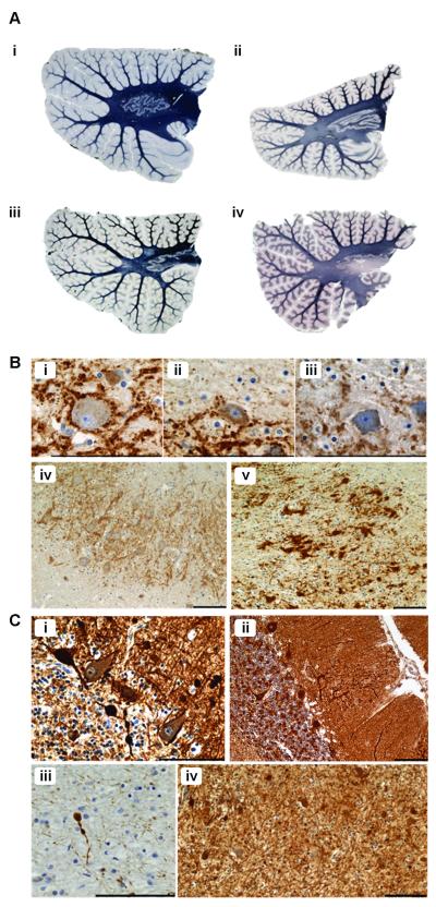
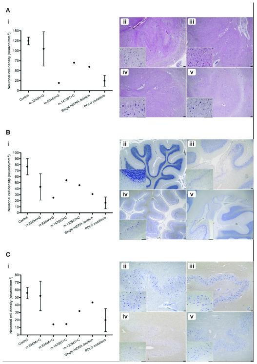
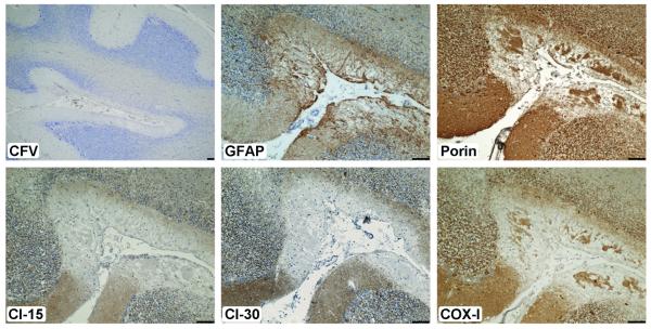
Cerebellar ataxia is a prominent clinical symptom in patients with mitochondrial DNA (mtDNA) disease. This is often progressive with onset in young adulthood. We performed a detailed neuropathologic investigation of the olivary-cerebellum in 14 genetically and clinically well-defined patients with mtDNA disease. Quantitative neuropathologic investigation showed varying levels of loss of Purkinje cells and neurons of the dentate nucleus and inferior olivary nuclei. Typically, focal Purkinje cell loss was present in patients with the m.3243A>G mutation caused by the presence of microinfarcts, with relative preservation of neuronal cell populations in the olivary and dentate nuclei. In contrast, patients with the m.8344A>G mutation or recessive POLG mutations showed extensive and global neuronal cell loss in all 3 olivary-cerebellum areas examined. Molecular analysis of mutated mtDNA heteroplasmy levels revealed that neuronal cell loss occurred independently of the level of mutated mtDNA present within surviving neurons. High levels of neuronal respiratory chain deficiency, particularly of complex I, were detected in surviving cells; levels of deficiency were greater in regions with extensive cell loss. We found a relationship between respiratory deficiency and neuronal cell density, indicating that neuronal cell death correlates with respiratory deficiency. These findings highlight the vulnerability of the olivary-cerebellum to mtDNA defects.


Figures

Figure 1
Figure 1
Myelin loss, axonal and dendritic pathology in patients with mitochondrial DNA (mtDNA) defects are likely to be secondary to neuronal cell loss in all cases except Kearns-Sayre syndrome (KSS). (A) Relative to control (i; Loyez) there is mild myelin pallor in some patients with the m.3243A>G mutation (ii; Loyez, Patient 5); spongiform degeneration and myelin loss are seen in a single mtDNA deletion patient (iii; Loyez, Patient 11 with KSS). There is severe myelin loss of the dentate nucleus outflow tract in patients with POLG mutations (iv; Loyez, Patient 12). (B) There are markedly reduced presynaptic terminals around dentate nucleus neurons in m.3243A>G (ii; synaptophysin, Patient 4) and m.8344A>G (iii; synaptophysin, Patient 8) cases vs. controls (i; synaptophysin). Relative to control (iv; synaptophysin) there are large intensely labeled presynaptic-like structures lacking morphologically distinguishable post-synaptic dentate neurons in a patient with m.14709T>C mutation (v; synaptophysin, Patient 9), indicating relative increase of input from Purkinje cells when the dentate neuron loss exceeds Purkinje cell loss. (C) There are multiple axonal swellings in the granular cell layer of the cerebellum (i; SMI31; single mtDNA deletion, Patient 11) and thickened dendritic trees containing ‘trapped’ mitochondria in the molecular layer (ii; porin; single mtDNA deletion, Patient 11). There are also axonal spheroids in the deep white matter (iii and iv; synaptophysin; single mtDNA deletion, Patient 11). Scale bar: 100 μm.
Figure 2
Figure 2
Variation in neuron loss across the olivo-cerebellum in patients with mitochondrial DNA (mtDNA) mutations. (A) Inferior olivary neuronal densities vary remarkably according to the mtDNA defect vs. control tissues (n = 2) (i). Cell loss is most severe in association with the m.8344A>G (iii; H&E) and recessive POLG (iv; H&E) mutations with only moderate loss associated with the m.14709T>C mutation (v; H&E) vs. control tissues (ii; H&E). (B) Purkinje cell density is reduced in all patients with mtDNA defects vs. controls (n = 5) (i). This is most apparent in a patient with POLG mutations in which neuron loss is global and with the presence of microinfarcts (v; cresyl fast violet [CFV)]). Atrophy of the cerebellar cortex is severe in m.3243A>G (iv; CFV) and Purkinje cell loss often occurs focally in microinfarcts (iii; CFV) when compared to control tissue (ii; CFV). (C) Neuronal cell density in the dentate nucleus also shows wide variation in patients relative to controls (n = 8) (i). Typically, patients with the m.3243A>G mutation show preservation of neurons (iii; CFV) vs. control tissue (ii; CFV); cell loss is severe in m.8344A>G (iv; CFV), and minimal in single mtDNA deletion patients (v; CFV). Scale bar: 100 μm.
Figure 3
Figure 3
Astrogliosis in regions of microinfarcts with evidence of severe respiratory chain deficiency in a patient with m.3243A>G. There are numerous microinfarcts in the cerebellar cortex (cresyl fast violet, [CFV)]); typically, these are accompanied by marked proliferation of glial fibrillary acidic protein (GFAP)-positive astrocytes (in a serial section). Evaluation of the mitochondrial proteins shows that mitochondrial density is reduced due to the necrosis (porin), but expression of complex I (subunits Cl-15 and -30) is completely absent; cytochrome c oxidase-1 (COX-I) is diminished. These observations suggest that astrocytes may also harbor respiratory-deficient mitochondria. Scale bar: 100 μm.
Figure 4
Figure 4
High levels of complex I-deficient neurons can be detected in remaining neuronal populations. The inferior olivary nucleus shows an unusual distribution of mitochondria (m.3243A>G, first column) (porin), whereas mitochondria appear abundant throughout the remainder of the cerebellum in all cases as can be seen in the patient with the m.13094T>C mutation (middle column) and a patient with recessive POLG mutation (third column). High levels of complex I-deficient neurons are observed throughout the olivo-cerebellum, nearing 100% of inferior olivary nucleus in m.3243A>G patient but there is marked deficiency throughout the remainder of the cerebellum. Complex IV-deficiency was rarely detected and low in many cases; cytochrome c oxidase/succinic acid dehydrogenase (COX/SDH) histochemistry often revealed intact neuronal COX activity. Scale bar: 100 μm.

References

    1. McFarland R, Taylor RW, Turnbull DM. A neurological perspective on mitochondrial disease. Lancet Neurol. 2010;9:829–40. - PubMed
    1. Taylor RW, Turnbull DM. Mitochondrial DNA mutations in human disease. Nature Rev. 2005;6:389–402. - PMC - PubMed
    1. Bristow EA, Griffiths PG, Andrews RM, Johnson MA, Turnbull DM. The distribution of mitochondrial activity in relation to optic nerve structure. Arch Ophthalmol. 2002;120:791–6. - PubMed
    1. Erecinska M, Silver IA. Ions and energy in mammalian brain. Prog Neurobiol. 1994;43:37–71. - PubMed
    1. Hollenbeck PJ, Saxton WM. The axonal transport of mitochondria. J Cell Sci. 2005;118:5411–9. - PMC - PubMed

Publication types

MeSH terms