. 2012 Feb;71(2):162-77.
doi: 10.1097/NEN.0b013e318244b635.

Nuclear localization of human SOD1 and mutant SOD1-specific disruption of survival motor neuron protein complex in transgenic amyotrophic lateral sclerosis mice

Affiliations

Nuclear localization of human SOD1 and mutant SOD1-specific disruption of survival motor neuron protein complex in transgenic amyotrophic lateral sclerosis mice

Barry Gertz et al. J Neuropathol Exp Neurol. 2012 Feb.

Abstract

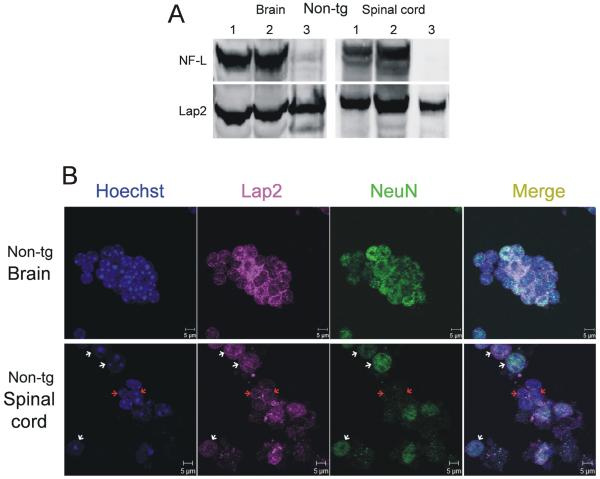
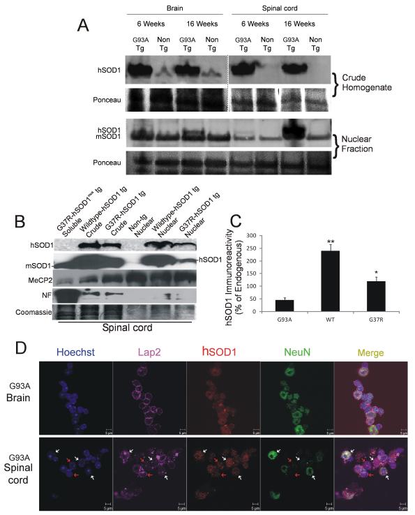
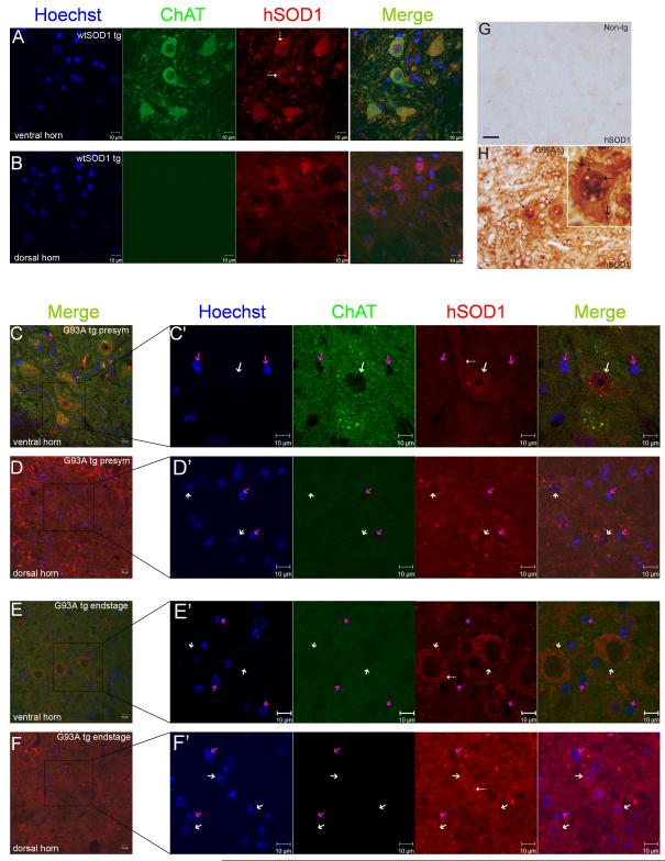
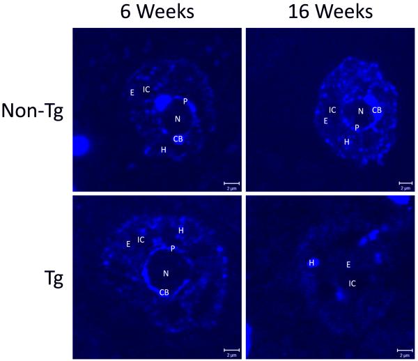
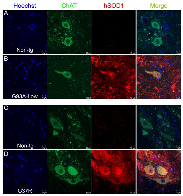
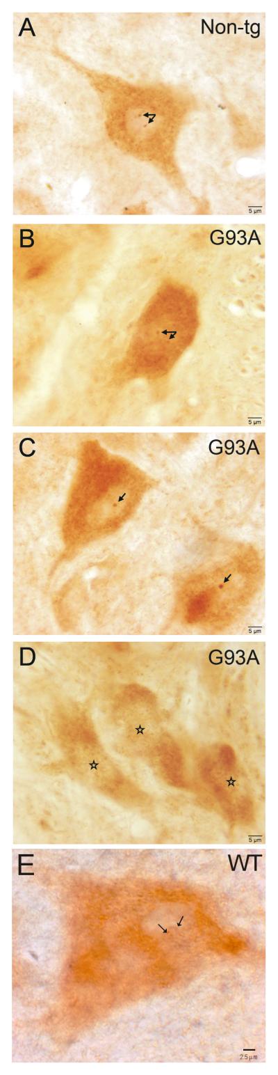
Amyotrophic lateral sclerosis (ALS) is a fatal adult-onset neurodegenerative disease that causes degeneration of motor neurons and paralysis. Approximately 20% of familial ALS cases have been linked to mutations in the copper/zinc superoxide dismutase (SOD1) gene, but it is unclear how mutations in the protein result in motor neuron degeneration. Transgenic (tg) mice expressing mutated forms of human SOD1 (hSOD1) develop clinical and pathological features similar to those of ALS. We used tg mice expressing hSOD1-G93A, hSOD1-G37R, and hSOD1-wild-type to investigate a new subcellular pathology involving mutant hSOD1 protein prominently localizing to the nuclear compartment and disruption of the architecture of nuclear gems. We developed methods for extracting relatively pure cell nucleus fractions from mouse CNS tissues and demonstrate a low nuclear presence of endogenous SOD1 in mouse brain and spinal cord, but prominent nuclear accumulation of hSOD1-G93A, -G37R, and -wild-type in tg mice. The hSOD1 concentrated in the nuclei of spinal cord cells, particularly motor neurons, at a young age. The survival motor neuron protein (SMN) complex is disrupted in motor neuron nuclei before disease onset in hSOD1-G93A and -G37R mice; age-matched hSOD1-wild-type mice did not show SMN disruption despite a nuclear presence. Our data suggest new mechanisms involving hSOD1 accumulation in the cell nucleus and mutant hSOD1-specific perturbations in SMN localization with disruption of the nuclear SMN complex in ALS mice and suggest an overlap of pathogenic mechanisms with spinal muscular atrophy.


Figures

Figure 1
Figure 1
Generation and validation of a highly enriched nuclear fraction from adult nontransgenic (tg) mouse brain and spinal cord. (A) Validation of nuclear purity of fractions. Fractions were assayed by immunoblotting to determine the extent of nuclear marker enrichment. The crude homogenate (lane 1) contains relatively equal levels of cytoplasmic proteins, as judged by the presence of light neurofilament (NF-L, a highly expressed cytoplasmic neuronal protein) and Lap2 (a ubiquitously expressed nuclear envelope protein). Incubation with proteinase K (lane 2) degrades contaminating soluble proteins but does not appreciatively degrade the nuclear proteins. The final fraction (lane 3) is highly enriched for nuclei, as determined by Lap2 immunoreactivity with almost no detectable NF-L. (B) Microscopic validation of nuclear purity of fractions by immunofluorescence. The fractions from non-tg mouse brain and spinal cord show high enrichment for nuclei, as demonstrated by DNA staining with Hoechst-33342 and Lap2 immunostaining. The brain fractions consist almost exclusively of neuronal nuclei (NeuN staining); the spinal cord fractions contain neuronal (white arrows) and non-neuronal nuclei (red arrows). Additionally, as determined by morphology (size and appearance of chromatin organization), putative motor neuron nuclei from the spinal cord constitute a small percentage of total nuclei. Data are representative of 5 mice.
Figure 2
Figure 2
Human superoxide dismutase 1 (hSOD1) protein is present in transgenic (tg) mouse CNS nuclear fractions. (A) hSOD1 protein is not detected in crude homogenates of non-tg mouse spinal cord (top), demonstrating the specificity of the antibody for the hSOD1 without detection of endogenous mouse SOD1 (mSOD1). In brain crude homogenate there is weak detection of a band at the same molecular weight as mouse SOD1 with the hSOD1 antibody, possibly indicating a small amount of cross-reactivity when high amounts of protein are loaded. The crude fractions of pre-symptomatic and late-stage disease tg mice showed relatively equal amounts of hSOD1 protein levels in both the brain and spinal cord, indicating that protein expression throughout disease progression remains fairly constant. In nuclear fractions (bottom), endogenous mouse SOD1, detected with an antibody that can recognize mouse SOD1 and hSOD1, is found in nuclear fractions; levels remain fairly constant throughout disease. hSOD1 is also present in the nuclear fractions of brain and spinal cord of hSOD1-G93A tg mice. The amount of hSOD1 in the nuclear fraction of the brain shows a moderate increase during disease progression; hSOD1 levels in the nuclear fraction of spinal cord increase dramatically. The Ponceau S-stained membrane for each immunoblot shows protein loading. (B) Western blot analyses of spinal cords from 6-week-old tg mice expressing hSOD1-G37R and hSOD1-wild type. Lanes loaded with crude extract and pure nuclear fractions are shown. Wild type- and G37R-hSOD1 are present in nuclei; endogenous mouse SOD1 (mSOD1) is seen at low or undetectable levels in pure nuclear fractions. The purity of the nuclear fraction is confirmed by exclusion of neurofilament (NF) immunoreactivity and enrichment of methyl-CpG-DNA-binding protein 2 (MeCP2) relative to crude fractions. A tg mouse negative control for hSOD1 in spinal cord as hSOD1-G37R tg mice with restricted expression of transgene in skeletal muscle (G37R-hSOD1mus), which have endogenous mouse SOD1 enriched in the soluble fraction but no hSOD1 in spinal cord (45). Another negative control is non-tg mouse spinal cord nuclear fraction. Post-transfer Coomassie-stained gel shows protein loading (image was uniformly darkened by digital enhancement due to light staining). (C) Graph showing the level of hSOD1-G93A, -G37R, -wild type (WT) immunoreactivity in spinal cord nuclear fractions of 6-week-old tg mice (n = 4/genotype). Values are mean ± SD of optical densities (**p < 0.01 or *p < 0.05 compared to endogenous). (D) Quadruple fluorescence labeling of nuclear fractions show the presence of hSOD1-G93A in virtually all nuclei from the tg mouse brain, many of which are positive for NeuN, but only some nuclei of the spinal cord contain the hSOD1. Immunophenotyping for the neuron nuclear marker showed that NeuN-positive nuclei (white arrows) and NeuN-negative nuclei (red arrows) are positive for hSOD1. DNA is labeled with Hoechst dye. Lap2 (nuclear envelope), hSOD1, and NeuN (neuron nucleus) are detected by immunofluorescence using far red, red, and green secondary antibodies and viewed on a Zeiss LSM 510 Meta confocal microscope. Data are representative of n ≥ 5 mice per group.
Figure 3
Figure 3
Human superoxide dismutase 1 (hSOD1) localization in the nucleus of motor neurons in hSOD1-wild type and hSOD1-G93A transgenic (tg) mouse spinal cord in situ. (A, B) In hSOD1-wild type tg mouse spinal cord, hSOD1 immunoreactivity localizes to the cytoplasm and nucleus. hSOD1-wild type immunoreactivity appears to be stronger in the cytoplasm compared to the nucleus. This result mimics the distribution of endogenous mouse SOD1 and is characteristic of motor neurons in the ventral horn, identified by choline acetyltransferase (ChAT) immunostaining (A) and sensory neurons in the dorsal horn (B) of spinal cord. Motor neurons expressing hSOD1-wild type show cytoplasmic aggregates of hSOD1 (A, hatched white arrows), consistent with previous observations (16). (C-F) Spinal cords of hSOD1-G93A tg mice. In pre-symptomatic mouse spinal cord viewed at low magnification (C, D) and high magnification (C’, D’), hSOD1 immunoreactivity shows robust presence in the nucleus of motor neurons (as seen by ChAT immunostaining, a motor neuron cytoplasmic marker) in the ventral horn (C, C’), but hSOD1 is not present in nuclei of neurons in the dorsal horn (D, D’, white arrows). Some cytoplasmic hSOD1 aggregates are seen in motor neurons (C’, hatched white arrow). In spinal cords of hSOD1-G93A tg mice at near end-stage disease, motor neurons viewed at low magnification (E, F) and high magnification (E’, F’) have lost ChAT immunoreactivity and the nuclei show nuclear structure abnormalities and a loss of hSOD1 immunoreactivity (E, E’; white arrows). This is in stark contrast to the nuclei of the neurons of the dorsal horn that show enhanced immunoreactivity for hSOD1 (F, F’; white arrows). Nonneuronal cells, in both the dorsal and ventral horn, do not show the presence of hSOD1 (pink arrows) in their nuclei. Cytoplasmic aggregates of hSOD1-G93A are prominent in motor neurons (E’, hatched white arrow) and dorsal horn cells (F’, hatched white arrow) at end-stage disease. G, H: hSOD1 localization in mouse spinal cord using immunoperoxidase staining and diaminobenzidine as chromogen. Non-tg mouse spinal cord sections show no immunostaining for hSOD1 (G); same batch-stained spinal cord sections from pre-symptomatic hSOD1-G93A tg mice show marked immunolabeling of motor neurons (H, hatched arrows and inset) with marked accumulation of hSOD-G93A1 in the nucleus (H, white asterisks). Pre-symptomatically, there is prominent aggregation of hSOD1-G93A in the nucleus (H, inset, hatched black arrows) and only slight to modest aggregation of hSOD1 in the cytoplasm (H, inset solid black arrows). Scale bar in G (same for H) = 8 μm. Data are representative of 5 mice per genotype, ≥ 6 sections per mouse, and ≥10 neurons analyzed per section by confocal microscopy.
Figure 4
Figure 4
Nuclear architecture abnormalities in motor neurons in human superoxide dismutase 1 (hSOD1)-G93A transgenic (tg) mice late-stage disease. In non-tg and tg mice, the spinal cord motor neuron nuclei at 6 weeks of age (pre-symptomatic) exhibit a patterned nuclear architecture when observed by Hoechst staining for DNA (blue). There is a central located nucleolus (N) with prominent Cajal bodies (CB) and a fainter annulus of heterochromatin (P). The “body” of the nucleus is composed of areas of interchromatin (IC) surrounded by euchromatin (E) interspersed with heterochromatin (H) in speckled manner. The same is true for motor neuron nuclei of non-tg mice at 16 weeks of age. By contrast, in the tg mice at 16 weeks (late stage), the motor neuron nuclei show distinctly abnormal architecture. There is loss of the central nucleolus, Cajal bodies, and surrounding annulus. Most of the nuclear space appears to be composed of euchromatin (E) with a few densely staining areas of heterochromatin (H) and interchromatin (IC). Sections are stained with Hoechst-33342 and images were captured on a Zeiss LSM 510 Meta inverted confocal microscope using LSM software. Data are representative of 5 mice per genotype, ≥6 sections per mouse, and ≥10 neurons analyzed per section by confocal microscopy.
Figure 5
Figure 5
Human superoxide dismutase 1 (hSOD1) immunofluorescent localization in spinal cord motor neurons of transgenic (tg) mice expressing hSOD1-G93Alow and hSOD1-G37R. (A, B) Choline acetyltransferase (ChAT)-positive motor neurons in age-matched non-tg littermate control mice for hSOD1-G93Alow tg mice do not stain for hSOD1 (A), while motor neurons in the hSOD1-G93Alow tg mice have conspicuous nuclear localization of hSOD1 (B). (C, D) ChAT-positive motor neurons in age-matched non-tg littermate control mice for hSOD1-G37R tg mice do not stain for hSOD1 (C), whereas motor neurons in hSOD1-G37R tg mice are strongly positive for hSOD1, which is present in the cytoplasm and nucleus (D). hSOD1 expression is not ubiquitously uniform in all motor neurons and some appear free of hSOD1-G37R nuclear immunoreactivity. Each column of panels is labeled at the top for the marker shown. Data are representative of 2 mice per genotype, 4 sections per mouse, and ≥10 neurons analyzed per section by confocal microscopy.
Figure 6
Figure 6
Immunolocalization of survival motor neuron protein (SMN) complex in motor neurons of spinal cord in non-transgenic (tg) and human superoxide dismutase 1 (hSOD1) tg mice. (A) Normal motor neuron nuclei of a 6-week-old non-tg mouse shows prominent double or single SMN-positive complex (arrows) next to the nucleolus. (B-D) Typical motor neuron nuclei from 6-week-old hSOD1-G93Ahigh tg mice. Some motor neurons display normal prominent SMN complexes next to the nucleolus (B), but others possessed small SMN complexes with diffuse/punctate SMN immunoreactivity in the nucleus (C) or had nuclei lacking SMN complexes (D). Stars mark the nucleolus in D. (E) Motor neuron nucleus of a 6-week-old hSOD1-wild type tg mouse shows the normal prominent double or single SMN-positive complex (arrows) next to the nucleolus. Data are representative of 3 mice per genotype, 4 sections per mouse, and ≥20 neurons analyzed per section by bright-field microscopy.
Figure 7
Figure 7
Survival motor neuron protein (SMN) immunofluorescent localization in nontransgenic (tg) and human superoxide dismutase 1 (hSOD1) tg mouse spinal cord motor neurons. A-C: SMN localization in 12-week-old non-tg and hSOD1-G93A tg spinal cord motor neurons. Results were similar to that seen with the immunohistochemistry (Fig. 6). All non-tg mouse (A) and hSOD1-wild type tg mouse (see Fig. 6E) motor neurons observed had prominent nuclear SMN complexes. Spinal cords of hSOD1-G93A tg mice contained a mix of SMN complex-positive (B) and -negative motor neurons (C). Note the peripheral nuclear membrane localization of SMN in the motor neuron with the SMN complex-negative nucleus (C, white arrow). (D-F) Control 6 mo old non-tg littermates for hSOD1-G37R mice show spinal cord motor neurons all with nuclear SMN complexes (D), whereas age-matched G37R tg mouse motor neurons show depletion of nuclear SMN complexes (E, F) and SMN-positive cytoplasmic spherical structures in motor neurons without nuclear SMN complexes (F, white arrows). Data are representative of 3 mice per genotype, 6 sections per mouse, and ≥10 neurons analyzed per section by confocal microscopy.

Similar articles

Cited by

References

    1. Chou SM. Pathology-light microscopy of amyotrophic lateral sclerosis. In: Smith RA, editor. Handbook of Amyotrophic Lateral Sclerosis. Marcel Deckker, Inc.; New York, NY: 1992. pp. 133–81.
    1. Rowland LP, Shneider NA. Amyotrophic lateral sclerosis. N Engl J Med. 2001;344:1688–1700. - PubMed
    1. Bendotti C, Carrì MT. Lessons from models of SOD1-linked familial ALS. Trends Mol Med. 2004;10:393–400. 2004. - PubMed
    1. Kabashi E, Valdmanis PN, Dion P, et al. TARDBP mutations in individuals with sporadic and familial amyotrophic lateral sclerosis. Nat Genet. 2008;40:572–4. - PubMed
    1. Turner BJ, Talbot K. Transgenics, toxicity and therapeutics in rodent models of mutant SOD1-mediated familial ALS. Prog Neurobiol. 2008;85:94–134. - PubMed

Publication types