. 2012;67(1):61-7.
doi: 10.6061/clinics/2012(01)10.

Reduced occurrence of programmed cell death and gliosis in the retinas of juvenile rabbits after shortterm treatment with intravitreous bevacizumab

Affiliations

Reduced occurrence of programmed cell death and gliosis in the retinas of juvenile rabbits after shortterm treatment with intravitreous bevacizumab

Maria Alice Fusco et al. Clinics (Sao Paulo). 2012.

Abstract

Objective: Bevacizumab has been widely used as a vascular endothelial growth factor antagonist in the treatment of retinal vasoproliferative disorders in adults and, more recently, in infants with retinopathy of prematurity. Recently, it has been proposed that vascular endothelial growth factor acts as a protective factor for neurons and glial cells, particularly in developing nervous tissue. The purpose of this study was to investigate the effects of bevacizumab on the developing retinas of juvenile rabbits.

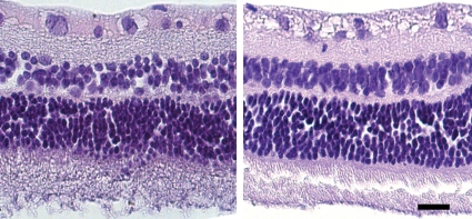
Methods: Juvenile rabbits received bevacizumab intravitreously in one eye; the other eye acted as an untreated control. Slit-lamp and fundoscopic examinations were performed both prior to and seven days after treatment. At the same time, retina samples were analyzed using immunohistochemistry to detect autophagy and apoptosis as well as proliferation and glial reactivity. Morphometric analyses were performed, and the data were analyzed using the Mann-Whitney U test.

Results: No clinical abnormalities were observed in either treated or untreated eyes. However, immunohistochemical analyses revealed a reduction in the occurrence of programmed cell death and increases in both proliferation and reactivity in the bevacizumab-treated group compared with the untreated group.

Conclusions: Bevacizumab appears to alter programmed cell death patterns and promote gliosis in the developing retinas of rabbits; therefore, it should be used with caution in developing eyes.


Conflict of interest statement

No potential conflict of interest was reported.

Figures

Figure 1
Figure 1
Fundus photography of a control (A) and treated (C) eyes before the intravitreal injection of bevacizumab. A repeat photograph taken seven days after treatment did not reveal abnormalities in either the control (B) or treated (D) eyes.
Figure 2
Figure 2
Photomicrograph of hematoxylin and eosin-stained control (left) and treated (right) retinas seven days after the experiment.
Figure 3
Figure 3
First row: Immunohistochemical staining of beclin1 revealing positive staining in control retinas (A) that is visibly stronger than that observed in the bevacizumab-treated animals (B). Box plot of the staining area for beclin1 showing a significant difference (p<0.05) between groups (C). Second row: TUNEL staining showing that the number of apoptotic nuclei (brown) in the control retinas (D) is significantly higher compared with the bevacizumab-treated retinas. (E). Box plot analysis revealed a significant difference between groups (p<0.05) (F). Third row: Bevacizumab increases cell proliferation in the retinas of juvenile rabbits. PCNA immunohistochemical analysis revealed more proliferating cells (brown nuclei) in the bevacizumab-treated retinas (H) than in the control group (G). The number of proliferating cells (PCNA-positive) in the bevacizumab-treated group was significantly greater (p<0.05) than that observed in the control group (I). Fourth row: GFAP immunohistochemical analysis revealed stronger staining in the bevacizumab-treated retinas (L) than in the control retinas (J), indicating that the number of glial cells in the retinas increased after treatment (p<0.05) (M).

Similar articles

Cited by

References

    1. Kim JH, Kim C, Kim JH, Lee BJ, Yu YS, Park KH, et al. Absence of intravitreal bevacizumab-induced neuronal toxicity in the retina. Neurotoxicology. 2008;29:1131–5. 10.1016/j.neuro.2008.06.006 - DOI - PubMed
    1. Gunther JB, Altaweel MM. Bevacizumab (Avastin) for the treatment of ocular disease. Surv Ophthalmol. 2009;54:372–400. 10.1016/j.survophthal.2009.02.004 - DOI - PubMed
    1. Michels S, Rosenfeld PJ, Puliafito CA, Marcus EN, Venkatraman AS. Systemic bevacizumab (Avastin) therapy for neovascular age-related macular degeneration twelve-week results of an uncontrolled open-label clinical study. Ophthalmology. 2005;112:1035–47. 10.1016/j.ophtha.2005.02.007 - DOI - PubMed
    1. Grisanti S, Ziemssen F. Bevacizumab: off-label use in ophthalmology. Indian J Ophthalmol. 2007;55:417–20. 10.4103/0301-4738.36474 - DOI - PMC - PubMed
    1. Saint-Geniez M, D'Amore PA. Development and pathology of the hyaloid, choroidal and retinal vasculature. Int J Dev Biol. 2004;48:1045–58. 10.1387/ijdb.041895ms - DOI - PubMed

Publication types

Substances