. 2012 Feb;139(2):135-44.
doi: 10.1085/jgp.201110672. Epub 2012 Jan 16.

Characterization of KCNQ1 atrial fibrillation mutations reveals distinct dependence on KCNE1

Affiliations

Characterization of KCNQ1 atrial fibrillation mutations reveals distinct dependence on KCNE1

Priscilla J Chan et al. J Gen Physiol. 2012 Feb.

Abstract

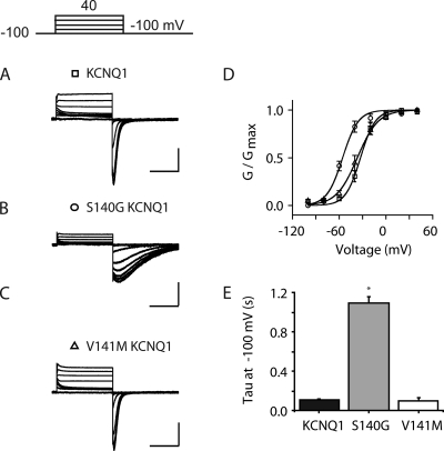
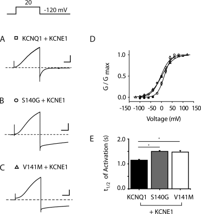
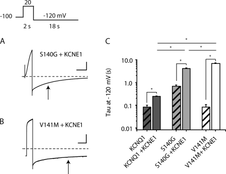
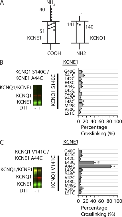
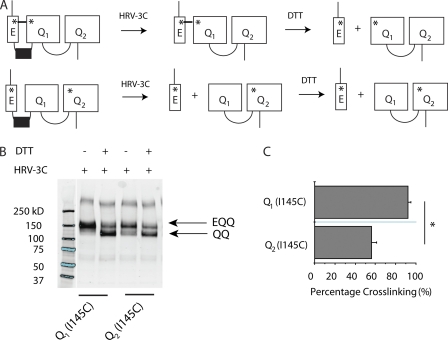
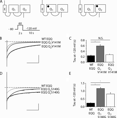
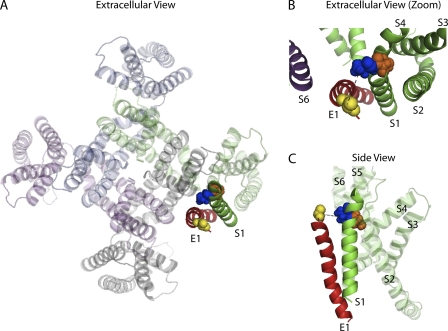
The I(Ks) potassium channel, critical to control of heart electrical activity, requires assembly of α (KCNQ1) and β (KCNE1) subunits. Inherited mutations in either I(Ks) channel subunit are associated with cardiac arrhythmia syndromes. Two mutations (S140G and V141M) that cause familial atrial fibrillation (AF) are located on adjacent residues in the first membrane-spanning domain of KCNQ1, S1. These mutations impair the deactivation process, causing channels to appear constitutively open. Previous studies suggest that both mutant phenotypes require the presence of KCNE1. Here we found that despite the proximity of these two mutations in the primary protein structure, they display different functional dependence in the presence of KCNE1. In the absence of KCNE1, the S140G mutation, but not V141M, confers a pronounced slowing of channel deactivation and a hyperpolarizing shift in voltage-dependent activation. When coexpressed with KCNE1, both mutants deactivate significantly slower than wild-type KCNQ1/KCNE1 channels. The differential dependence on KCNE1 can be correlated with the physical proximity between these positions and KCNE1 as shown by disulfide cross-linking studies: V141C forms disulfide bonds with cysteine-substituted KCNE1 residues, whereas S140C does not. These results further our understanding of the structural relationship between KCNE1 and KCNQ1 subunits in the I(Ks) channel, and provide mechanisms for understanding the effects on channel deactivation underlying these two atrial fibrillation mutations.


Figures

Figure 1.
Figure 1.
S140G, but not V141M, slows deactivation of homomeric channels. (A–C) Representative families of current traces in cells expressing KCNQ1 (A), S140G (B), and V141M (C) in the absence of KCNE1. Currents are in response to 2-s pulses from −100 mV to +40 mV in 20-mV increments from a −100-mV holding potential. (D) Normalized isochronal (2 s) activation curves for KCNQ1 (squares), S140G (circles), and V141M (triangles). (E) Deactivation time constant (tau) obtained at voltages from −80 to −120 mV from single exponential fits to tail currents after a common depolarization (+20 mV, 2 s). For all current traces, the vertical scale is 50 pA/pF and the horizontal scale is 1.0 s. Data are shown as mean ± SEM (error bars). *, P < 0.05.
Figure 2.
Figure 2.
S140G and V141M minimally affect KCNQ1/KCNE1 activation kinetics. (A–C) Representative current traces are shown for KCNQ1, S140G, and V141M, each coexpressed with KCNE1. Holding potential = −80 mV (KCNQ1) and −100 mV (S140G and V141M). Cells were pulsed to −120 mV (KCNQ1 and S140G) and −140 mV (V141M) to ensure that all channels were closed before the depolarizing pulse (+20 mV, 2 s). (D) Normalized isochronal (2 s) activation curves for KCNQ1 (squares), S140G (circles), and V141M (triangles). (E) Activation t1/2 for KCNQ1, S140G, and V141M. For all current traces, the vertical scale is 100 pA/pF and the horizontal scale is 0.5 s. Broken lines indicate zero current. Data are shown as mean ± SEM (error bars). *, P < 0.05.
Figure 3.
Figure 3.
KCNE1 slows deactivation of V141M heteromeric channels to a greater extent than S140G channels. (A) Representative current traces are shown for S140G and V141M, each coexpressed with KCNE1. Arrows indicate the tau for each subunit combination. (B) Deactivation time constant (tau) at −120 mV is represented in a log plot for KCNQ1, S140G, and V141M, each coexpressed with and without KCNE1 (n = 4–5). (C) Hatched bars represent KCNQ1, S140G, and V141M without KCNE1. For S140G/KCNE1 and V141M/KCNE1 current traces, the vertical scale is 50 pA/pF and 100 pA/pF, respectively; the horizontal scale is 2.5 s. Broken lines indicate zero current. Data are shown as mean ± SEM (error bars). *, P < 0.05.
Figure 4.
Figure 4.
Cross-linking of substituted cysteines in KCNQ1 and KCNE1 reveals the orientation of S140 and V141 relative to KCNE1. (A) Schematic illustration of S1 domain of KCNQ1 and KCNE1 indicating the region that was tested for cross-linking, as shown by green (KCNE1 residues) and red (KCNQ1 residues) dots. The numbers represent amino acid position. The dashed line above KCNQ1 indicates continuation of the channel. (B and C) Sample immunoblots are shown for the indicated pairs of Cys mutants of KCNQ1 and KCNE1. KCNQ1 is shown as a red signal and KCNE1 is shown as a green signal. The merged red and green signals indicate the cross-linked KCNQ1-KCNE1 band, which is shown as a yellow signal. The samples in the right lanes were reduced with 10 mM DTT in sample buffer. (B) Bar graph showing the percentage of spontaneous cross-linking for S140C with KCNE1 residues 40–51 (n = 3–6). (C) Bar graph showing the percentage of spontaneous cross-linking for V141C with KCNE1 residues 40–51 (n = 4–11). Calculations were based on the intensities of the bands, as described in Materials and methods. Data are shown as mean ± SEM (error bars). * and #, P < 0.05.
Figure 5.
Figure 5.
KCNE1 preferentially assembles next to proximal KCNQ1 subunit in tandem EQQ construct. (A) Schematic illustration of EQQ tandem constructs with engineered cysteine mutations (shown as asterisks). (B) Sample immunoblot is shown for the channels containing a Cys mutation, I145C, engineered into either Q1 or Q2 subunits. KCNE1 contains a Cys mutation, K41C. The samples were treated with HRV-3C protease to cleave between KCNE1 and KCNQ1, and were reduced with 10 mM DTT in sample buffer, as indicated above the blot. The molecular weight marker is taken from the same immunoblot but two lanes of the original blot were omitted, and hence the splicing of the gel is indicated by the line separating lanes 1 and 2. (C) Bar graph showing the percentage of spontaneous cross-linking for constructs Q1 (I145C) or Q2 (I145C) (n = 4). Calculations were based on the intensities of the bands, as described in Materials and methods. Data are shown as mean ± SEM (error bars). *, P < 0.05.
Figure 6.
Figure 6.
Intersubunit location of KCNE1 impacts functional consequences of KCNQ1 mutations. (A) Schematic illustration of EQQ tandem constructs: WT EQQ and AF mutations in the proximal (Q1) or distal (Q2) subunit (mutation represented as closed circles). (B) Representative tail current traces from cells expressing Q1 V141M and Q2 V141M EQQ constructs. The gray line indicates WT EQQ condition and the broken line indicates zero current. (C) Deactivation time constant (tau) obtained at −120 mV from single exponential fits to tail currents after conditioning pulses (+20 mV, 2 s). (D) Representative tail current traces from cells expressing Q1 S140G and Q2 S140G EQQ constructs. The gray line indicates WT EQQ condition and the broken line indicates zero current. (E) Deactivation time constant (tau) obtained at −120 mV from single exponential fits to tail currents after conditioning pulses (+20 mV, 2 s). For current traces (B and D), the vertical scale is 50 pA/pF and the horizontal scale is 0.5 s. Data are expressed as mean ± SEM (error bars). *, P < 0.05.
Figure 7.
Figure 7.
Predicted orientation of S1 KCNQ1 relative to KCNE1. (A) Extracellular view of KCNQ1 tetramer and KCNE1 transmembrane domain in the open state from Kang et al. (2008). (B) Extracellular view of KCNE1 (red) and S1 KCNQ1 (green) from Kang et al. (2008). V141 (blue) and S140 (orange) in S1 KCNQ1 are in space-fill representations. V141 points toward KCNE1, whereas S140 points toward S2–S4 domains of the same KCNQ1 subunit (gray). (C) Side view of KCNE1 and a single subunit of KCNQ1 taken from the Kang et al. (2008) open state model.

Similar articles

Cited by

References

    1. Barhanin J., Lesage F., Guillemare E., Fink M., Lazdunski M., Romey G. 1996. K(V)LQT1 and lsK (minK) proteins associate to form the I(Ks) cardiac potassium current. Nature. 384:78–80 10.1038/384078a0 - DOI - PubMed
    1. Bellocq C., van Ginneken A.C., Bezzina C.R., Alders M., Escande D., Mannens M.M., Baró I., Wilde A.A. 2004. Mutation in the KCNQ1 gene leading to the short QT-interval syndrome. Circulation. 109:2394–2397 10.1161/01.CIR.0000130409.72142.FE - DOI - PubMed
    1. Chen Y.H., Xu S.J., Bendahhou S., Wang X.L., Wang Y., Xu W.Y., Jin H.W., Sun H., Su X.Y., Zhuang Q.N., et al. 2003. KCNQ1 gain-of-function mutation in familial atrial fibrillation. Science. 299:251–254 10.1126/science.1077771 - DOI - PubMed
    1. Chung D.Y., Chan P.J., Bankston J.R., Yang L., Liu G., Marx S.O., Karlin A., Kass R.S. 2009. Location of KCNE1 relative to KCNQ1 in the I(KS) potassium channel by disulfide cross-linking of substituted cysteines. Proc. Natl. Acad. Sci. USA. 106:743–748 10.1073/pnas.0811897106 - DOI - PMC - PubMed
    1. Das S., Makino S., Melman Y.F., Shea M.A., Goyal S.B., Rosenzweig A., Macrae C.A., Ellinor P.T. 2009. Mutation in the S3 segment of KCNQ1 results in familial lone atrial fibrillation. Heart Rhythm. 6:1146–1153 10.1016/j.hrthm.2009.04.015 - DOI - PMC - PubMed

Publication types

MeSH terms