

ExoS and ExoT ADP ribosyltransferase activities mediate Pseudomonas aeruginosa keratitis by promoting neutrophil apoptosis and bacterial survival
- PMID: 22250085
- PMCID: PMC3273577
- DOI: 10.4049/jimmunol.1102148
ExoS and ExoT ADP ribosyltransferase activities mediate Pseudomonas aeruginosa keratitis by promoting neutrophil apoptosis and bacterial survival
Abstract
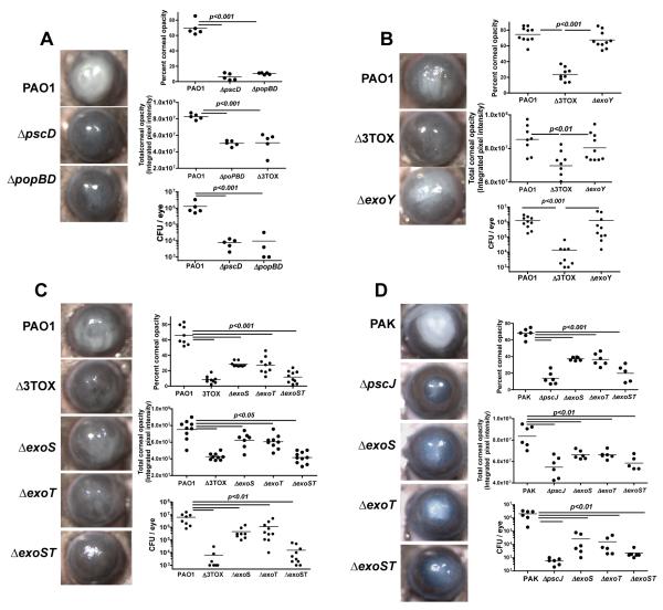
Pseudomonas aeruginosa is a leading cause of blinding corneal ulcers worldwide. To determine the role of type III secretion in the pathogenesis of P. aeruginosa keratitis, corneas of C57BL/6 mice were infected with P. aeruginosa strain PAO1 or PAK, which expresses ExoS, ExoT, and ExoY, but not ExoU. PAO1- and PAK-infected corneas developed severe disease with pronounced opacification and rapid bacterial growth. In contrast, corneas infected with ΔpscD or ΔpscJ mutants that cannot assemble a type III secretion system, or with mutants lacking the translocator proteins, do not develop clinical disease, and bacteria are rapidly killed by infiltrating neutrophils. Furthermore, survival of PAO1 and PAK strains in the cornea and development of corneal disease was impaired in ΔexoS, ΔexoT, and ΔexoST mutants of both strains, but not in a ΔexoY mutant. ΔexoST mutants were also rapidly killed in neutrophils in vitro and were impaired in their ability to promote neutrophil apoptosis in vivo compared with PAO1. Point mutations in the ADP ribosyltransferase (ADPR) regions of ExoS or ExoT also impaired proapoptotic activity in infected neutrophils, and exoST(ADPR-) mutants replicated the ΔexoST phenotype in vitro and in vivo, whereas mutations in rho-GTPase-activating protein showed the same phenotype as PAO1. Together, these findings demonstrate that the pathogenesis of P. aeruginosa keratitis in ExoS- and ExoT-producing strains is almost entirely due to their ADPR activities, which subvert the host response by targeting the antibacterial activity of infiltrating neutrophils.
Figures


Similar articles
-
Exotoxin S secreted by internalized Pseudomonas aeruginosa delays lytic host cell death.PLoS Pathog. 2022 Feb 7;18(2):e1010306. doi: 10.1371/journal.ppat.1010306. eCollection 2022 Feb. PLoS Pathog. 2022. PMID: 35130333 Free PMC article.
-
Translocon-independent intracellular replication by Pseudomonas aeruginosa requires the ADP-ribosylation domain of ExoS.Microbes Infect. 2012 Dec;14(15):1366-73. doi: 10.1016/j.micinf.2012.08.007. Epub 2012 Aug 30. Microbes Infect. 2012. PMID: 22981600 Free PMC article.
-
Type III Secretion System of Pseudomonas aeruginosa Affects Matrix Metalloproteinase 12 (MMP-12) and MMP-13 Expression via Nuclear Factor κB Signaling in Human Carcinoma Epithelial Cells and a Pneumonia Mouse Model.J Infect Dis. 2016 Sep 15;214(6):962-9. doi: 10.1093/infdis/jiw278. Epub 2016 Jul 4. J Infect Dis. 2016. PMID: 27377745
-
Pseudomonas aeruginosa ExoS and ExoT.Rev Physiol Biochem Pharmacol. 2004;152:79-92. doi: 10.1007/s10254-004-0031-7. Epub 2004 Aug 24. Rev Physiol Biochem Pharmacol. 2004. PMID: 15375697 Review.
-
Role of Pseudomonas aeruginosa type III effectors in disease.Curr Opin Microbiol. 2009 Feb;12(1):61-6. doi: 10.1016/j.mib.2008.12.007. Epub 2009 Jan 23. Curr Opin Microbiol. 2009. PMID: 19168385 Review.
Cited by
-
Cell-type-specific hypertranslocation of effectors by the Pseudomonas aeruginosa type III secretion system.Mol Microbiol. 2021 Feb;115(2):305-319. doi: 10.1111/mmi.14617. Epub 2020 Nov 5. Mol Microbiol. 2021. PMID: 33012037 Free PMC article.
-
Non-apoptotic toxicity of Pseudomonas aeruginosa toward murine cells.PLoS One. 2013;8(1):e54245. doi: 10.1371/journal.pone.0054245. Epub 2013 Jan 24. PLoS One. 2013. PMID: 23358229 Free PMC article.
-
Pseudomonas aeruginosa ExoS Induces Intrinsic Apoptosis in Target Host Cells in a Manner That is Dependent on its GAP Domain Activity.Sci Rep. 2018 Sep 19;8(1):14047. doi: 10.1038/s41598-018-32491-2. Sci Rep. 2018. PMID: 30232373 Free PMC article.
-
Intracellular replication of Pseudomonas aeruginosa in epithelial cells requires suppression of the caspase-4 inflammasome.mSphere. 2023 Oct 24;8(5):e0035123. doi: 10.1128/msphere.00351-23. Epub 2023 Aug 17. mSphere. 2023. PMID: 37589460 Free PMC article.
-
ExsB is required for correct assembly of the Pseudomonas aeruginosa type III secretion apparatus in the bacterial membrane and full virulence in vivo.Infect Immun. 2015 May;83(5):1789-98. doi: 10.1128/IAI.00048-15. Epub 2015 Feb 17. Infect Immun. 2015. PMID: 25690097 Free PMC article.
References
-
- Al-Hazzaa SA, Tabbara KF. Bacterial keratitis after penetrating keratoplasty. Ophthalmology. 1988;95:1504–1508. - PubMed
-
- Bharathi MJ, Ramakrishnan R, Meenakshi R, Kumar CS, Padmavathy S, Mittal S. Ulcerative keratitis associated with contact lens wear. Indian J Ophthalmol. 2007;55:64–67. - PubMed
-
- Green M, Apel A, Stapleton F. Risk factors and causative organisms in microbial keratitis. Cornea. 2008;27:22–27. - PubMed
-
- Willcox MD. Pseudomonas aeruginosa infection and inflammation during contact lens wear: a review. Optom Vis Sci. 2007;84:273–278. - PubMed
Publication types
MeSH terms
Substances
Grants and funding
LinkOut - more resources
Full Text Sources
Other Literature Sources
Molecular Biology Databases
