. 2012 Feb 15;188(4):1953-60.
doi: 10.4049/jimmunol.1102543. Epub 2012 Jan 16.

Activation of the NLRP3 inflammasome by group B streptococci

Affiliations

Activation of the NLRP3 inflammasome by group B streptococci

Alessandro Costa et al. J Immunol. .

Abstract

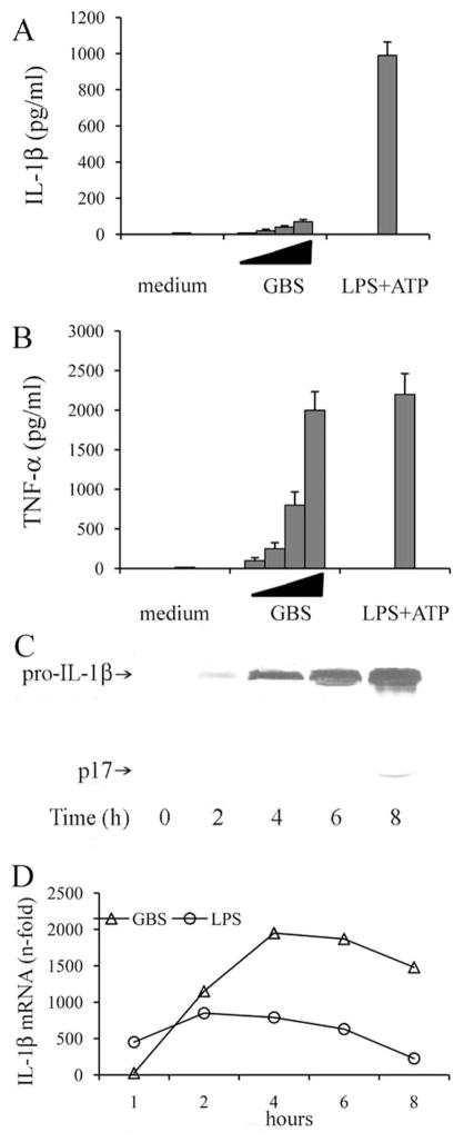
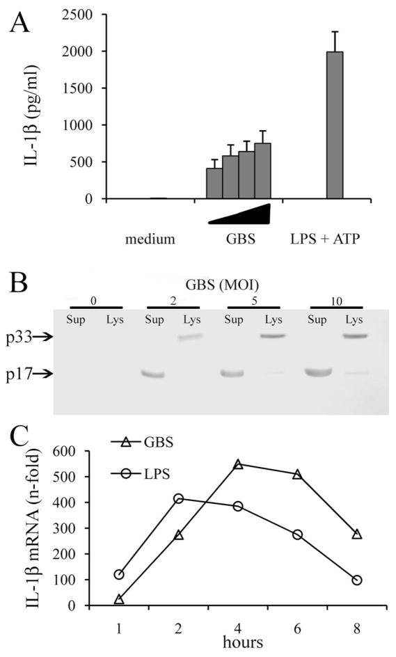
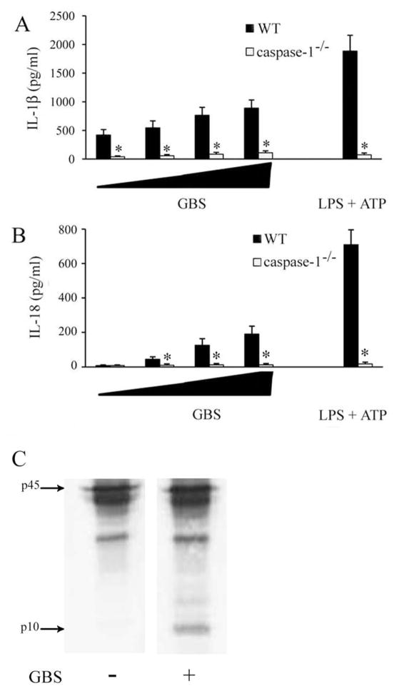
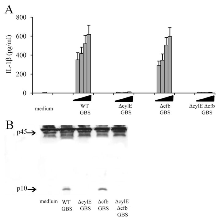
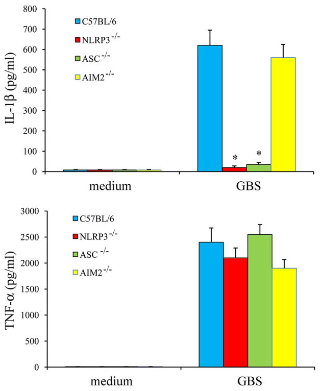
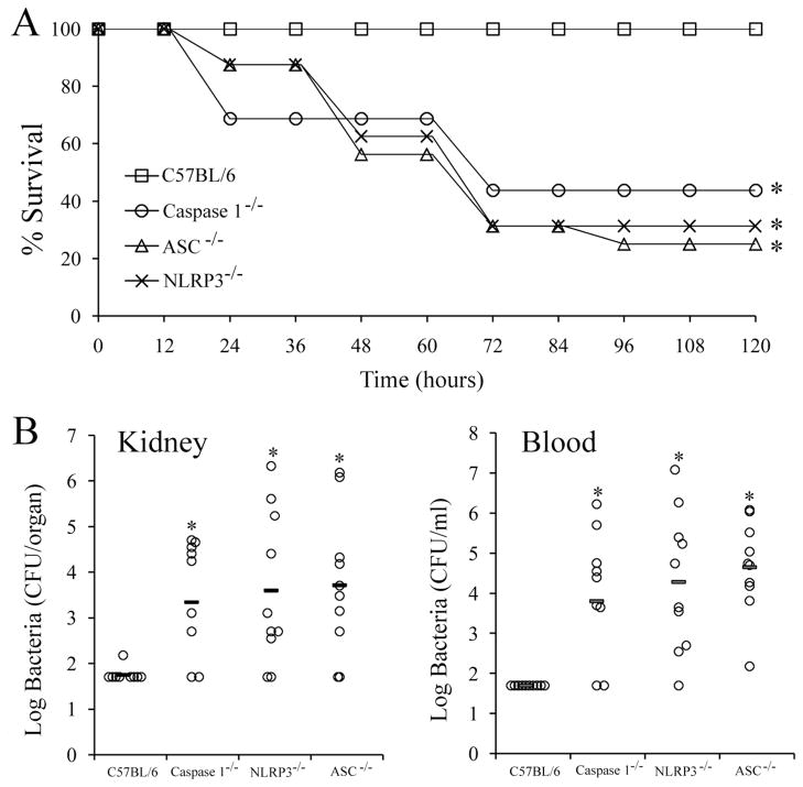
Group B Streptococcus (GBS) is a frequent agent of life-threatening sepsis and meningitis in neonates and adults with predisposing conditions. We tested the hypothesis that activation of the inflammasome, an inflammatory signaling complex, is involved in host defenses against this pathogen. We show in this study that murine bone marrow-derived conventional dendritic cells responded to GBS by secreting IL-1β and IL-18. IL-1β release required both pro-IL-1β transcription and caspase-1-dependent proteolytic cleavage of intracellular pro-IL-1β. Dendritic cells lacking the TLR adaptor MyD88, but not those lacking TLR2, were unable to produce pro-IL-1β mRNA in response to GBS. Pro-IL-1β cleavage and secretion of the mature IL-1β form depended on the NOD-like receptor family, pyrin domain containing 3 (NLRP3) sensor and the apoptosis-associated speck-like protein containing a caspase activation and recruitment domain adaptor. Moreover, activation of the NLRP3 inflammasome required GBS expression of β-hemolysin, an important virulence factor. We further found that mice lacking NLRP3, apoptosis-associated speck-like protein, or caspase-1 were considerably more susceptible to infection than wild-type mice. Our data link the production of a major virulence factor by GBS with the activation of a highly effective anti-GBS response triggered by the NLRP3 inflammasome.


Conflict of interest statement

Disclosures

The authors have no financial conflicts of interest

Figures

Fig. 1
Fig. 1. GBS induces the production and intracellular accumulation of high levels of pro-IL-1β in BMDMs
BMDMs were stimulated with GBS at MOIs of 2, 5, 10 and 20 for 8h. The positive control consisted of cells pretreated with LPS (0.1μg/ml) for 3h and then pulsed with ATP (5mM) for 30 min. IL-1β (A) and TNF-α (B) release in culture supernatants were measured by ELISA. Data are expressed as mean ± SD of three independent experiments. C, Lysates from BMDMs (1 × 10⁶/well) infected for the indicated times with GBS at a MOI of 10 were immunoblotted with anti-IL-1β. Results are representative of two independent experiments. D, RT-PCR measurements of pro-IL-1β mRNA expression in BMDMs at the indicated times after stimulation with GBS (MOI 10) or LPS (0.1 μg/ml). Data are presented as ‘fold’ increases in mRNA expression relative to uninfected cells, using a total of 3 × 10⁶ BMDMs. Data are from one experiment representative of three independent ones.
Fig. 2
Fig. 2. GBS induces the release of Il-1β in BMDCs
A, BMDCs were stimulated with GBS at MOIs of 2, 5, 10 and 20 for 8h. The positive control consisted of cells pretreated with LPS (0.1μg/ml) for 3h and then pulsed with ATP (5mM) for 30 min. IL-1β release in culture supernatants was measured by ELISA. Data are expressed as means ± SD of three independent experiments. B, Supernatants or lysates from BMDCs (1 × 10⁶/well) infected with GBS at the indicated MOIs were collected at 8h after infection, concentrated and immunoblotted with anti-IL-1β. Results are representative of two independent experiments. C, RT-PCR measurement of pro-IL-1β mRNA expression in BMDCs at different times after stimulation with GBS (MOI 10) or LPS (0.1 μg/ml). Data are presented as ‘fold’ increases in mRNA expression relative to uninfected cells, using a total of 3 × 10⁶ BMDCs. Data are from one experiment representative of three independent ones.
Fig. 3
Fig. 3. GBS-induced IL-1β production is dependent on MyD88 and phagocytosis
IL-1β (A, B) and TNF-α (D) protein secretion in culture supernatants of BMDCs lacking various TLR adaptors (A, D) or TLRs (B), after stimulation with GBS (MOI 10) or with LPS and ATP. C, RT-PCR analysis of the expression of pro-IL-1β mRNA in BMDCs, lacking TLR-2, TLR-9 or MyD88, exposed for various times (horizontal axis) to GBS (MOI 10), presented as ‘fold increase’ (vertical axis) in expression relative to uninfected cells. E, IL-1β and TNF-α concentrations in supernatants of BMDCs treated with cytochalasin D (5 μg/ml) or bafilomycin A (1μM) and stimulated with GBS (MOI 10) or LPS and ATP (inset). Data are expressed as means ± SD of three independent observations, each conducted on a different animal. *, p<0.05 vs untreated cells by one-way analysis of variance and the Student’s-Keuls-Newman test.
Fig. 4
Fig. 4. Caspase 1 is essential for IL-1β secretion by BMDCs in response to GBS
IL-1β (A) and IL-18 (B) protein secretion in culture supernatant of BMDCs from WT and caspase-1 deficient mice infected for 8h with GBS at MOI of 2, 5, 10 and 20. The positive control consisted of cells pretreated with LPS (0.1μg/ml) for 3h and then pulsed with ATP (5mM) for 30 min. *, p<0.05 vs untreated cells by one-way analysis of variance and the Student’s-Keuls-Newman test. Data are expressed as mean ± SD of three independent experiments. Western blot analysis (C) of BMDCs lysates from WT mice infected with GBS (MOI 10) for 8h, after treatment with anti-caspase-1. Results are representative of two independent experiments.
Fig. 5
Fig. 5. β-hemolysin is essential for GBS stimulated IL-1β release
IL-1β release (A) and Western blot analysis (B) of BMDCs infected for 8h with GBS (MOI 10). WT strain NEM316 serotype III or its isogenic mutants deficient in β-hemolysin (ΔcylE), in CAMP-factor (Δcfb)- or in both (ΔcylE Δcfb) were used. A, Data are expressed as means ± SD of three independent observations, each conducted on a different animal. B, Results are representative of two independent experiments
Fig. 6
Fig. 6. NLRP3 and ASC are essential for IL-1β release from BMDCs infected with GBS
IL-1β (A) and TNF-α (B) protein secretion in culture supernatants of BMDCs from WT, NLRP3-, ASC- and AIM2-deficient mice infected with GBS for 8h at a MOI of 10. Data are expressed as means ± SD of three independent observations, each conducted on a different animal. *, p<0.05 vs cells from WT mice by one-way analysis of variance and the Student’s-Keuls-Newman test.
Fig. 7
Fig. 7. Mice lacking caspase-1, ASC and NLP3 are highly susceptible to GBS infection
A, Survival of WT C57BL/6, caspase-1-, ASC-, NLRP3-deficient mice (16 per group) after i.p. challenge with 1 × 10⁵ CFU of GBS. *, p<0.05 vs WT mice by Kaplan-Meier survival plots. B, bacterial numbers in the kidney and blood after challenge, as above described. Log CFU are expressed as means ± SD of 10 determinations, each conducted on a different animal. *, p<0.05 vs WT mice by one-way analysis of variance and the Student’s-Keuls-Newman test.

Similar articles

Cited by

References

    1. Akira S, Uematsu S, Takeuchi O. Pathogen recognition and innate immunity. Cell. 2006;124:783–801. - PubMed
    1. Ishii KJ, Koyama S, Nakagawa A, Coban C, Akira S. Host innate immune receptors and beyond: making sense of microbial infections. Cell Host Microbe. 2008;3:352–363. - PubMed
    1. Ye Z, Ting JP. NLR, the nucleotide-binding domain leucine-rich repeat containing gene family. Curr Opin Immunol. 2008;20:3–9. - PubMed
    1. Franchi L, Park JH, Shaw MH, Marina-Garcia N, Chen G, Kim YG, Núñez G. Intracellular NOD-like receptors in innate immunity, infection and disease. Cell Microbiol. 2008;10:1–8. - PubMed
    1. Yu HB, Finlay BB. The caspase-1 inflammasome: a pilot of innate immune responses. Cell Host Microbe. 2008;4:198–208. - PubMed

Publication types

MeSH terms