Review
. 2012 Apr;121(1):4-27.
doi: 10.1111/j.1471-4159.2012.07664.x. Epub 2012 Feb 2.

Glial cells in (patho)physiology

Affiliations
Review

Glial cells in (patho)physiology

Vladimir Parpura et al. J Neurochem. 2012 Apr.

Abstract

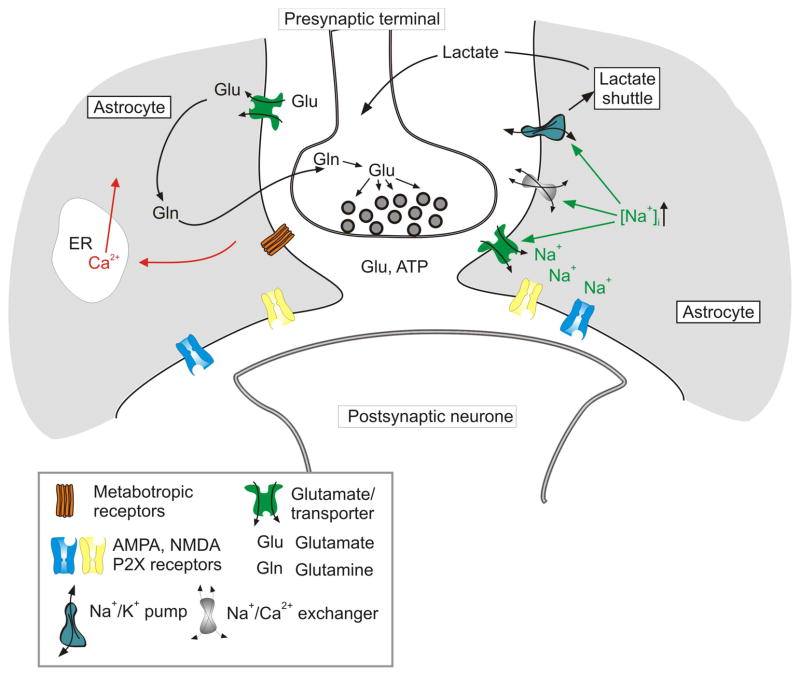
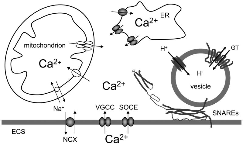
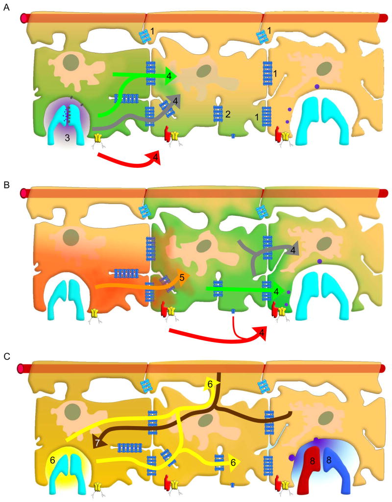
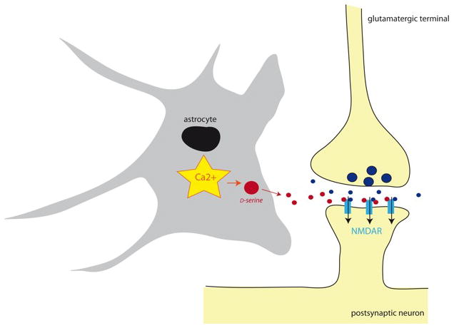
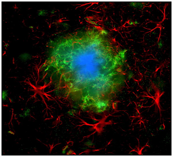
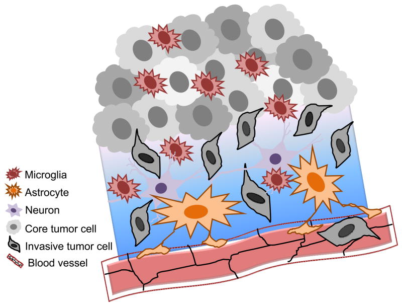
Neuroglial cells define brain homeostasis and mount defense against pathological insults. Astroglia regulate neurogenesis and development of brain circuits. In the adult brain, astrocytes enter into intimate dynamic relationship with neurons, especially at synaptic sites where they functionally form the tripartite synapse. At these sites, astrocytes regulate ion and neurotransmitter homeostasis, metabolically support neurons and monitor synaptic activity; one of the readouts of the latter manifests in astrocytic intracellular Ca(2+) signals. This form of astrocytic excitability can lead to release of chemical transmitters via Ca(2+) -dependent exocytosis. Once in the extracellular space, gliotransmitters can modulate synaptic plasticity and cause changes in behavior. Besides these physiological tasks, astrocytes are fundamental for progression and outcome of neurological diseases. In Alzheimer's disease, for example, astrocytes may contribute to the etiology of this disorder. Highly lethal glial-derived tumors use signaling trickery to coerce normal brain cells to assist tumor invasiveness. This review not only sheds new light on the brain operation in health and disease, but also points to many unknowns.


Figures

Figure 1
Figure 1
Schematic representation of the various types of co-existing glial domains, illustrated layered over two background drawings: i) a pyramidal cell (left) from Ramón y Cajal (Ramón y Cajal 1911); and ii) a figure from Vogt and Vogt (Vogt and Vogt 1937), showing the transition (vertical arrows) between the striate (center) and the occipital (right) cortex in a human brain. Glial cells form a specific functional organization at different levels from nanodomains up to superdomains for their interaction with neurons. Individual synapses are associated with their ensheathing glial microdomains, but parallel stimulation of related inputs may integrate (oligo-)cellular domains involving the whole glial cell (or a few of them) and their neuronal partners. Such cellular domains can be of columnar or spherical (center, n1) shape. Appropriate stimulation may then activate, via gap junctional coupling, networks (center, ni and nii), which are likely dynamic and consist of many astrocytes. Macrodomains of variable size may form depending on neuronal activity. A further progression of integration will result in the generation of very large functional units, so-called superdomains, corresponding to entire cortical areas or gyri. Eventually, even a whole hemisphere associated with huge astrocytic populations may transiently be involved, putatively mediating events such as spreading depression and seizures.
Figure 2
Figure 2
Brain tissue is largely divided between domains of astrocytes. Mouse astrocytes, whose gap juncrions were closed, were filled with two different dyes to visualize their respective domains. In a human brain, a single astrocyte domain can encompass over million neuronal synapses. Reprinted from (Pekny and Wilhelmsson 2006).
Figure 3
Figure 3
Local signaling mediated by ionotropic receptors and transporters in astroglial perisynaptic processes. Synaptic release of neurotransmitters (glutamate and/or ATP) activates ionotropic receptors and glutamate transporter, which generate Na+ and Ca2+ influx. Increase in [Na+]i can assume a signaling role through modulating neurotransmitter transporters, switching the reverse mode of NCX and stimulating Na+/K+ pump. This in turn can influence synaptic transmission and plasticity by affecting the time kinetics of glutamate removal from the cleft and through stimulating local metabolic support via the lactate shuttle.
Figure 4
Figure 4
Ca2+- dependent vesicular release of gliotransmitters from astrocytes. The sources of Ca2+ for cytosolic Ca2+ increase are: the endoplasmic reticulum (ER) and the extracellular space (ECS). Cytosolic Ca2+ accumulation could be caused by the entry of Ca2+ from the ER store that possess inositol 1,4,5 trisphospate and ryanodine receptors. Store specific Ca2+-ATPase fills these stores with Ca2+. Ultimately, this (re)filling requires Ca2+ entry from the ECS through store-operated Ca2+ entry (SOCE). Additional Ca2+ entry from the ECS to the cytosol can be mediated by plasma membrane voltage -gated Ca2+ channels (VGCCs) and Na+/Ca2+ exchangers (NCXs), the latter operating in the reverse mode. Mitochondria represent a source/sink of cytosolic Ca2+. Mitochondrial Ca2+ uptake from the cytosol to its matrix is mediated by the Ca2+uniporter. Free Ca2+ exits the mitochondrial matrix through the Na+/Ca2+ exchanger and also by brief openings of the mitochondrial permeability transition pore. Increase of cytosolic Ca2+ is sufficient and necessary to cause vesicular fusions and release of gliotransmitters (GT). This process requires the activity of the ternary SNARE complex (SNAREs) consisting of: i) synaptobrevin 2 and/or cellubrevin located at vesicular membrane and ii) the binary cis complex pre-formed at the plasma membrane and composed of syntaxin (shown in an open form) and SNAP-23. Astrocytic vesicles are filled by various vesicular GT transporters, which for this action utilize the proton gradient generated by the vacuolar type H+-ATPase. Drawing is not to scale.
Figure 5
Figure 5
Astrocyte gap junction mediated pathways for modulation of synaptic transmission. A) Gap junctions between astrocytes (1), or reflexive gap junctions between processes of a cell (2) facilitate movement of ions, second messengers, and nutrients within and between astrocytes. Synaptic activity (3) stimulates a Ca2+ wave (4) that is transmitted through gap junctions by Ca2+ (green), InsP3 (gray), and primarily through pannexin 1 channels (yellow channel) and, under some conditions, unpaired connexon channel opening that allows release of ATP (red arrow). B) The Ca2+ wave (4) spreads to a third astrocyte to the right. Na+ spikes are buffered and spread to adjacent cells (5). C) Increased K+ is taken up from areas of neuronal activity and spatially buffered by gap junctions (6). Metabolic fuel (7) is delivered through gap junctions in a directional manner, from blood vessels to areas of synaptic activity. Gliotransmitter release and other changes in astrocyte activity lead to modulation of the synaptic activity due to gap junction communication between astrocytes (8). Note at the top of each panel of this figure the presence of the blood vessel (illustrated as a horizontal red rod) which is enwrapped by the astroglial endfeet. Astroglia play active roles in so-called “neurovascular coupling” both by inducing the blood-brain barrier and through release of vasoactive agents.
Figure 6
Figure 6
Increase in intracellular Ca2+ levels triggers exocytotic release of D-serine from astrocytes. As D-serine (red) reaches the synaptic cleft, it binds to post-synaptic NMDA receptors (NMDARs), which also binds glutamate (blue) released from the pre-synaptic terminal. This co-incidental NMDAR detection favors NMDAR activation and induction of the most common forms of synaptic plasticity. Astrocytic nucleus is drawn as a black oval.
Figure 7
Figure 7
Astroglia assembling around a plaque in an amyloid precursor protein/presenilin 1 transgenic mouse. Astrocytes (red) are labeled using indirect immunochemistry and antibody against glial fibrillary acidic protein, while the core and periphery of the β-amlyoid (Aβ) plaque are stained using methoxy-XO4 [binding Aβ fibrils; (Klunk et al. 2002)] and monoclonal antibody IC16 [binding Aβ dimer, with the epitope at residues 2–8 of Aβ; (Muller-Schiffmann et al. 2011)], respectively. Courtesy of Drs. Markus Kummer and Michael Heneka.
Figure 8
Figure 8
Intricate interactions of glioblastoma multiforme (GBM) tumor edge with the brain microenvironment. Cells within the core GBM mass are heterogeneous with ~30% microglia residing within. Tumor cells secrete various motility factors that maintain the process of invasion and express receptors for factors produced by normal brain cells to smooth the progress of tumor infiltration, e.g., bradykinin concentration gradient formed predominantly from endothelial cells attracts tumor cells and increases their motility directing them towards blood vessels. Glial cells in surrounding also respond to bradykinin, i.e., astrocytes by releasing a number of signaling molecules, and increasing matrix metalloproteinase (MMP)-9 expression and cell migration, while microglia by increasing mobility. Microglia further facilitates glioma invasion by releasing membrane type 1 MMP/MMP-14 essential for activation of an inactive MMP-2 pro-form secreted by glioma cells. Migration of glioma cells away from the core GBM mass may lead to the formation of the satellite tumors. Drawing is not to scale.

Similar articles

Cited by

References

    1. Agulhon C, Fiacco TA, McCarthy KD. Hippocampal short- and long-term plasticity are not modulated by astrocyte Ca2+ signaling. Science. 2010;327:1250–1254. - PubMed
    1. Agulhon C, Petravicz J, McMullen AB, Sweger EJ, Minton SK, Taves SR, Casper KB, Fiacco TA, McCarthy KD. What is the role of astrocyte calcium in neurophysiology? Neuron. 2008;59:932–946. - PMC - PubMed
    1. Andriezen WL. The neuroglia elements of the brain. Brit Med J. 1893;2:227–230. - PMC - PubMed
    1. Anlauf E, Derouiche A. Astrocytic exocytosis vesicles and glutamate: a high-resolution immunofluorescence study. Glia. 2005;49:96–106. - PubMed
    1. Araque A, Parpura V, Sanzgiri RP, Haydon PG. Glutamate-dependent astrocyte modulation of synaptic transmission between cultured hippocampal neurons. Eur J Neurosci. 1998;10:2129–2142. - PubMed

Publication types