. 2012 Feb 13;13(2):342-9.
doi: 10.1021/bm201763n. Epub 2012 Feb 1.

Promoting nerve cell functions on hydrogels grafted with poly(L-lysine)

Affiliations

Promoting nerve cell functions on hydrogels grafted with poly(L-lysine)

Lei Cai et al. Biomacromolecules. .

Abstract

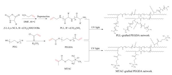
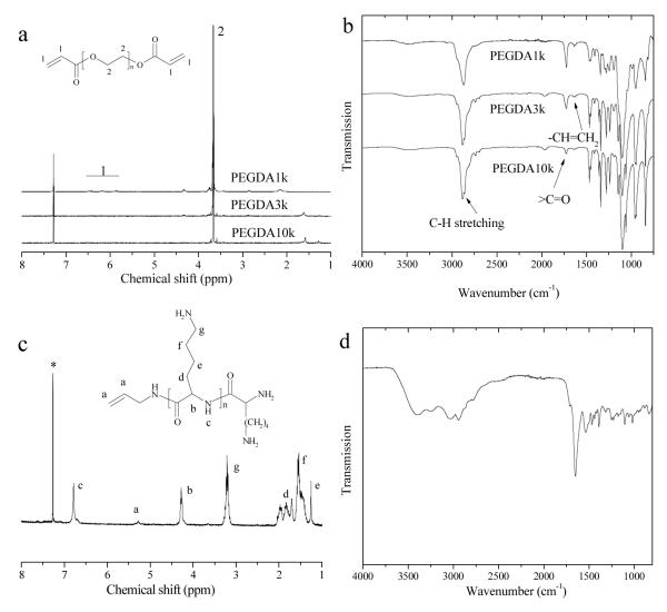
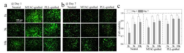
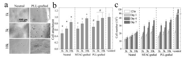
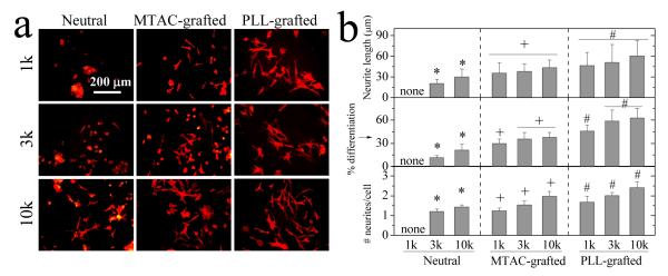
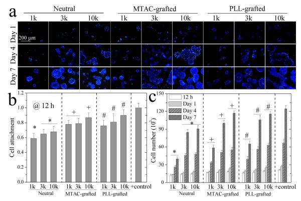
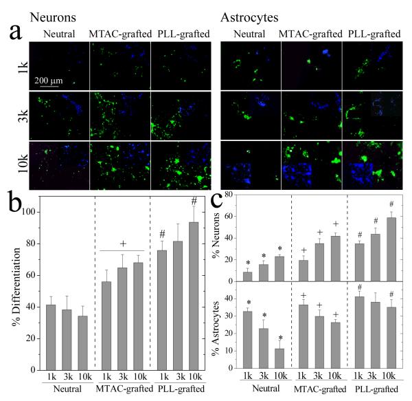
We present a novel photopolymerizable poly(L-lysine) (PLL) and use it to modify polyethylene glycol diacrylate (PEGDA) hydrogels for creating a better, permissive nerve cell niche. Compared with their neutral counterparts, these PLL-grafted hydrogels greatly enhance pheochromocytoma (PC12) cell survival in encapsulation, proliferation, and neurite growth and also promote neural progenitor cell proliferation and differentiation capacity, represented by percentages of both differentiated neurons and astrocytes. The role of efficiently controlled substrate stiffness in regulating nerve cell behavior is also investigated and a polymerizable cationic small molecule, [2-(methacryloyloxy)ethyl]-trimethylammonium chloride (MTAC), is used to compare with this newly developed PLL. The results indicate that these PLL-grafted hydrogels are promising biomaterials for nerve repair and regeneration.


Figures

Figure 1
Figure 1
Synthesis of polymerizable PLL and PEGDA, and preparation of PEGDA networks grafted with PLL or MTAC via photo-crosslinking.
Figure 2
Figure 2
(a) NMR and (b) IR spectra of PEGDA1k, 3k, and 10k, (c) NMR and (d) IR spectra of photo-crosslinkable PLL. *: CDCl3 peak.
Figure 3
Figure 3
PC12 cells encapsulated in neutral, MTAC-grafted, and PLL-grafted PEGDA hydrogels for (a) 1 and (b) 7 days. Scale bar of 100 μm is applicable to all. (c) Cell viability after encapsulation in the hydrogels for 1 and 7 days. p < 0.05 between neutral and PLL-grafted hydrogels made from the same PEGDAs at day 7; between hydrogels made from PEGDA1k and 3k or 10k; between day 1 and day 7.
Figure 4
Figure 4
PC12 cell attachment and proliferation on the hydrogels. (a) Phase contrast images of PC12 cells cultured for 7 days. Scale bar of 100 μm is applicable to all. (b) PC12 cell attachment at 12 h post-seeding. *: p < 0.05 between two marked samples. +,#: p < 0.05 between two marked samples and compared to the corresponding data on the neutral hydrogels. Positive (+) control: TCPS. (c) PC12 cell proliferation at 12 h, days 1, 4, and 7 post-seeding. p < 0.05 between hydrogels made from the same PEGDAs at the same time, except for between PLL-grafted hydrogels of PEG3k and 10k at 12 h and day 1. Positive (+) control: TCPS.
Figure 5
Figure 5
PC12 cell differentiation on the hydrogels. (a) Fluorescent images of PC12 neurites induced by NGF at day 7. Stained with rhodamine-phalloidin. Scale bar of 200 μm is applicable to all. (b) Neurite length, percentage of differentiated cells, and the number of neurites per cell for PC12 cells at day 7. *: p < 0.05 between two marked samples. +,#: p < 0.05 between two marked samples and relative to neutral hydrogels made from the same PEGDAs.
Figure 6
Figure 6
NPC proliferation on the hydrogels. (a) Nuclei (blue) of NPCs for 1, 4, and 7 days. Scale bar of 200 μm is applicable to all. (b) NPC attachment at 12 h post-seeding. Positive (+) control: TCPS. (c) NPC cell number at 12 h, days 1, 4, and 7 post-seeding. *: p < 0.05 between two marked samples at the same time. +,#: p < 0.05 between two marked samples and relative to the neutral hydrogels made from the same PEGDAs at the same time. Positive (+) control: TCPS.
Figure 7
Figure 7
NPC differentiation on the hydrogels. (a) NPC differentiated neurons stained with anti-β-tubulin-III (green, left side) and astrocytes stained with anti-GFAP (green, right side) at day 7. Inset is DAPI nuclear staining (blue) of the same field. Scale bar of 200 μm is applicable to all. (b) Percentage of differentiated NPCs. +: p < 0.05 relative to neutral and PLL-grafted hydrogels made from the same PEGDAs. #: p < 0.05 between two marked samples. (c) Percentages of differentiated neurons and astrocytes. *: p < 0.05 between two marked samples. +,#: p < 0.05 between two marked samples and relative to neutral hydrogels made from the same PEGDAs.

References

    1. Schmidt CE, Leach JB. Annu. Rev. Biomed. Eng. 2003;5:293. - PubMed
    1. Madigan NN, McMahon S, O’Brien T, Yaszemski MJ, Windebank AJ. Respir. Physiol. Neurobiol. 2009;169:183. - PMC - PubMed
    1. Wang S, Cai L, Kulshrestha AS, Mahapatro A, Henderson LA. Biomaterials. Vol. 1054. American Chemical Society; Washington, DC: 2010. pp. 43–63.
    1. Wang S, Cai L. Int. J. Polym. Sci. 2010:138686.
    1. Wang S, Kempen DH, Simha NK, Lewis JL, Windebank AJ, Yaszemski MJ, Lu L. Biomacromolecules. 2008;9:1229. - PMC - PubMed

Publication types

MeSH terms