. 2012 Mar;279(6):1000-13.
doi: 10.1111/j.1742-4658.2012.08489.x. Epub 2012 Feb 27.

Role of α-synuclein penetration into the membrane in the mechanisms of oligomer pore formation

Affiliations

Role of α-synuclein penetration into the membrane in the mechanisms of oligomer pore formation

Igor F Tsigelny et al. FEBS J. 2012 Mar.

Abstract

Parkinson's disease (PD) and dementia with Lewy bodies are common disorders of the aging population and characterized by the progressive accumulation of α-synuclein (α-syn) in the central nervous system. Aggregation of α-syn into oligomers with a ring-like appearance has been proposed to play a role in toxicity. However, the molecular mechanisms and the potential sequence of events involved in the formation of pore-like structures are unclear. We utilized computer modeling and cell-based studies to investigate the process of oligomerization of wild-type and A53T mutant α-syn in membranes. The studies suggest that α-syn penetrates the membrane rapidly, changing its conformation from α-helical towards a coiled structure. This penetration facilitates the incorporation of additional α-syn monomers in the complex, and the subsequent displacement of phospholipids and the formation of oligomers in the membrane. This process occurred more rapidly, and with a more favorable energy of interaction, for mutant A53T compared with wild-type α-syn. After 4 ns of simulation of the protein-membrane model, α-syn had penetrated through two-thirds of the membrane. By 9 ns, the penetration of the annular α-syn oligomers can result in the formation of pore-like structures that fully perforate the lipid bilayer. Experimental incubation of recombinant α-syn in synthetic membranes resulted in the formation of similar pore-like complexes. Moreover, mutant (A53T) α-syn had a greater tendency to accumulate in neuronal membrane fractions in cell cultures, resulting in greater neuronal permeability, as demonstrated with the calcein efflux assay. These studies provide a sequential molecular explanation for the process of α-syn oligomerization in the membrane, and support the role of formation of pore-like structures in the pathogenesis of the neurodegenerative process in PD.


Figures

Figure 1
Figure 1. Simulated penetration of α-syn into the membrane
(A) Initial position of α-syn before membrane penetration. Highlighted residues interact with the membrane upon the initial contact. This position was obtained using our program MAPAS. (B) Prediction of the possible transmembrane character of the α-syn primary sequence. Score of the possible transmembrane character of the initial α-syn helix. The existing transmembrane helices typically have a score above 500. A score of approximately 400 suggests that this helix has a tendency to be transmembrane and may insert into the membrane. (C) Evolution of α-syn α-helices during membrane penetration. (D) Evolution Rate of formation of α-syn π-helices during membrane penetration.
Figure 2
Figure 2. Time course and structural analysis of α-syn penetration into the membrane
(A) Snapshots of wild type α-syn at different time points during membrane penetration (no membrane in image), (B) Snapshot of wild type α-syn at different time points during membrane penetration (membrane present). (C) Close-up of the membrane penetrating region of wild type α-syn. (D) Snapshots of A53T mutant α-syn at different time points during membrane penetration (no membrane in image), (E) Snapshot of A53T mutant α-syn at different time points during membrane penetration (membrane present). (F) Close-up of the membrane penetrating region of the A53T mutant α-syn. In images A, B, D and E: 0 ns-cyan; 2 ns-violet; 4 ns-pink; 6 ns-yellow; 8 ns-red; 9 ns-magenta. For A53T the deepest penetration is achieved at 7 ns therefore the snapshot 9 ns is not shown. Arrows point to the A53 residues (CPK, red) and T53 (CPK, yellow).
Figure 3
Figure 3. Characterization of the amino acid residues interacting with lipids in the membrane in the membrane-penetrating region of wild type α-syn
(A) Initial position, (B) 3 ns, (C) 5 ns and (D) 7 ns snapshots of membrane penetration by wild type α-syn. The stick-like structures represent the residues that interact with the phospholipids and the insets represent an overview of the position of the α-syn interaction with the membrane.
Figure 4
Figure 4. Analysis of α-syn membrane penetration utilizing lateral projection for the wild type and A53T mutant α-syn
(A, B) Initial position on the membrane of wild type (yellow) and A53T (red) mutant α-syn monomers respectively, (C, D) 4 ns position of wild type and A53T mutant α-syn respectively and (E, F) 7 ns position of wild type and A53T mutant α-syn respectively. (G) Distance between the front penetrating residue of α-syn to the median of the membrane (red – wild type, blue – A53T mutant). (H) Van der Waals energies of α-syn during membrane penetration (red – wild type, blue – A53T mutant). Arrows point to the A53 residues (CPK, red) and T53 (CPK, yellow).
Figure 5
Figure 5. Analysis of β-synuclein and mutated α-syn embedding into the membrane utilizing lateral projection
(A) β-synuclein initial position on the membrane and progression after 4 and 7 ns. (B) Docking and MD simulatiosn with mutated (E57A, E61A, K58A,Q62A,N65A, V63A, V66A) α-syn. These aa substituations are predicted to reduce interactions with lipids in the membrane. Initial position on the membrane and progression after 4 and 7 ns
Figure 6
Figure 6. Model of α-syn octamer penetration into the membrane
(A, B, C) Initial position of α-syn octomer on the membrane in semi-transparent membrane view, side view and view from beneath respectively, (D, E, F) 4 ns snapshot of α-syn octomer on the membrane in semi-transparent membrane view, side view and view from beneath respectively and (G, H, I) 9 ns snapshot of α-syn octomer on the membrane in semi-transparent membrane view, side view and view from beneath respectively.
Figure 7
Figure 7. Structure of possible octamer constructed from the 9 ns conformers of α-syn
(A) The α-syn octomer viewed from above and (B) the side. The region highlighted in the yellow box is depicted in (C) which shows in greater detail the contact residues of neighboring α-syn molecules in the octamer. (D) Electrostatic energy changes during possible growth from the single wt α-syn molecule to octamer for the conformers taken at different times of MD of the single α-syn in the membrane. (E) Overlay of the model for the α-syn octomer with that obtained by incubating α-syn with synthetic lipid membrane and analysis by electron microscopy, merged image is on the right side.
Figure 8
Figure 8. Characterization of the effects of α-syn oligomers in a neuronal cell line
(A) Immunoblot analysis of α-syn species in the soluble (cytoplasmic) and membrane-bound (membrane) fractions from B103 cells infected with LV-wild type α-syn or LV-A53T α-syn, uninfected cells were used as a control. Analysis of α-syn monomers in the soluble and membrane-bound fractions from B103 cells infected with LV-wild type α-syn or LV-A53T α-syn. (B) Analysis of α-syn oligomers in the soluble and membrane-bound fractions from B103 cells infected with LV-wild type α-syn or LV-A53T α-syn. Co-localization of α-syn aggregates and the dendritic marker MAP2 in (C-F) control cells, (C) Native gel analysis of membrane fractions from control B103 cells and neurons infected with LV-wild type α-syn or LV-A53T α-syn (D-F) cells infected with LV-wild type α-syn and (J-L) cells infected with LV-A53T α-syn. (M, N) Calcein and calcium assays respectively in control cells or cells infected with LV-wild type α-syn or LV-A53T α-syn.
Figure 9
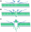
Figure 9. Schematic hypothesis of α-syn annular oligomer penetration into the membrane
(A) Hydrophilic residues of α-syn interact with polar heads on the surface of the membrane. (B) As α-syn begins to penetrate into the membrane, the hydrophobic residues interact with the non-polar tails of the lipids. (C) Finally, the hydrophilic residues participate in hydrogen bonding interactions with polar heads of lipids on the opposing side of the membrane bilayer allowing the molecule through the membrane.

References

    1. Wright PE, Dyson HJ. Intrinsically unstructured proteins: re-assessing the protein structure-function paradigm. J Mol Biol. 1999;293:321–331. - PubMed
    1. Lansbury PT., Jr Evolution of amyloid: what normal protein folding may tell us about fibrillogenesis and disease. Proc Natl Acad Sci U S A. 1999;96:3342–3344. - PMC - PubMed
    1. Chandra S, Gallardo G, Fernandez-Chacon R, Schluter OM, Sudhof TC. Alpha-synuclein cooperates with CSPalpha in preventing neurodegeneration. Cell. 2005;123:383–396. - PubMed
    1. Burre J, Sharma M, Tsetsenis T, Buchman V, Etherton MR, Sudhof TC. Alpha-synuclein promotes SNARE-complex assembly in vivo and in vitro. Science. 2010;329:1663–1667. - PMC - PubMed
    1. Lashuel HA, Petre BM, Wall J, Simon M, Nowak RJ, Walz T, Lansbury PT., Jr Alpha-synuclein, especially the Parkinson’s disease-associated mutants, forms pore-like annular and tubular protofibrils. J Mol Biol. 2002;322:1089–1102. - PubMed

Publication types