

Interleukin-1 receptor role in the viability of corneal myofibroblasts
- PMID: 22251454
- PMCID: PMC3296874
- DOI: 10.1016/j.exer.2011.12.022
Interleukin-1 receptor role in the viability of corneal myofibroblasts
Abstract
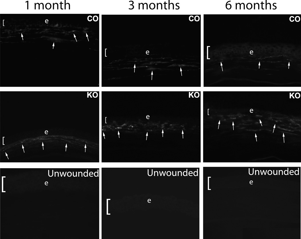
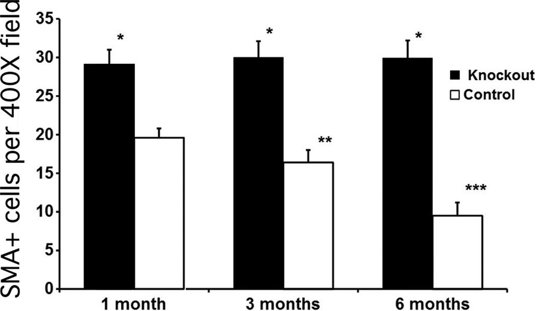
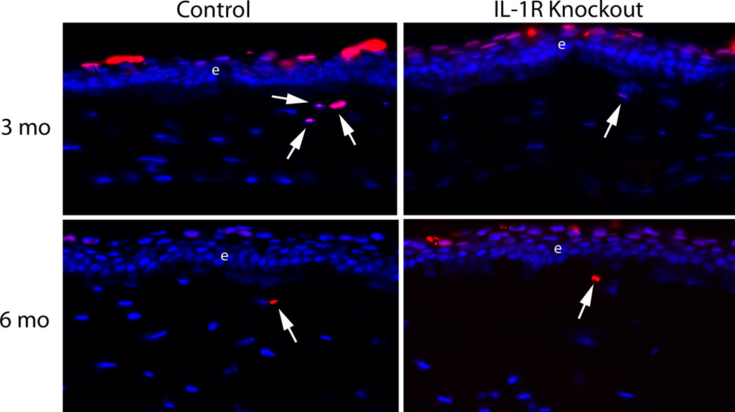
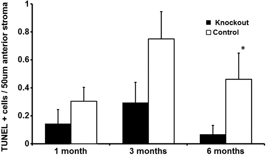
The purpose of this study was to investigate the role of interleukin-1 (IL-1) in modulating myofibroblast viability in mouse corneas with stromal opacity. Twenty-four female B6; 129S1-Il1r1tm1Roml/J homozygous IL-1RI knockout mice and 24 control B6129SF2/J mice were included in this study. Each mouse had opacity-generating irregular phototherapeutic keratectomy (PTK) performed with an excimer laser in one eye. Groups of 8 mice from each group were euthanized at one month, three months and six months after surgery and the eyes cryo-preserved. The contralateral eye served as unwounded control. Immunohistochemistry was performed for α-smooth muscle actin (SMA) in central sections of all corneas. The TUNEL assay for apoptosis was performed on 8 sections of four eyes from each group. No SMA+ cells were detected in the stroma of unwounded control or knockout corneas. SMA+ myofibroblast density was significantly higher (p < 0.001) in the IL-1RI knockout group than in the control group at one month, three and six months after irregular PTK. Mean TUNEL+ stromal cells in the anterior 50 μm of stroma was significantly lower in the IL-1RI knockout group compared to the control group at six months after irregular PTK (p = 0.04). These results corroborate the findings of recent in vitro work that demonstrated an antagonistic effect of TGFβ and IL-1 on myofibroblast viability, and found that IL-1-triggered myofibroblast apoptosis was suppressed by TGFβ. Thus, IL-1 is an important modulator of myofibroblast viability during corneal wound healing.
Copyright © 2012 Elsevier Ltd. All rights reserved.
Figures
Similar articles
-
A novel method for generating corneal haze in anterior stroma of the mouse eye with the excimer laser.Exp Eye Res. 2008 Feb;86(2):235-40. doi: 10.1016/j.exer.2007.10.014. Epub 2007 Nov 5. Exp Eye Res. 2008. PMID: 18068702 Free PMC article.
-
Stromal haze, myofibroblasts, and surface irregularity after PRK.Exp Eye Res. 2006 May;82(5):788-97. doi: 10.1016/j.exer.2005.09.021. Epub 2005 Nov 21. Exp Eye Res. 2006. PMID: 16303127 Free PMC article.
-
Corneal myofibroblast generation from bone marrow-derived cells.Exp Eye Res. 2010 Jul;91(1):92-6. doi: 10.1016/j.exer.2010.04.007. Epub 2010 Apr 24. Exp Eye Res. 2010. PMID: 20417632 Free PMC article.
-
The corneal fibrosis response to epithelial-stromal injury.Exp Eye Res. 2016 Jan;142:110-8. doi: 10.1016/j.exer.2014.09.012. Exp Eye Res. 2016. PMID: 26675407 Free PMC article. Review.
-
Two-phase mechanism in the treatment of corneal stromal fibrosis with topical losartan.Exp Eye Res. 2024 May;242:109884. doi: 10.1016/j.exer.2024.109884. Epub 2024 Apr 1. Exp Eye Res. 2024. PMID: 38570181 Review.
Cited by
-
Corneal myofibroblast biology and pathobiology: generation, persistence, and transparency.Exp Eye Res. 2012 Jun;99(1):78-88. doi: 10.1016/j.exer.2012.03.018. Epub 2012 Apr 20. Exp Eye Res. 2012. PMID: 22542905 Free PMC article. Review.
-
Cell Biology of Spontaneous Persistent Epithelial Defects After Photorefractive Keratectomy in Rabbits.Transl Vis Sci Technol. 2023 May 1;12(5):15. doi: 10.1167/tvst.12.5.15. Transl Vis Sci Technol. 2023. PMID: 37184499 Free PMC article.
-
Corneal wound healing.Exp Eye Res. 2020 Aug;197:108089. doi: 10.1016/j.exer.2020.108089. Epub 2020 Jun 15. Exp Eye Res. 2020. PMID: 32553485 Free PMC article. Review.
-
Injury and defective regeneration of the epithelial basement membrane in corneal fibrosis: A paradigm for fibrosis in other organs?Matrix Biol. 2017 Dec;64:17-26. doi: 10.1016/j.matbio.2017.06.003. Epub 2017 Jun 15. Matrix Biol. 2017. PMID: 28625845 Free PMC article. Review.
-
Corneal myofibroblasts and fibrosis.Exp Eye Res. 2020 Dec;201:108272. doi: 10.1016/j.exer.2020.108272. Epub 2020 Sep 30. Exp Eye Res. 2020. PMID: 33010289 Free PMC article. Review.
References
-
- Jester JV, Huang J, Barry-Lane PA, Kao WW, Petroll WM, Cavanagh HD. Transforming growth factor (beta)-mediated corneal myofibroblast differentiation requires actin and fibronectin assembly. Invest. Ophthalmol. Vis. Sci. 1999;40:1959–1967. - PubMed
Publication types
MeSH terms
Substances
Grants and funding
LinkOut - more resources
Full Text Sources
Molecular Biology Databases
