. 2012 Feb;122(2):674-92.
doi: 10.1172/JCI60070. Epub 2012 Jan 17.

Crk1/2-dependent signaling is necessary for podocyte foot process spreading in mouse models of glomerular disease

Affiliations

Crk1/2-dependent signaling is necessary for podocyte foot process spreading in mouse models of glomerular disease

Britta George et al. J Clin Invest. 2012 Feb.

Abstract

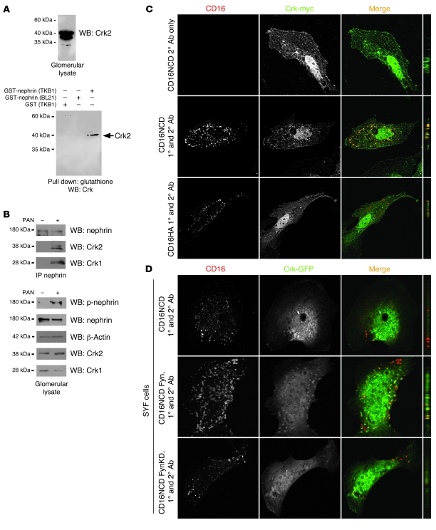
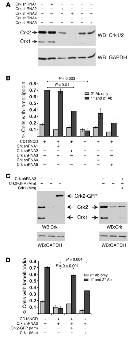
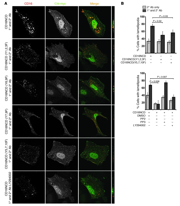
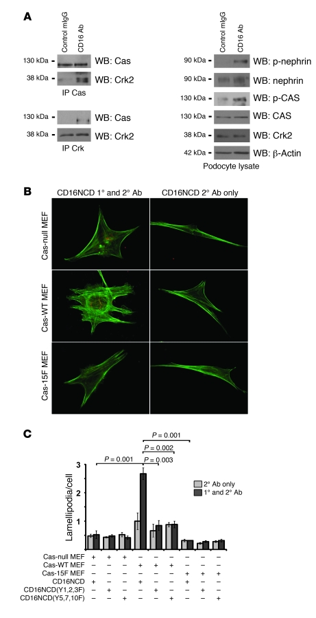
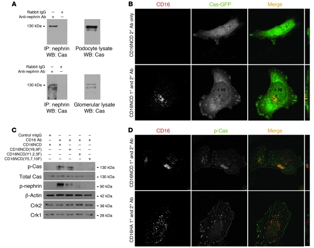
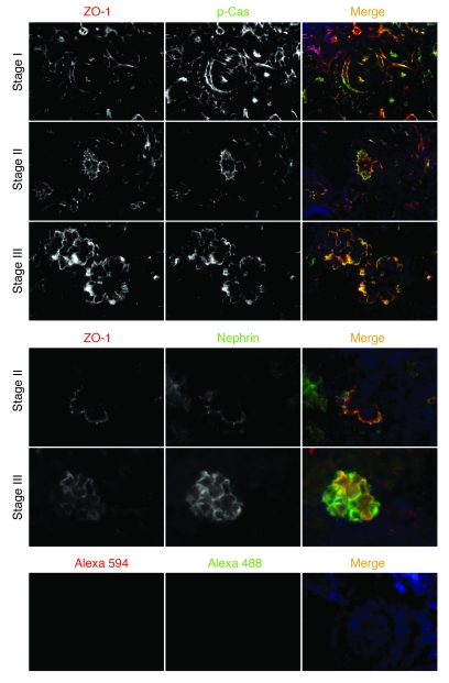
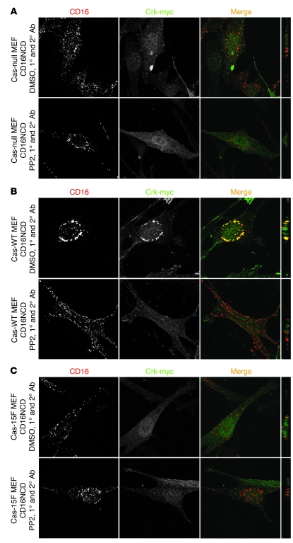
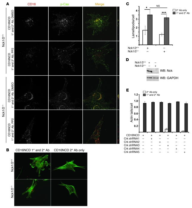
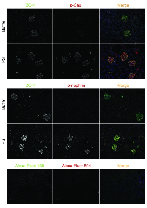
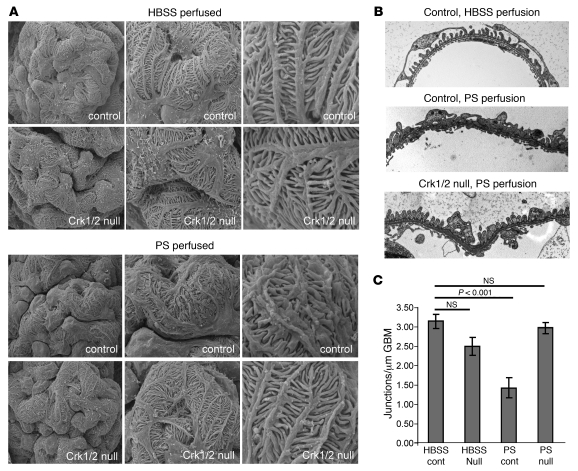
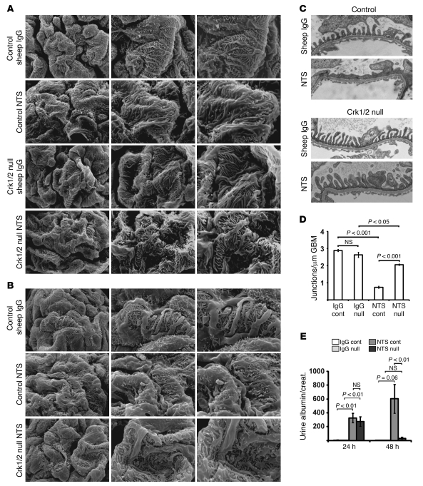
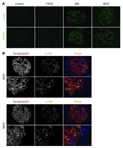
The morphology of healthy podocyte foot processes is necessary for maintaining the characteristics of the kidney filtration barrier. In most forms of glomerular disease, abnormal filter barrier function results when podocytes undergo foot process spreading and retraction by remodeling their cytoskeletal architecture and intercellular junctions during a process known as effacement. The cell adhesion protein nephrin is necessary for establishing the morphology of the kidney podocyte in development by transducing from the specialized podocyte intercellular junction phosphorylation-mediated signals that regulate cytoskeletal dynamics. The present studies extend our understanding of nephrin function by showing that nephrin activation in cultured podocytes induced actin dynamics necessary for lamellipodial protrusion. This process required a PI3K-, Cas-, and Crk1/2-dependent signaling mechanism distinct from the previously described nephrin-Nck1/2 pathway necessary for assembly and polymerization of actin filaments. Our present findings also support the hypothesis that mechanisms governing lamellipodial protrusion in culture are similar to those used in vivo during foot process effacement in a subset of glomerular diseases. In mice, podocyte-specific deletion of Crk1/2 prevented foot process effacement in one model of podocyte injury and attenuated foot process effacement and associated proteinuria in a delayed fashion in a second model. In humans, focal adhesion kinase and Cas phosphorylation - markers of focal adhesion complex-mediated Crk-dependent signaling - was induced in minimal change disease and membranous nephropathy, but not focal segmental glomerulosclerosis. Together, these observations suggest that activation of a Cas-Crk1/2-dependent complex is necessary for foot process effacement observed in distinct subsets of human glomerular diseases.


Figures

Figure 1
Figure 1. Crk1/2 interacts with nephrin in a tyrosine phosphorylation–dependent fashion.
(A) Purified recombinant GST-nephrinCD expressed in BL21 or TKB1 E. coli (to produce nonphosphorylated or tyrosine-phosphorylated nephrin, respectively) or purified GST alone was mixed with rat glomerular lysate, pulled down with glutathione agarose, and immunoblotted with monoclonal anti-Crk1/2 antibody. (B) Glomerular lysates from rats injected with PBS (control) or PAN were immunoprecipitated and/or immunoblotted using the indicated antibodies. (C) Cultured human podocytes expressing CD16/7-nephrinCD (CD16NCD) or CD16/7-HA (CD16HA) and Crk2-myc were activated by clustering: namely, addition of monoclonal anti-CD16 primary antibody (1°) and/or goat anti-mouse IgG Texas Red–conjugated secondary antibody (2°), as indicated, to the media of live cells. Crk2-myc was detected with rabbit polyclonal anti-myc primary antibody and Alexa Fluor 488–labeled secondary antibody. Cells were analyzed by confocal microscopy. CD16/7-nephrinCD (red) and Crk2-myc (green) colocalized in the plane of the plasma membrane. (D) SYF MEFs transiently expressing CD16/7-nephrinCD and Crk-GFP were activated by clustering. Although colocalization of nephrin and Crk was not observed in SYF MEFs, nephrin-Crk association was rescued by reexpressing Fyn, but not by expressing a kinase-dead variant of Fyn (FynKD). (C and D) Original magnification, ×630. yz plane reconstructions are shown at far right.
Figure 2
Figure 2. Clustered, activated CD16/7-nephrinCD and CD16/7-Neph1CD induce lamellipodia formation in cultured human podocytes.
(A) CD16/7-nephrinCD or CD16/7-Neph1CD was coexpressed with actin-GFP in human podocytes, which were activated by clustering as in Figure 1. Transfected cells with structures typical of lamellipodia on more than 30% of their circumferences were counted as cells with lamellipodial protrusions. (B) Using the same system as in A, cells were treated with PP2 or PP3 before activation. Transfected cells with actin tails longer than 5 μm or lamellipodia extending over more than 30% of the cell circumference were counted positive for these features. 20 transfected cells total were counted per experiment. Data (mean ± SEM) are from single experiments and are representative of 3 unique experiments.
Figure 3
Figure 3. Crk1/2 knockdown attenuates nephrin-induced lamellipodial protrusive activity.
(A) Immunoblot demonstrating attenuation of Crk1/2 expression in human podocyte cell lines stably expressing 1 of 5 different shRNA constructs targeting the Crk1/2 gene. GAPDH protein expression served as loading control. (B) Podocytes stably expressing the indicated Crk1/2 shRNA constructs were transfected with CD16/7-nephrinCD and actin-GFP and were activated by clustering as in Figure 1. The fraction of cells exhibiting lamellipodial protrusions was evaluated after fixation. (C) Immunoblot showing that Crk2-GFP or mouse Crk1 were reexpressed in stably knocked down human Crk shRNA3 podocytes. Mm, Mus musculus. (D) Crk shRNA3 podocytes were transfected with CD16/7-nephrinCD and actin-GFP and activated by clustering. After fixation, the fraction of cells exhibiting lamellipodial protrusions was determined. Results (mean ± SEM) are representative of 3 independent experiments.
Figure 4
Figure 4. Nephrin-Crk association and nephrin-induced lamellipodial protrusions rely on nephrin tyrosine residues.
(A) CD16/7-nephrinCD plasmids expressed in cultured podocytes were activated by clustering as in Figure 1 and analyzed by confocal microscopy. Podocytes pretreated with LY294002 before clustering are also shown. Crk was stained with anti-myc antibody and Alexa Fluor 488–conjugated secondary antibody. Original magnification, ×630. yz plane reconstructions are shown at far right. (B) Podocytes expressing CD16/7-nephrinCD or the indicated mutants and actin-GFP were activated with or without PP2, PP3, or LY294002, and the fraction of cells with lamellipodia was determined. Cells treated without primary CD16 antibody served as controls. Data (mean ± SEM) are representative of 3 experiments.
Figure 5
Figure 5. Nephrin engagement leads to phosphorylation of Cas, induces Cas-Crk complex formation, and results in Cas-Crk interaction–dependent lamellipodia formation.
(A) Coimmunoprecipitation and immunoblots using indicated antibodies demonstrated tyrosine phosphorylation of Cas and increased Cas-Crk complex formation after CD16/7-nephrinCD clustering in cultured podocytes. (B and C) Cas-null, Cas-WT, and Cas-15F MEFs were stably transduced to express CD16/7-nephrinCD or the indicated mutants. Cells were activated by clustering as in Figure 1. Actin was stained with Alexa Fluor 488–labeled phalloidin. (B) Cells imaged by fluorescence microscopy. Original magnification, ×630. (C) Lamellipodia per cell were counted. Data (mean ± SEM) are representative of 3 independent experiments.
Figure 6
Figure 6. Cas is recruited to clustered nephrin in a PI3K-dependent fashion, while its phosphorylation depends on Y5,7,10.
(A) Nephrin coimmunoprecipitated with Cas from cultured podocytes transiently transfected with plasmid encoding CD16/7-nephrinCD (top) and from isolated rat glomerular cell lysate (bottom). (B) Human podocytes expressing CD16/7-nephrinCD (red) and Cas-GFP (green) were activated by clustering as in Figure 1, fixed, and imaged by confocal microscopy. (C) Cultured NIH3T3 cells expressing CD16/7-nephrinCD from the indicated plasmids were activated as indicated. Immunoblots shown are representative of 3 independent experiments. (D) Cultured human podocytes were transfected with CD16/7-nephrinCD or CD16/7-HA (red) and activated, and endogenous p-Cas was identified using a specific p-Cas antibody and Alexa Fluor 488–conjugated secondary antibody. Cells were analyzed by confocal microscopy. (B and D) Original magnification, ×630. yz plane reconstructions are shown at far right.
Figure 7
Figure 7. p-Cas is targeted to podocyte precursor intercellular junctions in newborn mice.
Mouse newborn kidney sections were stained with anti–p-Cas (green) or anti-nephrin antibody (green) as indicated. Note that p-Cas was detected at the podocyte precursor intercellular junction as early as the s-shape body stage (stage I), whereas nephrin expression was first seen at the capillary loop stage (stage II). ZO-1 staining was used as a junction marker (red). Secondary antibody controls are shown below. Stage III, maturing glomerulus. Original magnification, ×630.
Figure 8
Figure 8. Crk is recruited to nephrin indirectly via Cas.
Cas-null (A), Cas-WT (B), and Cas-15F (C) MEFs were transfected with CD16/7-nephrinCD and Crk-myc as indicated. Cells were activated by clustering as in Figure 1 and analyzed by confocal microscopy. Crk-myc was stained with myc antibody and labeled with secondary antibody (Alexa Fluor 488). Cells treated with PP2 prior to clustering are also shown. (AC) Original magnification, ×630. yz plane reconstructions are shown at far right.
Figure 9
Figure 9. Nephrin-induced actin filament polymerization and lamellipodia formation are mediated by distinct signaling pathways.
(A) Nck1/2+/+ and Nck1/2–/– MEFs stably expressing CD16/7-nephrinCD were activated as in Figure 1, and endogenous p-Cas was stained with p-Cas antibody and Alexa Fluor 488–labeled secondary IgG antibody and imaged by confocal microscopy. Cells treated with PP2 prior to clustering are also shown. (B and C) Nephrin-induced lamellipodia formation was independent of Nck. Nck1/2+/+ and Nck1/2–/– MEFs expressing CD16/7-nephrinCD were evaluated for lamellipodial protrusion activity 30 minutes after clustering. Actin was stained with green-labeled phalloidin. (D) Cell lysates from Nck1/2+/+ and Nck1/2–/– MEFs were analyzed by immunoblotting with Nck antibody or GAPDH antibody as loading control. (E) Nephrin-induced local actin filament polymerization was independent of Crk1/2. Human podocyte cell lines stably expressing 1 of 5 different shRNA targeting human Crk1/2 were transfected with CD16/7-nephrinCD. Actin tails per podocyte were counted after activation by clustering. (A and B) Original magnification, ×630. (C and E) Data (mean ± SEM) are from 3 independent experiments. *P < 0.05; ***P < 0.0005.
Figure 10
Figure 10. Selective deletion of Crk1/2 in mouse podocytes.
(A) Crk1/2fl/fl mice with loxP sites flanking exon 1 were crossed with podocin-CreTg/+ mice to generate Crk1/2fl/flpodocin-CreTg/+ mice (deleted of Crk1/2 in podocytes). (B) Paraffin-embedded mouse kidney sections of Crk1/2fl/fl (control) and Crk1/2fl/flpodocin-CreTg/+ (Crk1/2 null) mice were double stained for Crk1/2 (green) and podocin as a podocyte marker (red). Higher-magnification images of portions of a glomerulus are shown below. Note the absence of Crk1/2 staining in podocytes (arrows) of Crk1/2fl/flpodocin-CreTg/+, whereas Crk1/2 was still expressed in mesangial cells. Original magnification, ×600 (top); ×4,200 (bottom).
Figure 11
Figure 11. Enhanced nephrin and Cas phosphorylation after protamine sulfate–induced foot process spreading.
WT mice were perfused with either buffer or protamine sulfate (PS), and paraffin-embedded kidney sections were stained with either anti–p-nephrin (red) or anti–p-Cas (red) and anti–ZO-1 (green) antibodies. Cas and nephrin phosphorylation was enhanced after protamine sulfate–induced podocyte injury. Original magnification, ×200.
Figure 12
Figure 12. Foot process spreading in Crk1/2fl/flpodocin-CreTg/+ mice is prevented by protamine sulfate perfusion.
(A and B) Scanning EM (A) and transmission EM (B) of Crk1/2fl/fl and Crk1/2fl/flpodocin-CreTg/+ mice perfused with HBSS or protamine sulfate. Results are representative of 3–5 mice per group. Original magnification, ×3,000 (A, left); ×7,000 (A, middle); ×20,000 (A, right); ×15,000 (B). (C) Number of junctions per micron glomerular basement membrane (GBM), as seen by transmission EM. Data are mean ± SEM.
Figure 13
Figure 13. Podocyte-specific Crk1/2 deletion attenuates the glomerular phenotype induced by NTS.
(AC) Scanning EM (A and B) and transmission EM (C) of Crk1/2fl/fl and Crk1/2fl/flpodocin-CreTg/+ mouse glomeruli, either 72 (A and C) or 24 (B) hours after injection of NTS or control sheep IgG. Original magnification, ×3,000 (A and B, left); ×7,000 (A and B, middle); ×10,000 (A and B, right); ×25,000 (C). (D) Number of junctions per micron glomerular basement membrane, as seen by transmission EM 72 hours after injection (mean ± SEM). (E) Albumin/creatinine ratios of Crk1/2fl/fl and Crk1/2fl/flpodocin-CreTg/+ mouse urines 24 and 48 hours after injection with NTS or sheep IgG. Data (mean ± SEM) are representative of 3 (AD) or 3–9 (E) mice per group.
Figure 14
Figure 14. Enhanced Cas and FAK phosphorylation in membranous nephropathy and minimal change disease.
(A) Representative images showing anti–p-Cas (green) and anti–p-FAK (green) antibody staining of frozen kidney sections from control subjects and from patients with focal segmental glomerulosclerosis (FSGS), membranous nephropathy (MN), or minimal change disease (MCD). (B) Coimmunofluorescence study of frozen kidney sections of patients with minimal change disease with anti–p-Cas (green) or anti–p-FAK antibody (green) and the podocyte slit diaphragm marker synaptopodin (red). Higher-magnification images are shown below. Note that p-Cas and p-FAK almost completely colocalized with synaptopodin. Original magnification, ×400 (A and B, top); ×1,000 (B, bottom).

References

    1. Johnstone DB, Holzman LB. Clinical impact of research on the podocyte slit diaphragm. Nat Clin Pract Nephrol. 2006;2(5):271–282. doi: 10.1038/ncpneph0180. - DOI - PubMed
    1. Faul C, Asanuma K, Yanagida-Asanuma E, Kim K, Mundel P. Actin up: regulation of podocyte structure and function by components of the actin cytoskeleton. Trends Cell Biol. 2007;17(9):428–437. doi: 10.1016/j.tcb.2007.06.006. - DOI - PubMed
    1. Jones N, et al. Nck adaptor proteins link nephrin to the actin cytoskeleton of kidney podocytes. Nature. 2006;440(7085):818–823. doi: 10.1038/nature04662. - DOI - PubMed
    1. Verma R, Kovari I, Soofi A, Nihalani D, Patrie K, Holzman LB. Nephrin ectodomain engagement results in Src kinase activation, nephrin phosphorylation, Nck recruitment, and actin polymerization. J Clin Invest. 2006;116(5):1346–1359. doi: 10.1172/JCI27414. - DOI - PMC - PubMed
    1. Kaplan JM, et al. Mutations in ACTN4, encoding alpha-actinin-4, cause familial focal segmental glomerulosclerosis. Nat Genet. 2000;24(3):251–256. doi: 10.1038/73456. - DOI - PubMed

Publication types

MeSH terms

Substances