. 2012 Feb;122(2):529-37.
doi: 10.1172/JCI58847. Epub 2012 Jan 17.

IFN-α inhibits HBV transcription and replication in cell culture and in humanized mice by targeting the epigenetic regulation of the nuclear cccDNA minichromosome

Affiliations

IFN-α inhibits HBV transcription and replication in cell culture and in humanized mice by targeting the epigenetic regulation of the nuclear cccDNA minichromosome

Laura Belloni et al. J Clin Invest. 2012 Feb.

Abstract

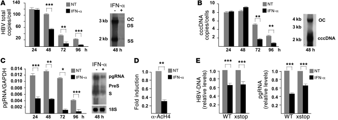
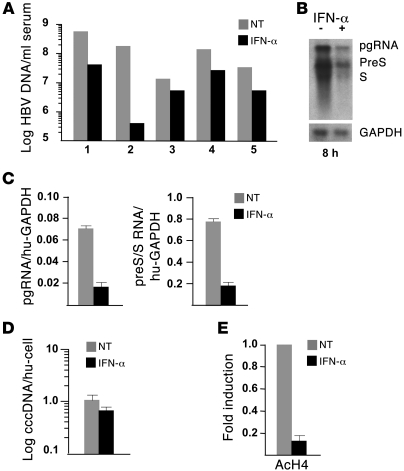
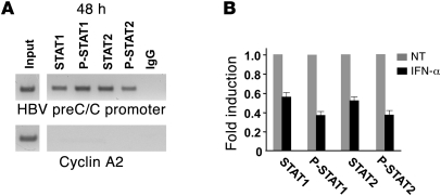
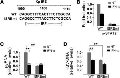
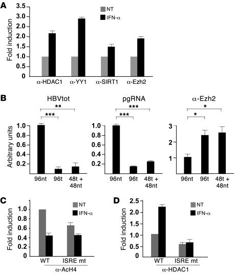
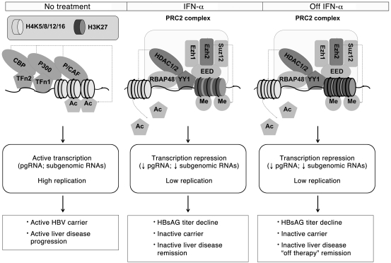
HBV infection remains a leading cause of death worldwide. IFN-α inhibits viral replication in vitro and in vivo, and pegylated IFN-α is a commonly administered treatment for individuals infected with HBV. The HBV genome contains a typical IFN-stimulated response element (ISRE), but the molecular mechanisms by which IFN-α suppresses HBV replication have not been established in relevant experimental systems. Here, we show that IFN-α inhibits HBV replication by decreasing the transcription of pregenomic RNA (pgRNA) and subgenomic RNA from the HBV covalently closed circular DNA (cccDNA) minichromosome, both in cultured cells in which HBV is replicating and in mice whose livers have been repopulated with human hepatocytes and infected with HBV. Administration of IFN-α resulted in cccDNA-bound histone hypoacetylation as well as active recruitment to the cccDNA of transcriptional corepressors. IFN-α treatment also reduced binding of the STAT1 and STAT2 transcription factors to active cccDNA. The inhibitory activity of IFN-α was linked to the IRSE, as IRSE-mutant HBV transcribed less pgRNA and could not be repressed by IFN-α treatment. Our results identify a molecular mechanism whereby IFN-α mediates epigenetic repression of HBV cccDNA transcriptional activity, which may assist in the development of novel effective therapeutics.


Figures

Figure 1
Figure 1. IFN-α inhibits HBV replication and cccDNA transcription in HCC cells.
(A) Left panel: HepG2 cells were transfected with monomeric linear full-length WT HBV adw (genotype A) genomes. HBV core particles were isolated from untreated and IFN-α–treated cells at the indicated time points after transfection. Results are expressed as number of HBV DNA copies per transfected cell. Right panel: Southern blot hybridization. OC, open circular duplex HBV DNA; DS, double-stranded HBV DNA replicative intermediates; SS, single-stranded HBV DNA replicative intermediates. (B) Left panel: cccDNA levels (copies per transfected cell) were analyzed by qPCR with selective cccDNA primers and β-globin primers (DNA sample normalization). Right panel: Southern blot analysis. (C) Left panel: pgRNA levels were analyzed by qPCR using pgRNA selective primers and GAPDH primers (equal loading of each RNA sample). Right panel: Northern blot analysis. pgRNA, HBV pregenomic RNA. (D) Cross-linked chromatin was immunoprecipitated with a relevant control IgG or specific anti-AcH4 antibody and analyzed by qPCR with HBV cccDNA selective primers. Results are expressed as fold induction relative to the untreated cells using the comparative Ct method. (E) HepG2 cells were transfected with monomeric linear full-length WT or HBx mutant HBV genomes (4). Core particles HBV-DNA (left panel) and pgRNA (right panel) results are expressed as in Figure 1, A and C, respectively. All histograms show mean values from 3 independent experiments; bars indicate SD. P values were determined using Student’s t test. *P < 0.05, **P < 0.01, ***P < 0.001.
Figure 2
Figure 2. IFN-α inhibits cccDNA transcription and HBV replication in chimeric uPA/SCID mice.
Uninfected and chronically HBV infected (median, 2 × 10⁸ serum HBV-DNA copies/ml serum) chimeric uPA/SCID mice transplanted with thawed human hepatocytes received daily injections of human IFN-α (1,300 IU/g body weight) for 5 days or saline. (A) HBV viremia (log HBV-DNA copies/ml serum) were determined in individual animals (n = 5) at baseline, shortly before IFN treatment, and 8 hours after the last administration of IFN-α. (B) Representative Northern blot analysis from one IFN-α–treated mouse and one untreated mouse. (C) Steady-state levels of pregenomic RNA (left panel) and preS/S RNAs (right panel). Intrahepatic preS/S RNAs were determined by subtracting pgRNA amounts from total HBV RNAs (pgRNA + preS/S RNA) estimated in the same RNA preparation as described in Methods, and values were normalized using human-specific GAPDH primers. (D) Intrahepatic cccDNA loads in IFN-α–treated (n = 5) and control (saline) HBV-infected mice (n = 5). Real-time qPCR analysis was performed using selective cccDNA primers, and β-globin primers were used to normalize cccDNA copies (median + SD) per human hepatocyte (expressed as human genome equivalents) determined in chimeric livers. (E) Cross-linked chromatin from liver samples of chronically HBV-infected untreated and IFN-α–treated (48 hours) uPA chimeric mice was immunoprecipitated with a relevant control IgG or specific anti-AcH4 antibody. Immunoprecipitated chromatin was analyzed by qPCR as in Figure 1D. All histograms show mean values from 3 independent experiments; bars indicate SD.
Figure 3
Figure 3. STAT1 and STAT2 transcription factors are recruited on the cccDNA.
(A) Cross-linked chromatin from HepG2 cells transfected with monomeric linear full-length HBV DNA was immunoprecipitated with a relevant control IgG or specific anti-STAT1, anti–phospho-STAT1, anti-STAT2, and anti–phospho-STAT2 antibodies. Immunoprecipitated chromatin samples were analyzed by real-time PCR with either HBV cccDNA selective primers (upper panel) or primers specific for the cyclin A2 coding region as a negative control (lower panel). (B) Chromatin was prepared from untreated and IFN-α–treated HepG2 cells transfected with WT HBV genomes. Immunoprecipitated chromatin was analyzed and results expressed as in Figure 1D. All histograms show mean values from 3 independent experiments; bars indicate SD.
Figure 4
Figure 4. HBV ISRE mediates IFN-α transcriptional repression.
(A) Sequence of the HBV enhancer 1/X gene promoter around the HBV ISRE. ISRE mutations are shown. The nucleotide substitutions do not alter the HBV polymerase polypeptide sequence. (B) Chromatin prepared from untreated and IFN-α–treated HepG2 cells transfected with WT or ISREmt HBV genomes was immunoprecipitated with a relevant control IgG or specific anti-STAT2 antibodies. Immunoprecipitated chromatin was analyzed by qPCR and results expressed as in Figure 1D. (C) mRNAs were prepared from untreated and IFN-α–treated HepG2 cells transfected with WT and ISREmt HBV genomes, and HBV pregenomic RNA was quantified by qPCR using specific primers. GAPDH amplification was used to normalize for equal loading of each RNA sample. (D) Cytoplasmic HBV core particles were isolated from untreated and IFN-α–treated HepG2 cells 48 hours after transfection with monomeric linear full-length WT or ISREmt genomes. Results are expressed as in Figure 1A. Results are shown as mean values from 3 independent experiments; bars indicate SD. P values were determined using Student’s t test. **P < 0.01.
Figure 5
Figure 5. IFN-α modulates the epigenetic control of cccDNA function by affecting the recruitment of chromatin-modifying enzymes.
(A) Cross-linked chromatin from untreated and IFN-α–treated HepG2 cells transfected with WT HBV genomes was immunoprecipitated with a relevant control IgG or anti-HDAC1, anti-YY1, anti-hSirt1 and anti-EzH2 antibodies and analyzed as in Figure 1D. (B) HepG2 cells, transfected as in A, were either left untreated (96nt), or treated for 96 hours after transfection with IFN-α (96t), or treated with IFN-α for 48 hours and then left untreated for 48 hours (48t + 48nt). Left panel: cross-linked chromatin immunoprecipitated with a relevant control IgG or anti-Ezh2 antibody was analyzed as in Figure 1D. pgRNA (middle panel) and cytoplasmic core particles HBV-DNA (right panel) were quantified by real-time qPCR. Results are expressed as in Figure 1. (C and D) Chromatin was prepared from untreated and IFN-α–treated HepG2 cells transfected with WT or ISREmt HBV genomes and immunoprecipitated with a relevant control IgG or anti-AcH4 (B) or anti-HDAC1 (C) antibodies. Immunoprecipitated chromatin was analyzed by qPCR and results expressed as in Figure 1D. All histograms show the mean from 3 independent experiments; bars indicate SD. P values were determined using Student’s t test. *P < 0.05; **P < 0.01; ***P < 0.001.
Figure 6
Figure 6. Schematic representation of cccDNA chromatin changes in response to IFN-α treatment.
cccDNA-bound histone acetylation status and the recruitment of chromatin-modifying enzymes onto the viral minichromosome change in relation to viral replication and IFN-α treatment. The drawings reflect what is observed in an in vitro replication system. The translation into the clinical scenario as it is observed in patients (lower boxes) is inferred, and it awaits to be confirmed by ex vivo experiments. In the context of high HBV replication and in the absence of IFN-α treatment, cccDNA-bound histones are hyperacetylated, cccDNA-associated chromatin is in an open configuration, pgRNA is actively transcribed, and HBV replication is unrestricted (left drawing). The clinical correlate is an active HBV carrier with high HBV viremia, reflecting high levels of intrahepatic viral replication and liver disease progression. In response to IFN-α, HDACs (HDAC1 and Sirt1) substitute HAT enzymes (p300, CBP, and P/CAF) on the cccDNA, and a PRC2-repressor complex is recruited. This leads to histone deacetylation/methylation at specific lysine residues, a “closed” chromatin configuration, and a striking reduction of pgRNA transcription, HBsAg synthesis, and HBV replication (middle drawing). In the clinical setting, this would translate into a rapid serum HBsAg decline and viral suppression (inactive carrier) with disease remission. When treatment is stopped, the chromatin changes imposed by IFN-α tend to persist (right drawing) resulting in the “off-therapy” maintenance of the virological suppression and clinical improvement (achieved in 30%–35% of HBeAg-positive patients and 20%–25% of HBeAg-negative patients). Darker forms in the middle and right drawings indicate components of the PRC2 complex whose recruitment has been directly investigated in this paper.

References

    1. Nassal M. Hepatitis B viruses: reverse transcription a different way. Virus Res. 2008;134(1–2):235–249. doi: 10.1016/j.virusres.2007.12.024. - DOI - PubMed
    1. Levrero M, Pollicino T, Petersen J, Belloni L, Raimondo G, Dandri M. Control of cccDNA function in hepatitis B virus infection. J Hepatol. 2009;51(3):581–592. doi: 10.1016/j.jhep.2009.05.022. - DOI - PubMed
    1. Pollicino T, et al. Hepatitis B virus replication is regulated by the acetylation status of hepatitis B virus cccDNA-bound H3 and H4 histones. Gastroenterology. 2006;130(3):823–837. doi: 10.1053/j.gastro.2006.01.001. - DOI - PubMed
    1. Belloni L, et al. Nuclear HBx binds the HBV minichromosome and modifies the epigenetic regulation of cccDNA function. Proc Natl Acad Sci U S A. 2009;106(47):19975–19979. - PMC - PubMed
    1. J Hepatol. 2009;50(2):227–242. doi: 10.1016/j.jhep.2008.10.001. - DOI - PubMed

Publication types