. 2012 Feb;122(2):575-85.
doi: 10.1172/JCI61034. Epub 2012 Jan 17.

Imiquimod clears tumors in mice independent of adaptive immunity by converting pDCs into tumor-killing effector cells

Affiliations

Imiquimod clears tumors in mice independent of adaptive immunity by converting pDCs into tumor-killing effector cells

Barbara Drobits et al. J Clin Invest. 2012 Feb.

Abstract

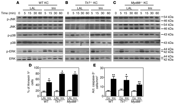
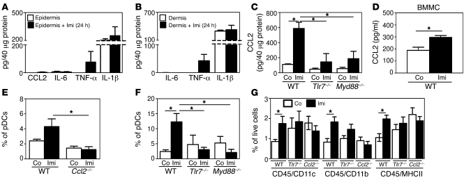
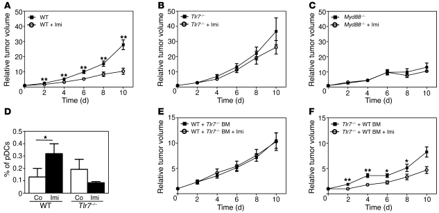
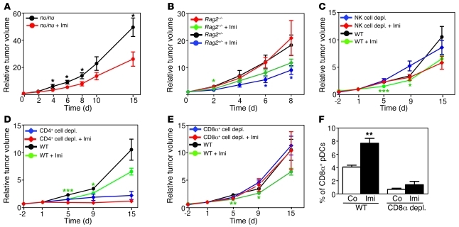
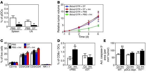
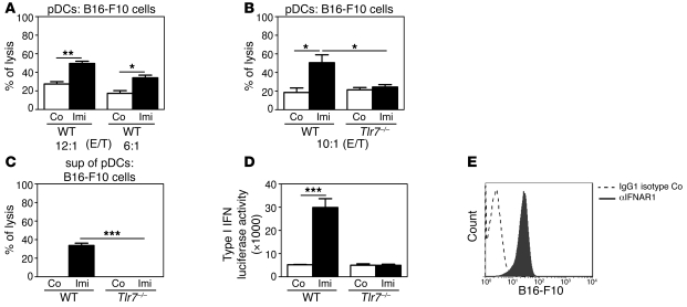
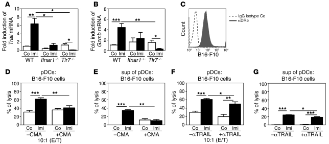
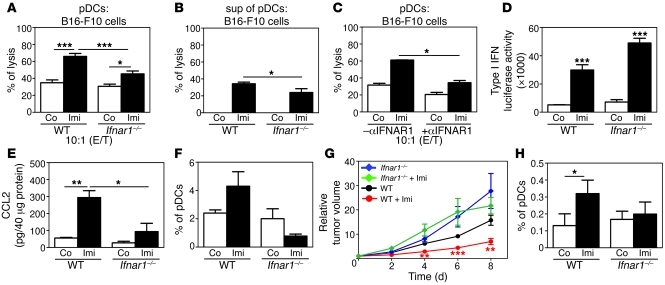
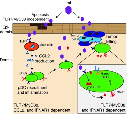
Imiquimod is a synthetic compound with antitumor properties; a 5% cream formulation is successfully used to treat skin tumors. The antitumor effect of imiquimod is multifactorial, although its ability to modulate immune responses by triggering TLR7/8 is thought to be key. Among the immune cells suggested to be involved are plasmacytoid DCs (pDCs). However, a direct contribution of pDCs to tumor killing in vivo and the mechanism of their recruitment to imiquimod-treated sites have never been demonstrated. Using a mouse model of melanoma, we have now demonstrated that pDCs can directly clear tumors without the need for the adaptive immune system. Topical imiquimod treatment led to TLR7-dependent and IFN-α/β receptor 1-dependent (IFNAR1-dependent) upregulation of expression of the chemokine CCL2 in mast cells. This was essential to induce skin inflammation and for the recruitment of pDCs to the skin. The recruited pDCs were CD8α+ and induced tumor regression in a TLR7/MyD88- and IFNAR1-dependent manner. Lack of TLR7 and IFNAR1 or depletion of pDCs or CD8α+ cells from tumor-bearing mice completely abolished the effect of imiquimod. TLR7 was essential for imiquimod-stimulated pDCs to produce IFN-α/β, which led to TRAIL and granzyme B secretion by pDCs via IFNAR1 signaling. Blocking these cytolytic molecules impaired pDC-mediated tumor killing. Our results demonstrate that imiquimod treatment leads to CCL2-dependent recruitment of pDCs and their transformation into a subset of killer DCs able to directly eliminate tumor cells.


Figures

Figure 1
Figure 1. TLR7/MyD88-independent effects of Imi in the skin.
Western blot analysis of primary keratinocytes (KCs) isolated from (A) C57BL/6 (WT), (B) Tlr7–/–, and (C) Myd88–/– mice treated with 12 μg/ml Imi or LAL reagent water (control) for the indicated time points. Results are representative of at least 3 independent batches. White lines indicate that samples were run on the same gel but were noncontiguous. (D) Flow cytometric analysis showing annexin V+ cells in primary keratinocyte cultures stimulated with Imi (12 μg/ml) or LAL water for 40 hours (n = 3–4 per group). (E) Apoptosis was measured by active (act.) caspase-3 staining of epidermal sheets from ears of WT, Tlr7–/–, and Myd88–/– mice treated topically with Imi for 7 days or left untreated (Co). Caspase-3+ cells were counted in 10 randomly chosen fields of at least 3 independent samples. *P < 0.05, **P < 0.005.
Figure 2
Figure 2. TLR7/MyD88-dependent effects of Imi in the skin.
(AC) Mouse back skin was treated topically for 24 hours with Imi or left untreated. IL-1β, IL-6, TNF-α, or CCL2 levels were measured in protein lysates of (A) epidermis and (B and C) dermis by ELISA. (D) BM-derived mast cells (BMMCs) were stimulated with Imi (12 μg/ml) for 20 hours, and CCL2 was measured in supernatants by ELISA. (E and F) Histograms showing the percentage of pDCs (E, B220+Ly6C+CD11b; F, mPDCA1+B220+Gr-1+) among total DCs (CD45+CD11c+) in the dermis of (E) WT and Ccl2–/– and (F) Tlr7–/– and Myd88–/– mice after 7 days of topical Imi treatment. (G) Graph depicting the percentage of infiltrating immune cells in the dermis of mice after 7 days of topical Imi treatment. *P < 0.05.
Figure 3
Figure 3. The antitumor effect of Imi depends on the presence of TLR7 and MyD88 on BM cells.
(AC) Graphs showing the kinetics of tumor growth in (A) C57BL/6 (WT), (B) Tlr7–/–, and (C) Myd88–/– mice measured at the indicated time points after Imi treatment (n = 10–14 per group). (D) FACS analysis showing the percentage of pDCs (CD45+CD11c+B220+CD11b) present in melanomas 3 days after Imi treatment. (E and F) Relative tumor volume in BM chimeras of the indicated genotypes after Imi treatment. C57BL/6 (WT) and Tlr7–/– mice were lethally irradiated and reconstituted intravenously with BM cells of the indicated genotypes. After 12 weeks, reconstituted mice were injected intradermally with B16-F10 melanoma cells, and tumors treated with Imi or left untreated (n = 7–11 per group). *P < 0.05, **P < 0.005.
Figure 4
Figure 4. Adaptive immune cells are not required for the tumoricidal effect of Imi.
(A and B) Graphs showing the relative tumor volume of (A) M3 melanomas intradermally induced in Foxn1nu mice (nu/nu mice) and (B) B16-F10 melanomas induced in Rag2–/– and Rag2+/– mice as controls (n = 11–13 per group). (CE) Relative tumor volume of M3 melanoma–bearing mice depleted of (C) NK cells by injection of anti-NK–specific antibodies (clone Asialo GM1); (D) CD4+ cells by anti-CD4 antibody injection (clone GK1.5); or (E) CD8α+ cells by anti-CD8α antibody injection (clone 53.6.7) before starting treatment with Imi (n = 8–9 per group). Untreated DBA/2 (WT) mice were used as controls. depl., depletion. (F) The percentage of CD8α+ pDCs (CD11bB220+) gated from CD45+CD11c+ cells present in melanomas of mice depleted of CD8α+ cells 3 days after starting Imi treatment. *P < 0.05, **P < 0.005, ***P < 0.0005.
Figure 5
Figure 5. pDCs are responsible for the tumoricidal effect of Imi.
(A) Graph demonstrating the absence of pDCs (CD11c+B220+SiglecH+) in spleen and tumor draining lymph nodes of Bdca2-DTR mice depleted of pDCs by DT injection (+DT). Control (not pDC-depleted) mice were injected with PBS. (B) Relative tumor growth in pDC-depleted mice treated with Imi. Bdca2-DTR transgenic mice were depleted of pDCs prior to intradermal injection of B16-F10 cells (n = 5–7 per group). (C and D) FACS analysis showing the percentage of monocytes/neutrophils (CD11b+), CD4+ T cells (CD3+CD4+), CD8α+ T cells (CD3+CD8α+), and NK cells (NK1.1+) in (C) tumor-draining lymph nodes and (D) tumor-infiltrating CD8α+ DCs in Bdca2-DTR mice treated as indicated. (E) Quantification of the active caspase-3–positive area (μm2 per mm2 of tumors) in tumor tissue of WT (n = 7–8 per group), pDC-depleted (pDCs depl.) (n = 8–9), and Tlr7–/– (n = 3) mice treated with Imi or left untreated. The active caspase-3+ area was analyzed in 10 randomly chosen fields of at least 3 independent samples. *P < 0.05, **P < 0.005.
Figure 6
Figure 6. Imi-stimulated pDCs can directly kill tumor cells.
(A and B) Killing assays measuring the percentage of lysis of B16-F10 melanoma cells occurring in pDCs that had been either (A) FACS sorted or (B) IMag sorted from Flt3L BM cultures of (A) WT and (B) Tlr7–/– mice and stimulated with Imi (2.5 μg/ml) for 16 hours. pDCs were cocultured with B16-F10 melanoma cells at indicated E/T ratios for 20 hours. (C) The killing assay was performed with the supernatant (sup) of the pDCs shown in B. (D) Type I IFN levels were determined by stimulating the IFN-luciferase reporter cell line LL171 (50) with supernatants of WT or Tlr7–/– pDCs triggered with Imi for 16 hours. (E) FACS analysis showing representative staining with anti-IFNAR1 antibodies (αIFNAR1) or isotype control (Co) rat IgG1 on B16-F10 cells. *P < 0.05, **P < 0.005, ***P < 0.0005.
Figure 7
Figure 7. pDCs kill tumor cells via TRAIL and granzyme B.
(A and B) qRT-PCR analysis for (A) Trail and (B) granzyme B (Gzmb) expression in IMag-sorted pDCs from Flt3L BM cultures of WT, Ifnar1–/–, and Tlr7–/– mice stimulated for 6 hours with Imi (2.5 μg/ml). (C) FACS analysis showing DR5 expression on B16-F10 melanoma cells. (DG) Killing assays performed with B16-F10 melanoma cells cocultured with (D and F) IMag-sorted pDCs from Flt3L cultures or (E and G) supernatants thereof previously treated with (D and E) 1 μg/ml CMA and 2.5 μg/ml Imi for 16 hours or with (F and G) anti-TRAIL antibody (αTRAIL) (25 μg/ml) for 2 hours and further stimulated with Imi (2.5 μg/ml) for 16 hours. Cytolytic assays were performed at a E/T ratio of 10:1. *P < 0.05, **P < 0.005, ***P < 0.0005.
Figure 8
Figure 8. pDCs require IFNAR1 signaling for their cytotoxic activity.
(A and B) Killing assay performed with B16-F10 melanoma cells cocultured with (A) IMag-sorted WT or Ifnar1–/– pDCs or (B) supernatants thereof. (C) Killing assay with pDCs pretreated with anti-IFNAR1 antibody (25 μg/ml) and simulated with Imi for 16 hours before coculturing with B16-F10 cells. Cytolytic assays were performed at a E/T ratio of 10:1. (D) Type I IFN levels were analyzed by culturing the IFN-luciferase reporter cell line LL171 with supernatants of WT or Ifnar1–/– pDCs that had been stimulated with Imi for 16 hours. (E) CCL2 levels measured in dermal protein lysates by ELISA after WT and Ifnar1–/– mouse back skin was treated for 24 hours with Imi or left untreated. (F) Graph showing the percentage of B220+Ly6C+CD11b pDCs among total CD45+CD11c+ DCs in the dermis after 7 days of topical Imi treatment. (G) Growth of intradermally injected B16-F10 melanomas in WT and Ifnar1–/– mice after Imi treatment (n = 8–9 mice per group). (H) FACS analysis showing the percentage of pDCs (CD45+CD11c+B220+CD11b) infiltrating into melanomas shown in G on day 3. *P < 0.05, **P < 0.005, ***P < 0.0005.
Figure 9
Figure 9. Mechanism of Imi-mediated tumor cell killing by pDCs.
Topical Imi treatment leads to increased apoptosis in keratinocytes independently of TLR7 and MyD88. Dermal mast cells secrete CCL2 after Imi stimulation in a TLR7/MyD88- and IFNAR1-dependent manner, resulting in skin inflammation and recruitment of pDCs to the treated sites. Imi-activated pDCs produce high amounts of type I IFNs, which act in an autocrine manner on pDCs to upregulate cytolytic molecules like granzyme B (Gzmb) and TRAIL via IFNAR1 signaling, thereby transforming pDCs into a subset of killer DCs able to directly eliminate tumor cells.

Comment in

Similar articles

Cited by

References

    1. Smits EL, Ponsaerts P, Berneman ZN, Van Tendeloo VF. The use of TLR7 and TLR8 ligands for the enhancement of cancer immunotherapy. Oncologist. 2008;13(8):859–875. doi: 10.1634/theoncologist.2008-0097. - DOI - PubMed
    1. Li VW, Li WW, Talcott KE, Zhai AW. Imiquimod as an antiangiogenic agent. J Drugs Dermatol. 2005;4(6):708–717. - PubMed
    1. Schon MP, et al. Death receptor-independent apoptosis in malignant melanoma induced by the small-molecule immune response modifier imiquimod. J Invest Dermatol. 2004;122(5):1266–1276. doi: 10.1111/j.0022-202X.2004.22528.x. - DOI - PubMed
    1. Hemmi H, et al. Small anti-viral compounds activate immune cells via the TLR7 MyD88-dependent signaling pathway. Nat Immunol. 2002;3(2):196–200. doi: 10.1038/ni758. - DOI - PubMed
    1. Diebold SS, Kaisho T, Hemmi H, Akira S, Reis e Sousa C. Innate antiviral responses by means of TLR7-mediated recognition of single-stranded RNA. Science. 2004;303(5663):1529–1531. doi: 10.1126/science.1093616. - DOI - PubMed

Publication types

MeSH terms