

Small cell and large cell neuroendocrine carcinomas of the pancreas are genetically similar and distinct from well-differentiated pancreatic neuroendocrine tumors
- PMID: 22251937
- PMCID: PMC3261427
- DOI: 10.1097/PAS.0b013e3182417d36
Small cell and large cell neuroendocrine carcinomas of the pancreas are genetically similar and distinct from well-differentiated pancreatic neuroendocrine tumors
Abstract
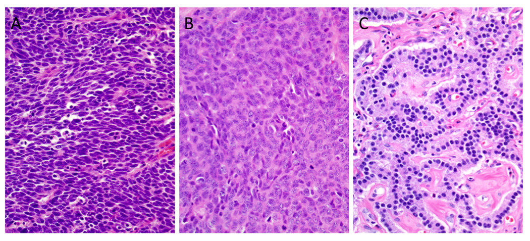
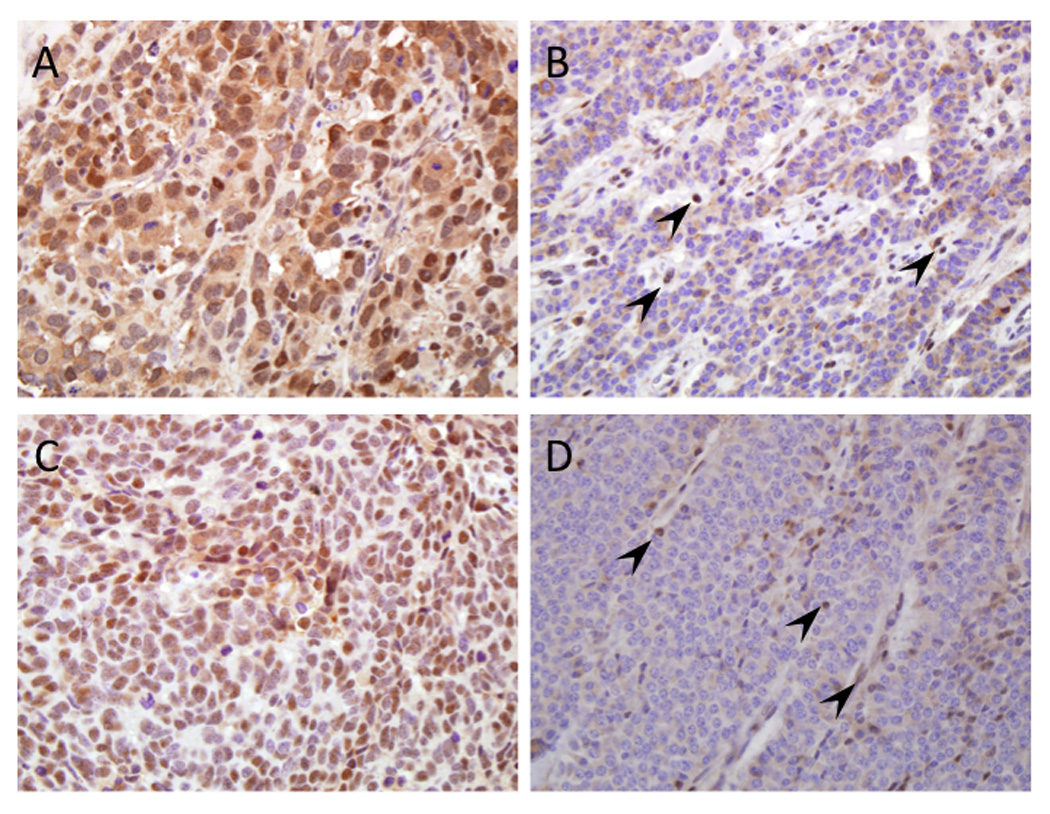
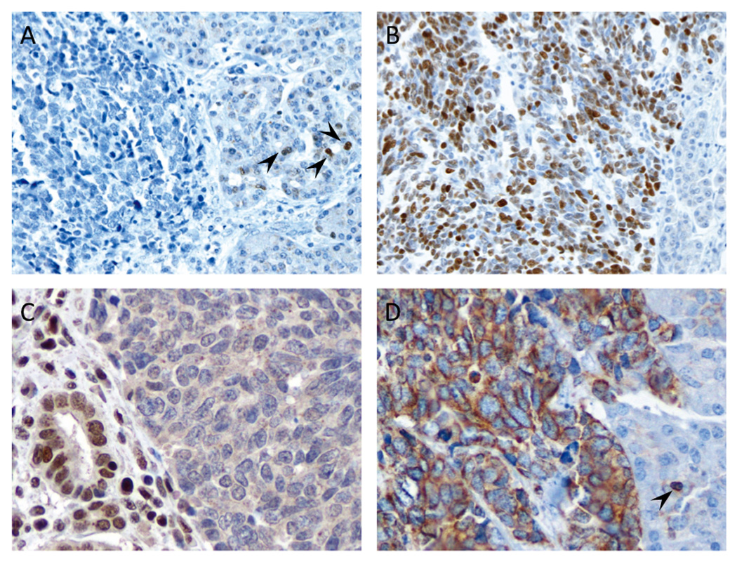
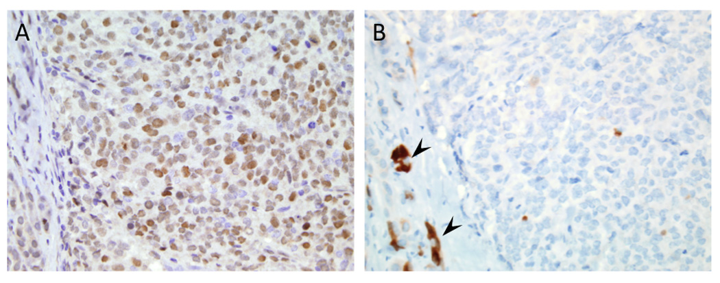
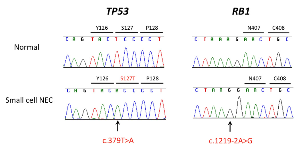
Poorly differentiated neuroendocrine carcinomas (NECs) of the pancreas are rare malignant neoplasms with a poor prognosis. The aim of this study was to determine the clinicopathologic and genetic features of poorly differentiated NECs and compare them with other types of pancreatic neoplasms. We investigated alterations of KRAS, CDKN2A/p16, TP53, SMAD4/DPC4, DAXX, ATRX, PTEN, Bcl2, and RB1 by immunohistochemistry and/or targeted exomic sequencing in surgically resected specimens of 9 small cell NECs, 10 large cell NECs, and 11 well-differentiated neuroendocrine tumors (PanNETs) of the pancreas. Abnormal immunolabeling patterns of p53 and Rb were frequent (p53, 18 of 19, 95%; Rb, 14 of 19, 74%) in both small cell and large cell NECs, whereas Smad4/Dpc4, DAXX, and ATRX labeling was intact in virtually all of these same carcinomas. Abnormal immunolabeling of p53 and Rb proteins correlated with intragenic mutations in the TP53 and RB1 genes. In contrast, DAXX and ATRX labeling was lost in 45% of PanNETs, whereas p53 and Rb immunolabeling was intact in these same cases. Overexpression of Bcl-2 protein was observed in all 9 small cell NECs (100%) and in 5 of 10 (50%) large cell NECs compared with only 2 of 11 (18%) PanNETs. Bcl-2 overexpression was significantly correlated with higher mitotic rate and Ki67 labeling index in neoplasms in which it was present. Small cell NECs are genetically similar to large cell NECs, and these genetic changes are distinct from those reported in PanNETs. The finding of Bcl-2 overexpression in poorly differentiated NECs, particularly small cell NEC, suggests that Bcl-2 antagonists/inhibitors may be a viable treatment option for these patients.
Conflict of interest statement
The authors have no financial conflicts of interest related to this work.
Figures
References
-
- Atasoy P, Bozdogan O, Ozturk S, et al. Bcl2 expression and its correlation with neuroendocrine differentiation in colon carcinomas. Tumori. 2004;90:233–238. - PubMed
-
- Baas IO, Mulder JW, Offerhaus GJ, et al. An evaluation of six antibodies for immunohistochemistry of mutant p53 gene product in archival colorectal neoplasms. The Journal of pathology. 1994;172:5–12. - PubMed
-
- Barton CM, McKie AB, Hogg A, et al. Abnormalities of the RB1 and DCC tumor suppressor genes: uncommon in human pancreatic adenocarcinoma. Molecular carcinogenesis. 1995;13:61–69. - PubMed
-
- Bosman FT, Carneiro F, Hruban RH, et al. WHO classification of tumours of the digestive system. Lyon: International Agency for Research on Cancer (IARC); 2010.
-
- Brennan SM, Gregory DL, Stillie A, et al. Should extrapulmonary small cell cancer be managed like small cell lung cancer? Cancer. 2010;116:888–895. - PubMed
Publication types
MeSH terms
Grants and funding
LinkOut - more resources
Full Text Sources
Medical
Research Materials
Miscellaneous
