. 2012 Mar 7;31(5):1062-79.
doi: 10.1038/emboj.2011.497. Epub 2012 Jan 17.

A novel pathway combining calreticulin exposure and ATP secretion in immunogenic cancer cell death

Affiliations

A novel pathway combining calreticulin exposure and ATP secretion in immunogenic cancer cell death

Abhishek D Garg et al. EMBO J. .

Abstract

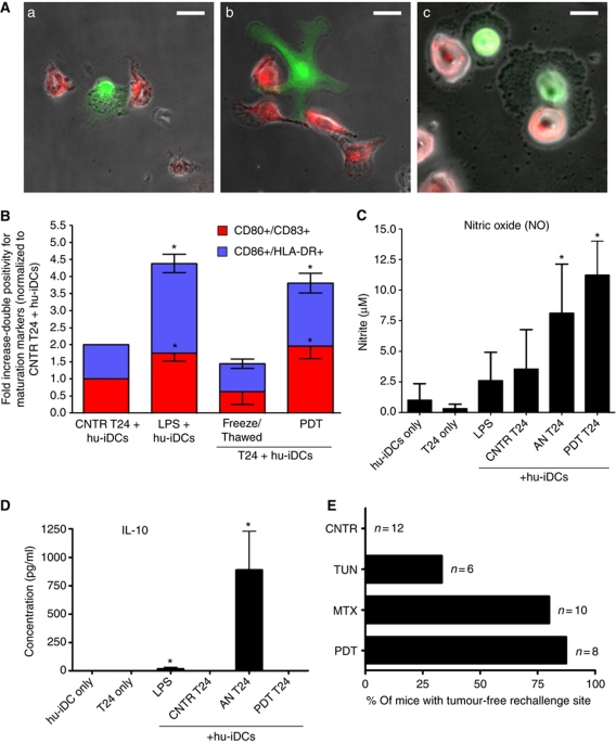
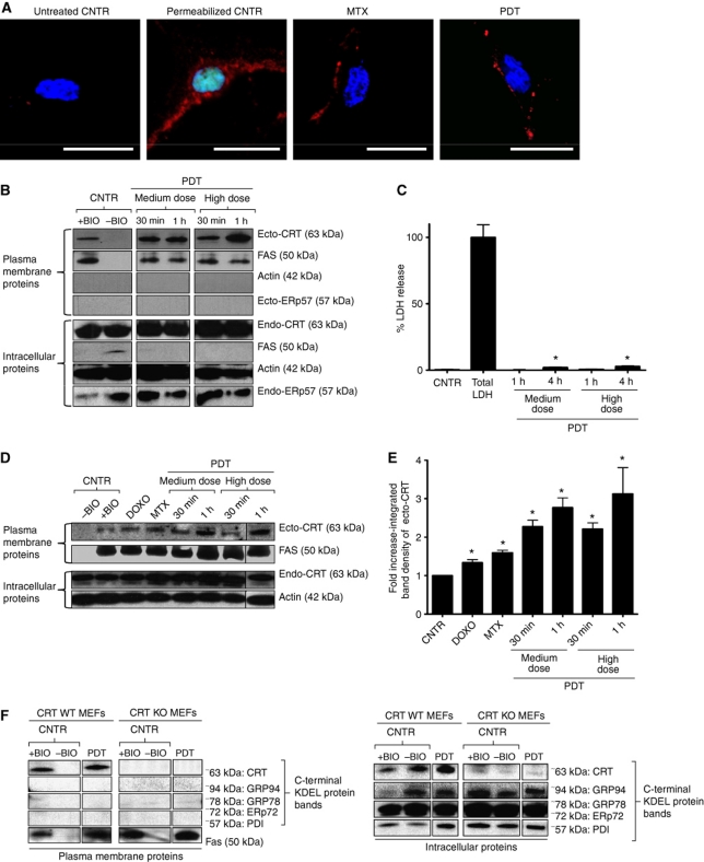
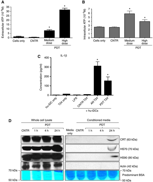
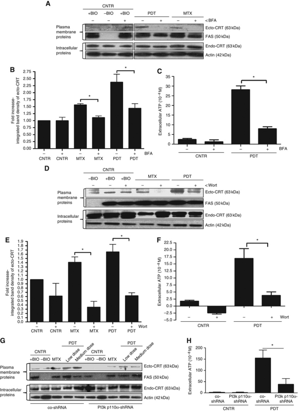
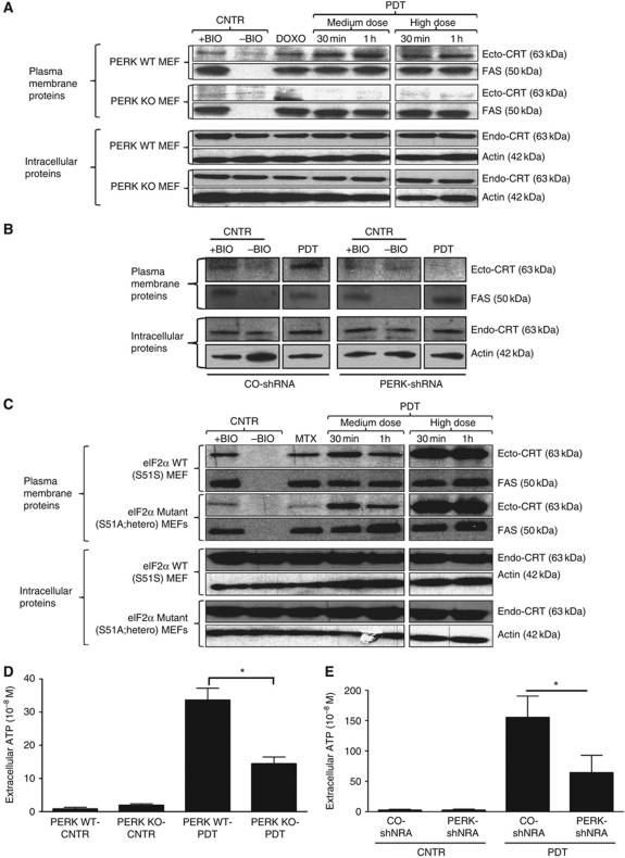
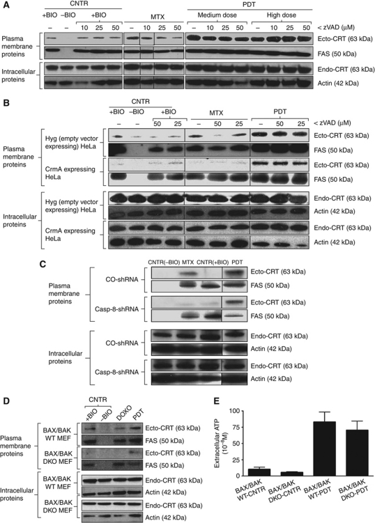
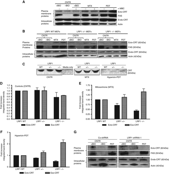
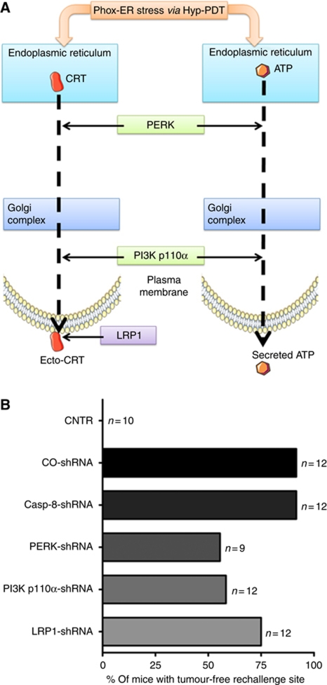
Surface-exposed calreticulin (ecto-CRT) and secreted ATP are crucial damage-associated molecular patterns (DAMPs) for immunogenic apoptosis. Inducers of immunogenic apoptosis rely on an endoplasmic reticulum (ER)-based (reactive oxygen species (ROS)-regulated) pathway for ecto-CRT induction, but the ATP secretion pathway is unknown. We found that after photodynamic therapy (PDT), which generates ROS-mediated ER stress, dying cancer cells undergo immunogenic apoptosis characterized by phenotypic maturation (CD80(high), CD83(high), CD86(high), MHC-II(high)) and functional stimulation (NO(high), IL-10(absent), IL-1β(high)) of dendritic cells as well as induction of a protective antitumour immune response. Intriguingly, early after PDT the cancer cells displayed ecto-CRT and secreted ATP before exhibiting biochemical signatures of apoptosis, through overlapping PERK-orchestrated pathways that require a functional secretory pathway and phosphoinositide 3-kinase (PI3K)-mediated plasma membrane/extracellular trafficking. Interestingly, eIF2α phosphorylation and caspase-8 signalling are dispensable for this ecto-CRT exposure. We also identified LRP1/CD91 as the surface docking site for ecto-CRT and found that depletion of PERK, PI3K p110α and LRP1 but not caspase-8 reduced the immunogenicity of the cancer cells. These results unravel a novel PERK-dependent subroutine for the early and simultaneous emission of two critical DAMPs following ROS-mediated ER stress.


Conflict of interest statement

The authors declare that they have no conflict of interest.

Figures

Figure 1
Figure 1
Tumour cells dying under phox-ER stress conditions induce DC maturation and activate the adaptive immune system. (A) In-vitro phagocytosis of T24 cells treated with Hyp-PDT (red) by human immature dendritic cells (hu-iDCs) (green). The confocal fluorescence images show various phagocytic interactions between dying T24 cells and hu-iDCs, such as tethering (a), initiation of engulfment by extending the pseudopodia (b), and final stages of engulfment (c); scale bar=20 μm. (B) Human DC maturation analysis. T24 cells were left untreated (CNTR), freeze/thawed (accidental necrosis=AN), or treated with a high PDT dose. They were then co-incubated with hu-iDCs. As a positive control, hu-iDCs were stimulated with LPS for 24 h. After co-incubation, the cells were immunostained in two separate groups for CD80/CD83 positivity and CD86/HLA-DR positivity and scored by FACS analysis. Data have been normalized to the ‘CNTR T24 + hu-iDCs’ values. Fold change values are means of two independent experiments (two replicate determinations in each)±s.e.m. (*P<0.05, versus ‘CNTR T24+hu-iDCs’). (C, D) Cytokine and respiratory burst patterns exhibited by human DCs. The T24-hu-iDC co-incubation conditioned media obtained during the experiments detailed in (B) were recuperated followed by analysis for concentrations of nitrite (solubilized form of nitric oxide or NO) (C), and IL-10 (D). Absolute concentrations are the means of two independent experiments (four replicate determinations in each)±s.d. (*P<0.05 versus hu-iDC only). (E) Priming of adaptive immune system by dead/dying CT26 cells. Following immunization with PBS (CNTR) or with CT26 cells treated with tunicamycin (TUN), mitoxantrone (MTX) and the highest PDT dose, the mice were rechallenged with live CT26 tumour cells. Subsequently, the percentage of mice with tumour-free rechallenge site was determined (n represents the number of mice).
Figure 2
Figure 2
Phox-ER-stressed cancer cells expose calreticulin on the surface (ecto-CRT). (A) Immunofluorescence analysis of ecto-CRT. T24 cells were treated with MTX (1 μM for 4 h) and a high PDT dose (recovered 1 h post PDT) or left untreated (CNTR). Alternatively, some cells were saponin permeabilized. This was followed by staining with Sytox Green (exclusion dye), fixation, and immunostaining for CRT and counterstaining with DAPI; scale bar=20 μm. (B) Surface biotinylation analysis of ecto-CRT following phox-ER stress. T24 cells were treated with indicated doses of PDT. They were recovered at the indicated intervals after PDT treatment. Surface proteins were biotinylated followed by immunoblotting. In (B), (D), and (F), ‘+BIO’ indicates controls exposed to buffer with biotin and ‘−BIO’ indicates controls exposed to buffer without biotin (negative control). (C) Plasma membrane permeabilization kinetics following phox-ER stress. T24 cells were treated with PDT and the resulting conditioned media derived at the indicated times post-PDT were analysed for the presence of cytosolic LDH. Total LDH content was determined following Triton-based permeabilization of cells. Data are presented as percent LDH release; values are means of five replicate determinations±s.d. (*P<0.05, versus CNTR). (D) Phox-ER stress induces more ecto-CRT than anthracyclines. T24 cells were treated with PDT, DOXO (25 μM for 4 h), and MTX (1 μM for 4 h). They were recovered at the indicated intervals after PDT treatment. Surface proteins were biotinylated as described for (B). (E) Integrated band densitometric analysis of ecto-CRT. T24 cells were treated with DOXO (25 μM for 4 h), MTX (1 μM for 4 h), and PDT (dose and recovery time points are indicated); and surface proteins were resolved as detailed in (B). Following this, the ecto-CRT protein bands were quantified for the integrated band density via Image J software. Data have been normalized to the CNTR values. Fold change values are means of three independent determinations±s.e.m. (*P<0.05, versus CNTR). (F) Surface biotinylation analysis for KDEL sequence detection following phox-ER stress. CRT WT and KO MEFs were treated with a low PDT dose and surface biotinylated as mentioned in (B). Immunoblotting was done to detect the C-terminal KDEL sequence of various ER proteins (expected molecular weights are indicated).
Figure 3
Figure 3
Cancer cells exposed to phox-ER stress actively secrete ATP, passively release HSP70, HSP90, CRT, and induce IL-1β production in DCs. (A, B) ATP secretion following phox-ER stress. T24 cells were treated with PDT or left untreated (CNTR), and the conditioned media derived from these cells (1 h post PDT in serum-free media) were analysed for the presence of ATP (A). Simultaneously, the corresponding cells were permeabilization with saponin (1 h post PDT) followed by determination of ATP content in the lysate (B). Absolute concentrations are mean values of six replicate determinations±s.d. (*P<0.05, versus CNTR). (C) Cancer cells subjected to phox-ER stress stimulate IL-1β production in hu-iDCs. T24 cells were treated to undergo accidental necrosis (AN), or treated with a high PDT dose (recovered 24 h post PDT), and then co-incubated with hu-iDCs for 24 h. Simultaneously hu-iDCs were stimulated with LPS. After co-incubation, the co-incubation conditioned media (CCM) were analysed for the presence of IL-1β. Cytokine concentrations (in pg/ml) are means of two independent experiments (four replicate determinations in each) ±s.e.m. (*P<0.05, versus CNTR T24). (D) Passive release of HSP70, HSP90, and CRT by dying T24 cells. T24 cells were treated with PDT and recovered at the indicated times. The conditioned media (CM) derived from these cells were concentrated followed by immunoblotting or Coomassie staining for BSA.
Figure 4
Figure 4
Induction of ecto-CRT and ATP secretion by phox-ER stress occurs via secretory pathway and PI3 kinase-dependent plasma membrane/extracellular trafficking. (A, B) Induction of ecto-CRT by phox-ER stress is sensitive to Brefeldin A (BFA). T24 cells were preincubated with 10 μM of BFA for 1 h followed by treatment with a medium PDT dose, MTX (1 μM for 4 h) or left untreated (CNTR) and recovered 1 h post PDT. Surface proteins were biotinylated and immunoblotted (A). In (A), (D), and (G), ‘+BIO’ indicates controls exposed to buffer with biotin and ‘−BIO’ indicates controls exposed to buffer without biotin (negative control). Also, the integrated density of the ecto-CRT protein band was quantified by Image J software (B). In (B) and (E), data have been normalized to CNTR values; fold change values are mean values of three independent experiments±s.e.m. (*P<0.05). (C) ATP secretion following phox-ER stress is sensitive to BFA. T24 cells were preincubated with 10 μM of BFA for 1 h and then treated with medium PDT dose. The resulting conditioned media (1 h post PDT in serum-free media) were analysed for the presence of ATP. Absolute concentrations are means of two independent experiments (five replicate determinations in each)±s.d. (*P<0.05). (D, E) Induction of ecto-CRT by phox-ER stress is sensitive to Wortmannin (Wort). T24 cells were preincubated with 100 nM of Wort for 1 h and then treated with a medium PDT dose or MTX (1 μM for 4 h). Cells were recovered 1 h post PDT. This was followed by biotinylation of the surface proteins (D) and measurement of the integrated density of ecto-CRT protein bands (E) as detailed in (A) and (B), respectively. (F) ATP secretion following phox-ER stress is sensitive to Wort. T24 cells were preincubated with 100 nM Wort for 1 h and then treated with medium PDT dose. The resulting conditioned media (1 h post PDT in serum-free media) were analysed for the presence of ATP as detailed in (C). (G) PI3K p110α shRNA decreases phox-ER stress-induced ecto-CRT. CO-shRNA CT26 cells and CT26 expressing PI3K p110α shRNA 3 were treated with indicated PDT doses or MTX (1 μM for 4 h). They were recovered 1 h post PDT followed by biotinylation of the surface proteins as detailed in (A). (H) ATP secretion following phox-ER stress is reduced by PI3K p110α shRNA. CO-shRNA CT26 cells and CT26 expressing PI3K p110α shRNA 3 were treated with medium PDT dose. The resulting conditioned media (1 h post PDT in serum-free media) were analysed for the presence of ATP. Absolute concentrations are means of five replicate determinations±s.d. (*P<0.05).
Figure 5
Figure 5
Induction of ecto-CRT and ATP secretion by phox-ER stress are PERK dependent. (A) Induction of ecto-CRT by phox-ER is reduced in the absence of PERK. MEF cells containing PERK (WT) or lacking it (KO) were treated with PDT, DOXO (25 μM for 4 h) or left untreated (CNTR). They were recovered at the indicated time points post-PDT. Surface proteins were biotinylated and immunoblotted. In (AC), ‘+BIO’ indicates controls exposed to buffer with biotin and ‘−BIO’ indicates controls exposed to buffer without biotin (negative control). (B) Induction of ecto-CRT by phox-ER is reduced by PERK shRNA. CO-shRNA CT26 cells and CT26 expressing PERK shRNA 3 were treated with medium PDT dose and recovered 1 h post PDT followed by surface biotinylation as detailed in (A). (C) Ecto-CRT induced by phox-ER stress is not affected by the presence of non-phosphorylable eIF2α. MEF cells expressing normal eIF2α (WT) or a non-phosphorylable mutant heterozygously (S51A knock-in mutation) were treated with PDT or MTX (1 μM for 4 h) and recovered at the indicated time points. This was followed by surface biotinylation as detailed in (A). (D) PERK deficiency decreases phox-ER stress-induced secreted ATP. MEF cells that were PERK WT or PERK KO were treated with a medium PDT dose. The resulting conditioned media (1 h post PDT in serum-free media) were analysed for the presence of ATP. Absolute concentrations are mean values of two independent experiments (five replicate determinations in each)±s.d. (*P<0.05). (E) PERK depletion decreases phox-ER stress-induced secreted ATP. CO-shRNA CT26 cells and CT26 expressing PERK shRNA 3 were treated with a medium PDT dose. The resulting conditioned media (1 h post PDT in serum-free media) were analysed for the presence of ATP. Absolute concentrations are mean values of five replicate determinations±s.d. (*P<0.05).
Figure 6
Figure 6
Ecto-CRT induced by phox-ER stress is caspase-8 independent but BAX/BAK dependent. (A) Ecto-CRT induced by phox-ER stress is caspase independent. T24 cells were preincubated with 0, 10, 25, or 50 μM of zVAD for 1 h and then treated with PDT, MTX (1 μM for 4 h) or left untreated (CNTR), and recovered 1 h post PDT. Surface proteins were biotinylated and immunoblotted. In (AD), ‘+BIO’ indicates controls exposed to buffer with biotin and ‘−BIO’ indicates controls exposed to buffer without biotin (negative control). (B) Ecto-CRT induced by phox-ER stress is caspase-8 independent. HeLa cells expressing empty vector Hyg or CrmA were preincubated with 0, 25, or 50 μM of zVAD for 1 h, then treated with a high PDT dose or MTX (1 μM for 4 h) and recovered 1 h post PDT. This was followed by biotinylation as explained in (A). (C) Induction of ecto-CRT by phox-ER is not affected by casp-8 shRNA. CO-shRNA CT26 cells and CT26 expressing casp-8 shRNA 1 were treated with medium PDT dose or MTX (1 μM for 4 h) and recovered 1 h post PDT followed by surface biotinylation as detailed in (A). (D) Ecto-CRT induced by phox-ER stress is BAX/BAK dependent. MEF cells either containing BAX/BAK (WT) or lacking it (DKO) were treated with a medium PDT dose or DOXO (25 μM for 4 h) and recovered 30 min post PDT. This was followed by surface biotinylation as detailed in (A). (E) BAX/BAK deficiency does not affect phox-ER stress-induced secreted ATP. MEF cells that were BAX/BAK WT or DKO were treated with a medium PDT dose. The resulting conditioned media (1 h post PDT in serum-free media) were analysed for the presence of ATP. Absolute concentrations are mean values of two independent experiments (five replicate determinations in each)±s.d.
Figure 7
Figure 7
Ecto-CRT induced by phox-ER stress docks on the surface via the LRP1 molecule. (A) Ecto-CRT induced by phox-ER stress is not dependent on lipid rafts. T24 cells were preincubated with 2 μM of MBC for 1 h and then treated with a medium PDT dose, MTX (1 μM for 1 h) or left untreated (CNTR). They were recovered 5 min post PDT. Surface proteins were biotinylated and immunoblotted. In (A), (B), and (G), ‘+BIO’ indicates controls exposed to buffer with biotin and ‘−BIO’ indicates controls exposed to buffer without biotin (negative control). (BF) Ecto-CRT retention on ER-stressed cancer cells is dependent on the level of LRP1. MEF cells containing levels of 100% LRP1 (LRP1 WT), 50% LRP1 (LRP1+/−), or 0% LRP1 (LRP−/−) were treated with a low PDT dose or MTX (1 μM for 4 h in serum-free media) followed by recovery in serum-free media after 1 h post PDT. Surface proteins were biotinylated and immunoblotted (B) as detailed in (A). Simultaneously, the conditioned media (CM) derived from these cells were concentrated and immunoblotted (C). The integrated density of CRT protein bands in (B) and (C) was quantified by Image J software for untreated (D), MTX-treated (E), and PDT-treated (F) conditions: where black is for ecto-CRT bands in the immunoblots of biotinylated surface proteins and grey is for ‘exo-CRT’ bands in the immunoblots of concentrated CM. Data have been normalized to the respective LRP1 WT values. Fold change values are means of three independent experiments±s.e.m. (G) Induction of ecto-CRT by phox-ER stress is reduced by LRP1 shRNA. CO-shRNA CT26 cells and CT26 expressing LRP1 shRNA 1 were treated with medium PDT dose and recovered 1 h post PDT followed by surface biotinylation as detailed in (A).
Figure 8
Figure 8
Depletion of PERK, PI3K p110α, and LRP1 but not caspase-8 reduces phox-ER stress-induced immunogenicity. (A) Hypothetical representation of main molecular players in phox-ER stress-induced ecto-CRT and ATP secretion pathways. In-vitro data suggested that while caspase-8 was dispensable for ecto-CRT induction after phox-ER stress; PERK and PI3K p110α were indispensable. In fact, both of these latter molecules were also required for ATP secretion. Moreover, LRP1 was observed to be crucial for surface tethering of ecto-CRT. (B) The mice were immunized either with PBS (CNTR) or with highest PDT dose treated CO-shRNA CT26 cells or with different CT26 cells expressing casp-8 shRNA 1, PERK shRNA 3, PI3K p110α shRNA 3, or LRP1 shRNA 1, also treated with the highest PDT dose. These ‘immunized’ mice were then rechallenged with live CT26 tumour cells. Subsequently, the percentage of mice with tumour-free rechallenge site was determined (n represents the number of mice).

Comment in

Similar articles

Cited by

References

    1. Abe M, Setoguchi Y, Tanaka T, Awano W, Takahashi K, Ueda R, Nakamura A, Goto S (2009) Membrane protein location-dependent regulation by PI3K (III) and rabenosyn-5 in Drosophila wing cells. PLoS One 4: e7306. - PMC - PubMed
    1. Agostinis P, Berg K, Cengel KA, Foster TH, Girotti AW, Gollnick SO, Hahn SM, Hamblin MR, Juzeniene A, Kessel D, Korbelik M, Moan J, Mroz P, Nowis D, Piette J, Wilson BC, Golab J (2011) Photodynamic therapy of cancer: an update. CA Cancer J Clin 61: 250–281 - PMC - PubMed
    1. Agostinis P, Vantieghem A, Merlevede W, de Witte PA (2002) Hypericin in cancer treatment: more light on the way. Int J Biochem Cell Biol 34: 221–241 - PubMed
    1. Aymeric L, Apetoh L, Ghiringhelli F, Tesniere A, Martins I, Kroemer G, Smyth MJ, Zitvogel L (2010) Tumor cell death and ATP release prime dendritic cells and efficient anticancer immunity. Cancer Res 70: 855–858 - PubMed
    1. Bajor A, Tischer S, Figueiredo C, Wittmann M, Immenschuh S, Blasczyk R, Eiz-Vesper B (2011) Modulatory role of calreticulin as chaperokine for dendritic cell-based immunotherapy. Clin Exp Immunol 165: 220–234 - PMC - PubMed

Publication types

MeSH terms