. 2012 Mar 7;31(5):1308-19.
doi: 10.1038/emboj.2011.496. Epub 2012 Jan 17.

Structure of a novel phosphotyrosine-binding domain in Hakai that targets E-cadherin

Affiliations

Structure of a novel phosphotyrosine-binding domain in Hakai that targets E-cadherin

Manjeet Mukherjee et al. EMBO J. .

Abstract

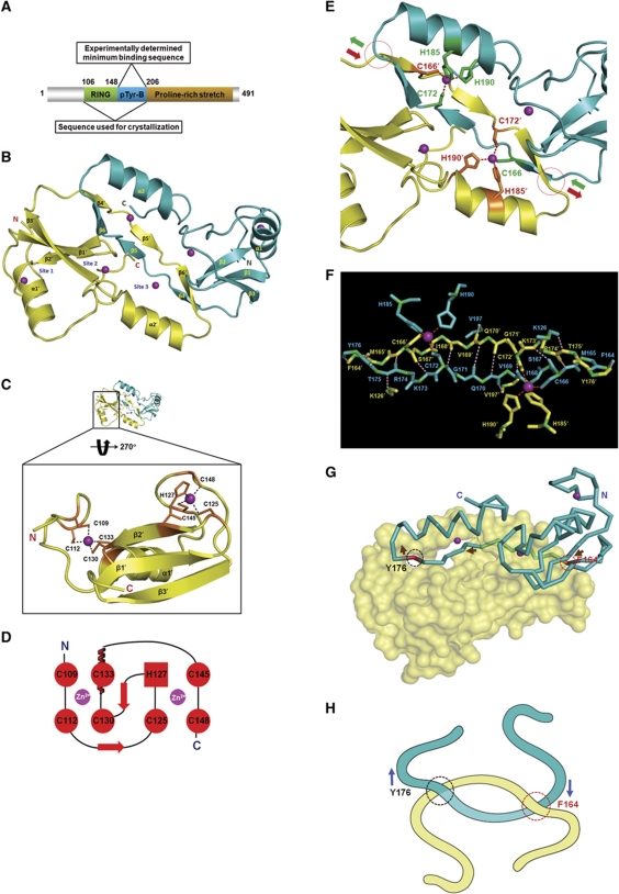
Phosphotyrosine-binding domains, typified by the SH2 (Src homology 2) and PTB domains, are critical upstream components of signal transduction pathways. The E3 ubiquitin ligase Hakai targets tyrosine-phosphorylated E-cadherin via an uncharacterized domain. In this study, the crystal structure of Hakai (amino acids 106-206) revealed that it forms an atypical, zinc-coordinated homodimer by utilizing residues from the phosphotyrosine-binding domain of two Hakai monomers. Hakai dimerization allows the formation of a phosphotyrosine-binding pocket that recognizes specific phosphorylated tyrosines and flanking acidic amino acids of Src substrates, such as E-cadherin, cortactin and DOK1. NMR and mutational analysis identified the Hakai residues required for target binding within the binding pocket, now named the HYB domain. ZNF645 also possesses a HYB domain but demonstrates different target specificities. The HYB domain is structurally different from other phosphotyrosine-binding domains and is a potential drug target due to its novel structural features.


Conflict of interest statement

The authors declare that they have no conflict of interest.

Figures

Figure 1
Figure 1
A novel protein fold in Hakai. (A) A schematic diagram of the Hakai protein. (B) The crystal structure of Hakai (aa 106–206) reveals a dimer in an anti-parallel configuration. Each monomer contains three zinc coordination sites. Sites 1 and 2 lie in the RING domain. Site 3 is shared between the two monomers. (C) The coordination of zinc ions (purple spheres) by the RING domain of Hakai is shown for one of the monomers. (D) A schematic diagram of the cross-brace arrangement of the Hakai RING domain as shown in (C). (E) The Hakai dimer forms an intertwined configuration spanning the points indicated in circles, with the entry and exit paths shown in green and brown arrows. The zinc-interacting side chains are shown as green and brown sticks. (F) The backbone of the Hakai (aa 106–206) residues involved in intermolecular main-chain H-bonding and the zinc-coordinating side chains of adjacent monomers at the dimer interface are shown in cyan and yellow. The pink dots indicate the main-chain H-bonds; the red dots indicate the zinc coordination bonds. (G) The monomers of the interlinked Hakai dimer are shown in surface representation and Cα trace, respectively. The Cα trace monomer enters and exits the other monomer at the red and black circles, respectively. Brown arrows show its entry and exit path. (H) A schematic diagram of the novel Hakai interlinked arrangement as shown in (G).
Figure 2
Figure 2
Hakai forms a dimer in solution. (A) A 3D 15N-NOESY spectrum showing the intermolecular NOE cross-peaks of amides corresponding to residues of Hakai (aa 106–206). (B) WT Hakai (aa 106–206) and four Hakai (aa 106–206) point mutants were each separately used for gel-filtration chromatography. Their respective elution profiles were overlain and compared. (C) HA- and FLAG-tagged Hakai were overexpressed in the presence of Src in HEK293 cells. FLAG immunoprecipitates were analysed for HA-tagged Hakai. (D) A schematic representation of the point mutations made to C166, C172, H185 and H190 in Hakai. (E) Cell lysates containing WT Hakai or Hakai mutants were used to analyse the effects of Hakai dimerization on E-cadherin recognition.
Figure 3
Figure 3
Hakai domain recognizes acidic residues. (A) Y753, Y754 and Y755 (red) of E-cadherin were mutated to phenylalanine (blue) in different combinations. (B) The WT E-cadherin and the mutants shown in (A) were overexpressed in HEK293 cells with v-Src to analyse their pTyr signals. (C) HEK293 cells were co-transfected with WT E-cadherin or its mutants together with Hakai to identify the tyrosine residues recognized by Hakai. (D) The Y754-phosphorylated and non-phosphorylated E-cadherin peptides were titrated against Hakai (aa 106–206) using ITC. The top panels show the heat release profiles after baseline correction and the lower panels indicate the binding isotherms for the interactions. The dissociation constant (Kd) and binding stoichiometry (N) are shown in the table. (E) The E-cadherin aa 747–758 were each substituted with alanine. (F) The E-cadherin mutants from (E) and Hakai were co-transfected into HEK293 cells to identify the target motif on E-cadherin. (G) E-cadherin and Hakai were co-transfected into HEK293 cells. Their interaction was analysed through immunoprecipitation of FLAG-tagged Hakai. (H) Cortactin was co-transfected into HEK293 cells with Hakai. The interaction between cortactin and Hakai was compared with that in (G). (I) Y482 and Y485 (red) were separately substituted with phenylalanine (blue). (J) WT and mutated cortactin were co-transfected into HEK293 cells with Hakai, in the absence or presence of v-Src. The pTyr signal of cortactin and its interaction with Hakai were analysed. (K) An alanine scan of cortactin aa 478–489. Each residue was substituted with alanine (blue). G484 was not mutated as glycine mutations affect the protein structure. (L) The cortactin mutants described in (K) were co-transfected into HEK293 cells with Hakai. The interaction between the cortactin mutants and Hakai was determined using immunoprecipitation.
Figure 4
Figure 4
DOK1 interacts with Hakai. (A) The sequence alignment of the different Src phosphorylation target sites in E-cadherin, cortactin and DOK1. The acidic amino-acid residues flanking the phosphorylated tyrosine are shown in blue. (B) Hakai and DOK1 were overexpressed in HEK293 cells in the absence or presence of Src. FLAG immunoprecipitates were analysed for DOK1 interaction. (C) DOK1 was co-transfected into HEK293 cells with Hakai to study its competition with endogenous cortactin for binding to Hakai. FLAG immunoprecipitates were immunoblotted for cortactin.
Figure 5
Figure 5
Target-binding amino acids of Hakai. (A) An overlay of the 1H–15N-HSQC spectra of Hakai (aa 106–206) in the absence (green) or the presence (red) of an tyrosine-phosphorylated E-cadherin peptide. (B) A graphical representation of the combined chemical shift perturbation (p.p.m.) plotted against all Hakai (aa 106–206) residues, with the cutoff at the combined chemical shift perturbation of 0.15 p.p.m. (C) The six potential E-cadherin-interacting residues in Hakai (aa 106–206) are highlighted as sticks in the ribbon representation of the crystal structure. (D) An electrostatic surface potential representation of Hakai (aa 106–206) shows that H127, Y176, H185 and R189 form part of the positively charged pocket. (E) The interaction between E-cadherin and the Hakai mutants of the residues identified in (C) was analysed by immunoprecipitating FLAG-tagged Hakai. (F) HEK293 cells were transfected with the identified Hakai mutants, and their interaction with endogenous cortactin was studied. (G) Immunoprecipitates of either WT Hakai or the Hakai zinc-coordinating mutants were tested for interaction with endogenous cortactin. (H) A schematic representation of the Hakai dimer and the HYB domain.
Figure 6
Figure 6
The HYB domain in other proteins. (A) A comparison of the Hakai protein from amino-acid residues 127–191 and the equivalent sequence in ZNF645. (B) E-cadherin and ZNF645 were analysed for their interaction using immunoprecipitation. Hakai was used as a positive control. The dotted arrow indicates a non-specific band; the solid arrow indicates the ZNF645 band. (C) ZNF645 was overexpressed in HEK293 cells and its interaction with endogenous cortactin was analysed using immunoprecipitation. (D) Sequence alignment of LNX1 and LNX2 with Hakai and ZNF645 based on the conserved zinc-coordinating residues from Hakai aa 106–206.
Figure 7
Figure 7
Novel structure of the HYB domain. (A) Representative structures of SH2 (PDB code 1SHB), PTB (PDB code 1SHC), PKCδ C2 (PDB code 1YRK) and PKM2 (PDB code 3BJF) in ligand-free forms are compared with the HYB domain. (B) The corresponding topologies of the domains in (A).

References

    1. Adams PD, Grosse-Kunstleve RW, Hung LW, Ioerger TR, McCoy AJ, Moriarty NW, Read RJ, Sacchettini JC, Sauter NK, Terwilliger TC (2002) PHENIX: building new software for automated crystallographic structure determination. Acta Crystallogr D Biol Crystallogr 58: 1948–1954 - PubMed
    1. Bellon SF, Rodgers KK, Schatz DG, Coleman JE, Steitz TA (1997) Crystal structure of the RAG1 dimerization domain reveals multiple zinc-binding motifs including a novel zinc binuclear cluster. Nat Struct Biol 4: 586–591 - PubMed
    1. Benes CH, Wu N, Elia AE, Dharia T, Cantley LC, Soltoff SP (2005) The C2 domain of PKCdelta is a phosphotyrosine binding domain. Cell 121: 271–280 - PubMed
    1. Borden KLB (2000) RING domains: master builders of molecular scaffolds? J Mol Biol 295: 1103–1112 - PubMed
    1. Brunger AT, Adams PD, Clore GM, DeLano WL, Gros P, Grosse-Kunstleve RW, Jiang JS, Kuszewski J, Nilges M, Pannu NS, Read RJ, Rice LM, Simonson T, Warren GL (1998) Crystallography & NMR system: a new software suite for macromolecular structure determination. Acta Crystallogr D Biol Crystallogr 54: 905–921 - PubMed

Publication types

Associated data