

The potential for Cerenkov luminescence imaging of alpha-emitting radionuclides
- PMID: 22252144
- PMCID: PMC5558792
- DOI: 10.1088/0031-9155/57/3/771
The potential for Cerenkov luminescence imaging of alpha-emitting radionuclides
Abstract
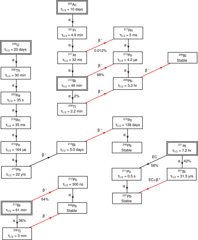
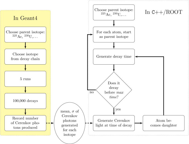
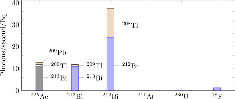
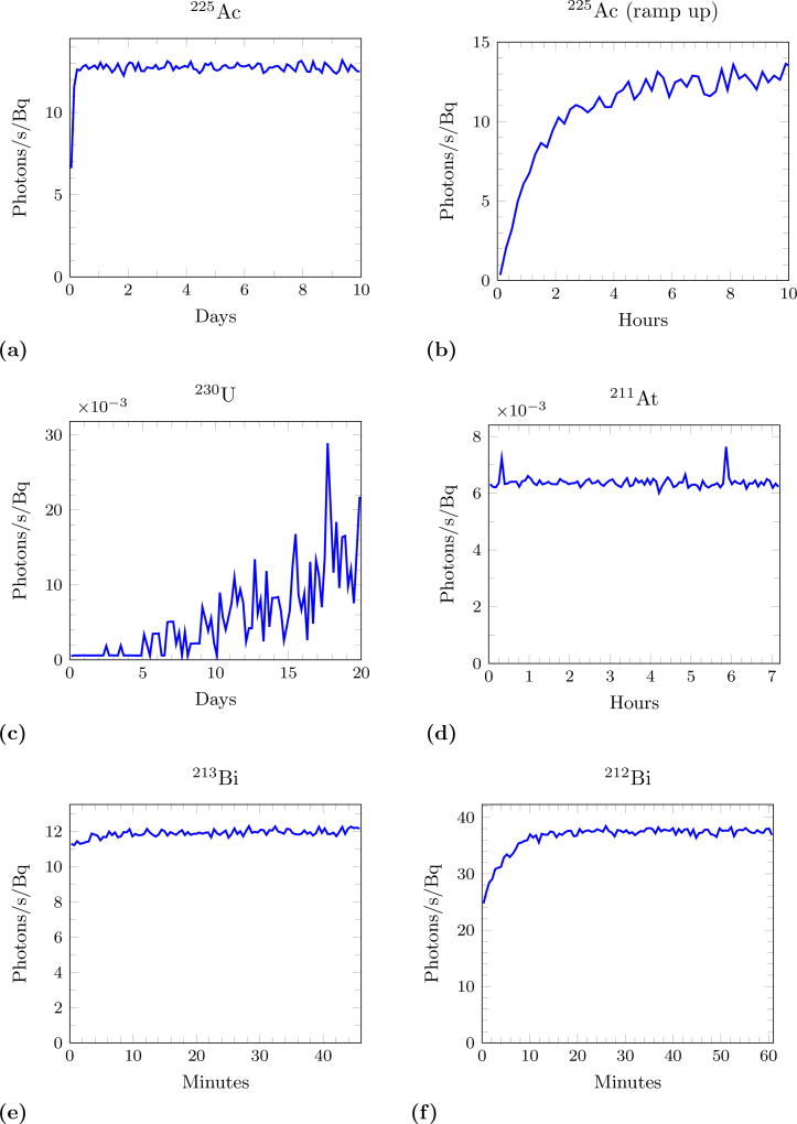
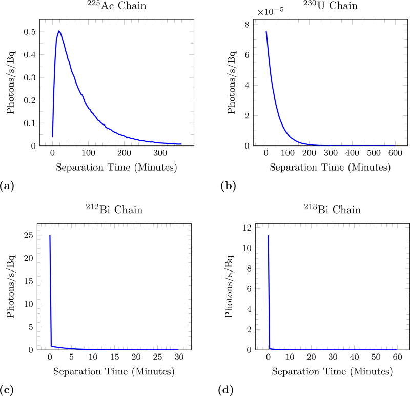
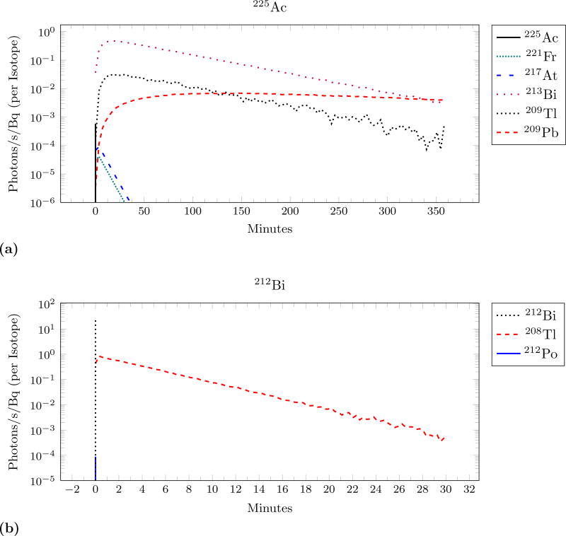
Targeted α-emitting drugs are promising for cancer therapy, but cannot be effectively imaged by conventional techniques. Cerenkov luminescence imaging (CLI) has previously been shown capable of imaging β(+)- and β(-)-emitting radionuclides in vivo and could have the potential to image α-emitters. Cerenkov light production from α-emitters is through Compton scattering and from farther down the decay chain. This causes the Cerenkov production to vary in time and depend on sample geometry, complicating the interpretation of CLI images. We used the simulation toolkit Geant4 to predict the Cerenkov light output from five α-emitting radionuclides that have therapeutic potential: (225)Ac, (230)U, (213)Bi, (212)Bi and (212)At. We found that (225)Ac, (213)Bi and (212)Bi produced an order of magnitude more Cerenkov light than (18)F. However, the light from (225)Ac is delayed from the initial decay, possibly decreasing the correlation of the drug and light source. This indicates that CLI will not be helpful in the development of some α-emitting drugs.
Figures
Similar articles
-
Computed Cerenkov luminescence yields for radionuclides used in biology and medicine.Phys Med Biol. 2015 Jun 7;60(11):4263-80. doi: 10.1088/0031-9155/60/11/4263. Epub 2015 May 14. Phys Med Biol. 2015. PMID: 25973972
-
Cerenkov luminescence imaging of medical isotopes.J Nucl Med. 2010 Jul;51(7):1123-30. doi: 10.2967/jnumed.110.076521. Epub 2010 Jun 16. J Nucl Med. 2010. PMID: 20554722 Free PMC article.
-
In vivo Cerenkov luminescence imaging: a new tool for molecular imaging.Philos Trans A Math Phys Eng Sci. 2011 Nov 28;369(1955):4605-19. doi: 10.1098/rsta.2011.0271. Philos Trans A Math Phys Eng Sci. 2011. PMID: 22006909 Free PMC article.
-
Quantitative Imaging of Alpha-Emitting Therapeutic Radiopharmaceuticals.Nucl Med Mol Imaging. 2019 Jun;53(3):182-188. doi: 10.1007/s13139-019-00589-8. Epub 2019 Feb 18. Nucl Med Mol Imaging. 2019. PMID: 31231438 Free PMC article. Review.
-
Cerenkov luminescence imaging (CLI) for image-guided cancer surgery.Clin Transl Imaging. 2016;4(5):353-366. doi: 10.1007/s40336-016-0183-x. Epub 2016 May 24. Clin Transl Imaging. 2016. PMID: 27738626 Free PMC article. Review.
Cited by
-
Radioluminescence in biomedicine: physics, applications, and models.Phys Med Biol. 2019 Feb 6;64(4):04TR01. doi: 10.1088/1361-6560/aaf4de. Phys Med Biol. 2019. PMID: 30524090 Free PMC article. Review.
-
Quantification of Cerenkov Luminescence Imaging (CLI) Comparable With 3-D PET Standard Measurements.Mol Imaging. 2018 Jan-Dec;17:1536012118788637. doi: 10.1177/1536012118788637. Mol Imaging. 2018. PMID: 30043654 Free PMC article.
-
Molecular targeted α-particle therapy for oncologic applications.AJR Am J Roentgenol. 2014 Aug;203(2):253-60. doi: 10.2214/AJR.14.12554. AJR Am J Roentgenol. 2014. PMID: 25055256 Free PMC article. Review.
-
212Pb in targeted radionuclide therapy: a review.EJNMMI Radiopharm Chem. 2025 Jul 1;10(1):34. doi: 10.1186/s41181-025-00362-7. EJNMMI Radiopharm Chem. 2025. PMID: 40591218 Free PMC article. Review.
-
Development of Targeted Alpha Particle Therapy for Solid Tumors.Molecules. 2019 Nov 26;24(23):4314. doi: 10.3390/molecules24234314. Molecules. 2019. PMID: 31779154 Free PMC article. Review.
References
-
- Agostinelli S, Allison J, Amako K, Apostolakis J, Araujo H, Arce P, Asai M, Axen D, Banerjee S, Barrand G, et al. G4–a simulation toolkit. Nuclear Instruments and Methods in Physics Research Section A: Accelerators, Spectrometers, Detectors and Associated Equipment. 2003;506:250–303.
-
- Allison J, Amako K, Apostolakis J, Araujo H, Dubois P, Asai M, Barrand G, Capra R, Chauvie S, Chytracek R, et al. Geant4 developments and applications. Nuclear Science, IEEE Transactions on. 2006;53:270–278.
-
- Brun R, Rademakers F. ROOT: An object oriented data analysis framework. Nucl. Instrum. Meth. 1997;A389:81–86.
-
- Hale GM, Querry MR, et al. Optical constants of water in the 200-nm to 200-um wavelength region. Appl. Opt. 1973;12:555–563. - PubMed
Publication types
MeSH terms
Substances
Grants and funding
LinkOut - more resources
Full Text Sources
Medical
Research Materials
Miscellaneous