Review
. 2012 Jul;32(7):1139-51.
doi: 10.1038/jcbfm.2011.197. Epub 2012 Jan 18.

Neurological diseases in relation to the blood-brain barrier

Affiliations
Review

Neurological diseases in relation to the blood-brain barrier

Gary A Rosenberg. J Cereb Blood Flow Metab. 2012 Jul.

Abstract

Disruption of the blood-brain barrier (BBB) has an important part in cellular damage in neurological diseases, including acute and chronic cerebral ischemia, brain trauma, multiple sclerosis, brain tumors, and brain infections. The neurovascular unit (NVU) forms the interface between the blood and brain tissues. During an injury, the cascade of molecular events ends in the final common pathway for BBB disruption by free radicals and proteases, which attack membranes and degrade the tight junction proteins in endothelial cells. Free radicals of oxygen and nitrogen and the proteases, matrix metalloproteinases and cyclooxgyenases, are important in the early and delayed BBB disruption as the neuroinflammatory response progresses. Opening of the BBB occurs in neurodegenerative diseases and contributes to the cognitive changes. In addition to the importance of the NVU in acute injury, angiogenesis contributes to the recovery process. The challenges to treatment of the brain diseases involve not only facilitating drug entry into the brain, but also understanding the timing of the molecular cascades to block the early NVU injury without interfering with recovery. This review will describe the molecular and cellular events associated with NVU disruption and potential strategies directed toward restoring its integrity.


Figures
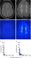
Figure 1
Figure 1
Density distribution of permeability values for white matter (WM) voxels of a control and a vascular cognitive impairment patient. (A) The color-coded permeability map shows normal permeability, which is below the threshold of 3 × 10⁻⁴ mL/g-min, which was established in 17 control subjects. (C) The histogram of permeability values for the control subject shown in panel A. (B) The permeability map of a VCI patient showing the regions of increased permeability in yellow and red. (D) Permeability histogram shows the shift to the right of permeability values for patient in panel B (modified from Taheri et al, 2011).
Figure 2
Figure 2
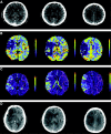
A 75-year-old right-handed man who presented with sudden onset of left hemiparesis and dysarthria. (A) Admission computed tomography (CT). (B) Admission cerebral blood volume (CBV) color maps showing hypoperfusion with right middle cerebral artery (MCA) acute ischemic stroke (AIS). (C) Admission permeability color maps showed permeability abnormality with right MCA AIS. (D) Subsequent head CT before hemicraniectomy showing malignant MCA infarction (Bektas et al, 2010) (permission obtained).
Figure 3
Figure 3
Hypoxic hypoperfusion in acute and chronic ischemia induces hypoxia-inducible factor-1α (HIF-1α), which induces the fur gene to transcribe the convertase, furin. Activation of proMMP-14 (membrane-type MMP) is necessary for the activation of proMMP-2, which attacks the tight junction proteins and basal lamina opening the blood–brain barrier (BBB). As a consequence of the activation of the MMPs, myelin is broken down. MMP, matrix metalloproteinases.
Figure 4
Figure 4
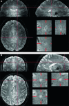
The 7-T T2*-weighted magnitude images were viewed in orthogonal planes. For each lesion, the presence or absence of a central vein was noted. The proportion of perivenous lesions in individual patients with multiple sclerosis (MS) (mean 80%, range 53% to 100%) was consistently much higher than in individual subjects without MS (mean 16%, range 0% to 34% (A). Perivenous lesion appearance was equally common in patients with clinically isolated syndrome, relapsing-remitting MS, primary progressive MS, and secondary progressive MS. (B) (Tallantyre et al, 2011) (permission obtained).
Figure 5
Figure 5
Fluid-attenuated inversion recovery (FLAIR) magnetic resonance imaging (MRI) and dynamic contrast-enhanced MRIs (DCEMRIs) from a patient with Binswanger's disease. Initial MRIs (A, C) show the extent of the white matter hyperintensities. Corresponding permeability measurements (B, D) show increased permeability in the regions with the lighter blue. Follow-up studies performed one and half years later show an increase in size of the white matter lesions (E, G). Permeability studies show persistent leakage of Gd-DTPA (F, H). The light blue, yellow, and red areas have increased permeability.
Figure 6
Figure 6
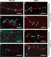
Representative confocal micrographs of cerebral blood vessels from aged Tg2576 and wild-type mice immunolabeled for either occludin or zonula occludens (ZO)-1 (red) and counterstained for DNA (blue) with TOTO-3. Blood vessels, imaged in the neocortex and hippocampus, which exhibited strong, continuous, and linear occludin (A, C) or ZO-1 (E, G) expression were considered normal, as demonstrated in the wild type. Abnormal occludin (B, F) and ZO-1 (D, H) staining displayed punctate (white arrowheads), discontinuous or interrupted (hollow white arrows), as seen in the Tg2576 cerebrovasculature. Results are representative from three mice per group from three separate experiments. Scale bar represents 20 μm (Biron et al, 2011) (permission obtained).

References

    1. Akiguchi I, Tomimoto H, Suenaga T, Wakita H, Budka H. Blood-brain barrier dysfunction in Binswanger's disease; an immunohistochemical study. Acta Neuropathol. 1998;95:78–84. - PubMed
    1. Bateman RJ, Siemers ER, Mawuenyega KG, Wen G, Browning KR, Sigurdson WC, Yarasheski KE, Friedrich SW, Demattos RB, May PC, Paul SM, Holtzman DM. A gamma-secretase inhibitor decreases amyloid-beta production in the central nervous system. Ann Neurol. 2009;66:48–54. - PMC - PubMed
    1. Bektas H, Wu T-C, Kasam M, Harun N, Sitton CW, Grotta JC, Savitz SI. Increased blood, ÄìBrain barrier permeability on perfusion CT might predict malignant middle cerebral artery infarction. Stroke. 2010;41:2539–2544. - PMC - PubMed
    1. Bell RD, Winkler EA, Sagare AP, Singh I, LaRue B, Deane R, Zlokovic BV. Pericytes control key neurovascular functions and neuronal phenotype in the adult brain and during brain aging. Neuron. 2010;68:409–427. - PMC - PubMed
    1. Biron KE, Dickstein DL, Gopaul R, Jefferies WA. Amyloid triggers extensive cerebral angiogenesis causing blood brain barrier permeability and hypervascularity in Alzheimer's disease. PLoS One. 2011;6:e23789. - PMC - PubMed

Publication types

MeSH terms