. 2012 Mar;32(6):1032-43.
doi: 10.1128/MCB.06021-11. Epub 2012 Jan 17.

Contribution of a TANK-binding kinase 1-interferon (IFN) regulatory factor 7 pathway to IFN-γ-induced gene expression

Affiliations

Contribution of a TANK-binding kinase 1-interferon (IFN) regulatory factor 7 pathway to IFN-γ-induced gene expression

Matthias Farlik et al. Mol Cell Biol. 2012 Mar.

Abstract

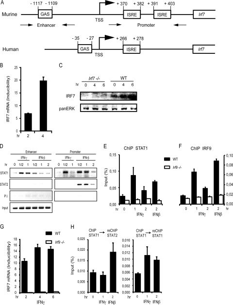
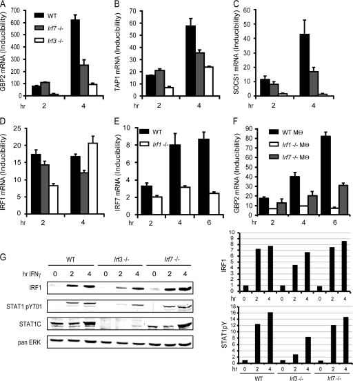
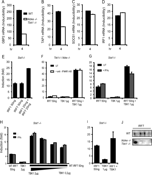
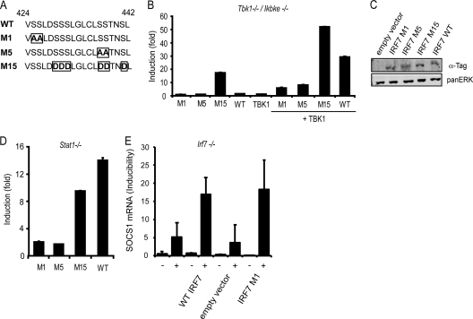
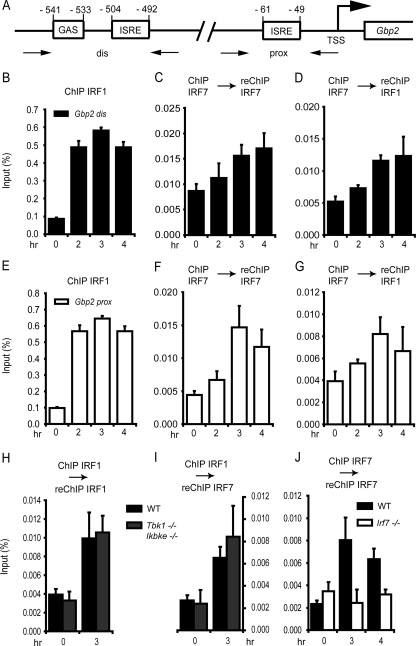
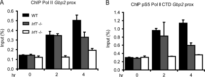
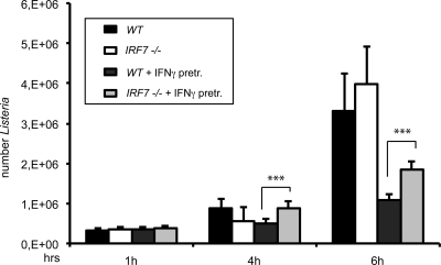
Signal transducers and activators of transcription (STATs) and interferon regulatory factors (IRFs) share common target genes. Here we show that the Irf7 gene is regulated by transcription factors STAT1 and IRF9 in response to the type II interferon (IFN) IFN-γ. IRF7 cooperated with STAT1 and IRF1 to stimulate the expression of a subset of IFN-γ-induced STAT1 target genes. IRF7-mediated control of the Gbp2 gene required the presence and basal activity of the S/T kinase TANK-binding kinase 1 (TBK1), whereas the binding of IRF7 to the Gbp2 promoter did not. Analysis of RNA polymerase II (Pol II) recruitment to the Gbp2 promoter revealed a role for IRF7 at later stages of the IFN-γ response. In support of the role of IRF7 in establishing an effective antibacterial response, IFN-γ-pretreated Irf7(-/-) macrophages showed an increased bacterial burden after infection with Listeria monocytogenes. Our data thus describe a biologically relevant basal activity of TBK1 and identify IRF7 as a novel player in the IFN-γ response.


Figures

Fig 1
Fig 1
Kinetics of IRF7 mRNA expression and STAT1 recruitment to the Irf7 promoter, determined by qPCR and ChIP, respectively. (A) Schematic drawing of the murine and human Irf7 promoters. ISRE sites are located in the 5′ UTR of the murine Irf7 gene, and the newly defined GAS is located in the enhancer region 1.1 kb upstream of the TSS. (B and C) IRF7 mRNA (B) and protein expression (C). mRNA expression after treatment of WT MEFs with IFN-γ for the indicated time points was determined by qPCR and normalized to GAPDH levels. IRF7 protein was determined by Western blotting with IRF7 antibodies. To increase sensitivity, IRF7 was stabilized by treatment with the proteasome inhibitor MG132. (D) WT MEFs were stimulated with IFN-γ or IFN-β and processed for ChIP at the indicated time points. Antibodies for ChIP are shown on the left. P. I. indicates controls performed with preimmune sera. The precipitates were amplified by PCR with primers flanking the enhancer (GAS) or promoter (ISRE) region as indicated in panel A and were analyzed by gel electrophoresis. (E and F) Bone marrow-derived macrophages (BMDMs) of WT and Irf9−/− mice were treated with IFN-γ or IFN-β for the times indicated and processed for ChIP with the antibodies indicated on the top of each panel. (G) BMDMs derived from WT or Irf9−/− mice were treated with IFN-γ for the indicated time periods. IRF7 mRNA expression was analyzed by qPCR and normalized to GAPDH levels. (H) BMDMs of WT mice were treated with IFN-γ or IFN-β for the times indicated and processed for ChIP-reChIP. Antibodies for ChIP are shown on top of each panel. The precipitates were amplified with primers flanking the Irf7 promoter as shown in panel A. qPCR measurements were made in triplicate. All experiments were repeated at least three times.
Fig 2
Fig 2
Regulation of IFN-γ-induced genes by IRFs. (A to D) WT, Irf3−/−, and Irf7−/− MEFs were treated with IFN-γ for the indicated time periods, followed by determination of GBP2, TAP1, SOCS1, and IRF1 mRNA expression by qPCR. (E) WT and Irf1−/− MEFs were treated with IFN-γ for the indicated time periods, followed by determination of IRF7 mRNA expression by qPCR. (F) WT, Irf1−/− and Irf7−/− bone marrow-derived macrophages (Mθ) were treated with IFN-γ for the indicated time periods, followed by determination of IRF7 mRNA expression by qPCR. GBP2, TAP1, SOCS1, IRF1, and IRF7 mRNA expression was determined by qPCR and normalized to GAPDH levels. (G) IRF1 protein expression and STAT1 tyrosine phosphorylation were detected by Western blot analysis. Differences in STAT1 expression levels between WT MEFs and MEFs deficient for IRF3 or IRF7 were analyzed by reprobing the blot with an antibody against the STAT1 C terminus. The Western blot was quantified by densitometry of the antibody-mediated signal (panel G, right), normalizing IRF1 and pYSTAT1 to the pan-ERK signal. qPCR measurements were made in triplicate. All experiments were repeated at least three times.
Fig 3
Fig 3
TBK1 is required for the regulation of IFN-γ-induced genes by IRF1 and IRF7. (A to D) Expression of GBP2, TAP1, SOCS1, and IRF1 mRNA in WT and Tbk1/Ikbke−/− MEFs after IFN-γ treatment for the times indicated was analyzed by qPCR and normalized to GAPDH mRNA levels. (E) Stat1−/− MEFs were transfected with either IRF1 or IRF7, and Gbp2-firefly luciferase reporter activity was measured. The values are expressed as fold induction relative to cells transfected only with reporter construct and normalization to a cotransfected, constitutively expressed, Renilla luciferase reporter. (F) Tbk1/Ikbke−/− MEFs were transfected with IRF7 or TBK1 alone or with a combination of both. MEFs were treated with IFNAR1 blocking antibody for the whole period of transfection or left untreated. (G) Stat1−/− MEFs were transfected with IRF7 or TBK1 alone or in combination. Transfected cells were stimulated overnight with IFN-γ or left untreated. (H) Stat1−/− MEFs were transfected with either WT IRF7 or TBK1 or a combination of both. IRF7 amounts were left constant, whereas TBK1 amounts were varied as shown. (I) Stat1−/− MEFs were transfected with IRF1 or TBK1 alone or in combination. In panels F to I, Gbp2 luciferase reporter activity was measured as described for panel A. (J) Requirement for TBK1/IKKε-mediated IRF7 phosphorylation determined by 2D gel electrophoresis. WT or Tbk1/Ikbke−/− MEFs were treated with IFN-γ for 4 h or left untreated. Nuclear extracts were prepared and subjected to 2D gel electrophoresis. IRF7 isoforms were analyzed by Western blotting. qPCR and luciferase measurements were made in triplicate. All experiments were repeated at least three times.
Fig 4
Fig 4
Transcriptional activity of IRF7 mutants. Transactivation of the Gbp2 promoter by different mutants of IRF7. Stat1−/− MEFs (D) or Tbk1/Ikbke−/− MEFs (B) were transfected with WT IRF7 or the IRF7 mutants indicated (A). Fifty nanograms of IRF7 constructs were transfected alone or cotransfected with 1 μg TBK1 (B) as indicated. Gbp2-firefly luciferase reporter gene expression is indicated as fold induction relative to cells transfected only with reporter construct and normalization to cotransfected, constitutively expressed Renilla luciferase reporter. (C) Protein expression of IRF7 M1, M5, M15, and WT in Stat1−/− MEFs was detected by Western blot analysis and probing the blot with anti-tag antibodies recognizing Flag-tagged IRF7 M1, M5, and M15 or HA-tagged WT IRF7. Pan-ERK levels were analyzed as a normalization control. (E) Irf7−/− MEFs were transfected with 2 μg of a WT IRF7 construct, an empty vector control, or the IRF7 M1 mutant and treated for 6 h with IFN-γ (+) or left without treatment (−), followed by determination of SOCS1 mRNA expression. qPCR and luciferase measurements were made in triplicate. All experiments were repeated at least six times.
Fig 5
Fig 5
IRF1 and IRF7 recruitment to the Gbp2 promoter after treatment with IFN-γ. (A) Schematic drawing of the GAS located in the distal region and ISRE sites located in both the distal and proximal promoter regions of the Gbp2 gene. (B to J) WT MEFs, Tbk1/Ikbke−/− MEFs (H and I), or Irf7−/− MEFs were treated with IFN-γ for the time periods indicated. The cells were processed for ChIP (B and E) or ChIP-reChIP (C, D, and F to J) with the antibodies shown on top of the panels. The precipitates were amplified with primers flanking the distal (B to D) or proximal (E to J) Gbp2 promoter and were analyzed by qPCR. Data are expressed as percentages of precipitate relative to input DNA. qPCR measurements were made in triplicate. All experiments were repeated at least three times.
Fig 6
Fig 6
Recruitment of RNA Pol II and of Pol II phosphorylated at serine 5 in its carboxy-terminal domain (pS5 RNA Pol II) to the proximal Gbp2 promoter after treatment with IFN-γ. WT, Irf7−/−, and Irf1−/− MEFs were treated with IFN-γ for the time periods indicated. The cells were processed for ChIP with the antibodies shown on top of the panels. The precipitates were amplified with primers flanking the proximal Gbp2 promoter and were analyzed by qPCR. Data are expressed as percentages of precipitate relative to input DNA. qPCR measurements were made in triplicate. All experiments were repeated at least three times.
Fig 7
Fig 7
Effect of Irf7−/− on intracellular growth of L. monocytogenes. Growth of L. monocytogenes LO28 was assessed by plating of serial dilutions of cellular lysates at the indicated time points. WT or Irf7−/− BMDMs were pretreated with IFN-γ for 15 h or left without pretreatment and infected with L. monocytogenes LO28 at a multiplicity of infection of 10. CFU counts represent mean values of six experimental values. The experiment was repeated three times. Data sets were analyzed by Student's t test (two tailed, equal variance). ***, P < 0.001.

Similar articles

Cited by

References

    1. Au WC, Pitha PM. 2001. Recruitment of multiple interferon regulatory factors and histone acetyltransferase to the transcriptionally active interferon A promoters. J. Biol. Chem. 276:41629–41637 - PubMed
    1. Au WC, Yeow WS, Pitha PM. 2001. Analysis of functional domains of interferon regulatory factor 7 and its association with IRF-3. Virology 280:273–282 - PubMed
    1. Baccarini M, Bistoni F, Lohmann-Matthes ML. 1985. In vitro natural cell-mediated cytotoxicity against Candida albicans: macrophage precursors as effector cells. J. Immunol. 134:2658–2665 - PubMed
    1. Bluyssen HA, et al. 1995. Combinatorial association and abundance of components of interferon-stimulated gene factor 3 dictate the selectivity of interferon responses. Proc. Natl. Acad. Sci. U. S. A. 92:5645–5649 - PMC - PubMed
    1. Briken V, et al. 1995. Interferon regulatory factor 1 is required for mouse Gbp gene activation by gamma interferon. Mol. Cell. Biol. 15:975–982 - PMC - PubMed

Publication types

MeSH terms

LinkOut - more resources