. 2012 Mar;32(6):1112-23.
doi: 10.1128/MCB.06511-11. Epub 2012 Jan 17.

snRNA 3' end formation requires heterodimeric association of integrator subunits

Affiliations

snRNA 3' end formation requires heterodimeric association of integrator subunits

Todd R Albrecht et al. Mol Cell Biol. 2012 Mar.

Abstract

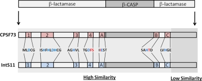
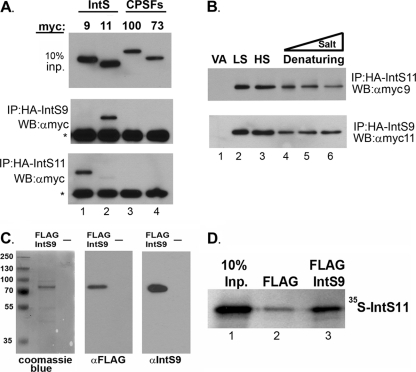
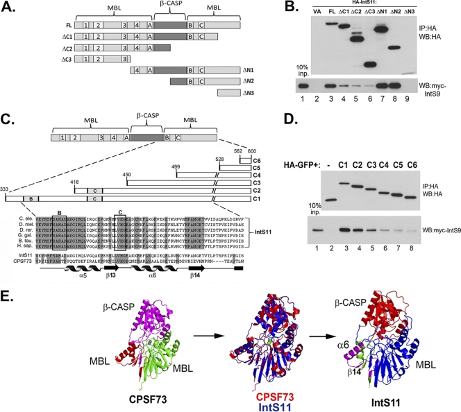
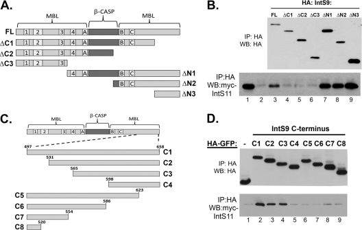
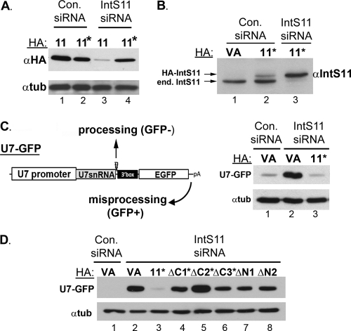
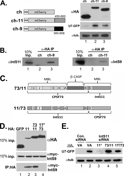
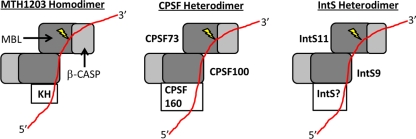
The Integrator Complex is a group of proteins responsible for the endonucleolytic cleavage of primary small nuclear RNA (snRNA) transcripts within the nucleus. Integrator subunits 9 and 11 (IntS9/11) are thought to contain the catalytic activity based on their high sequence similarity to CPSF100 and CPSF73, which have been shown to be components of both the poly(A)(+) and histone pre-mRNA cleavage complex. Here we demonstrate that the specific heterodimeric interaction between IntS9 and IntS11 is mediated by a discrete domain present at the extreme C terminus of IntS9 and within the C terminus of IntS11, adjacent to the predicted active site of this endonuclease. This domain is highly conserved within IntS11 but conspicuously absent in CPSF73. Using a cell-based complementation assay that measures Integrator activity, we determined that the IntS9 interaction domain within IntS11 is required for its ability to restore snRNA 3' end processing after RNA interference (RNAi)-mediated depletion of IntS11. Moreover, overexpression of these interaction domains alone elicits snRNA misprocessing through a dominant-negative titration of endogenous Integrator subunits. These data collectively explain the mechanism by which the IntS11/9 and, by analogy, the CPSF73/100 heterodimeric cleavage factors distinguish themselves from each other and demonstrate that the heterodimeric interaction is functionally required for snRNA 3' end formation.


Figures

Fig 1
Fig 1
Similarity between CPSF73 and IntS11. The schematic at the top represents the canonical organization of a β-CASP protein with the β-CASP domain inserted between an N-terminal and C-terminal MBL domains. The seven conserved motifs (motifs 1 to 4 and A to C) that are involved in the coordination of zinc and enzymatic catalysis are highlighted in red (CPSF73) and blue (IntS11). The amino acids that constitute these motifs are represented for CPFS73 and are identical in IntS11. Amino acids in black are identical in the two proteins, blue amino acids represent residues critical for function, and red amino acids distinguish those that are different in IntS11.
Fig 2
Fig 2
Integrator 9 and Integrator 11 form a specific and robust heterodimer. (A) Upper panel, Western blot (WB) analysis of cell lysates from HeLa cells transfected with myc-tagged IntS9, IntS11, CPSF100, or CPSF73; middle panel, Western blot analysis of immunoprecipitates (IP) by the use of anti-HA agarose beads from cells transfected with HA-tagged IntS9 along with each of the myc-tagged proteins shown in the upper panel; lower panel, same as the middle panel except that data represent cells cotransfected with HA-tagged IntS11. The asterisks represent the cross-reacting Ig heavy chain present after immunoprecipitation. (B) Western blot analysis of cells cotransfected with HA-IntS11 and myc-IntS9 or with HA-IntS9 and myc-IntS11 (lower panel). Lysates were subjected to immunoprecipitation with anti-HA antibodies under various lysis and wash conditions (LS, 150 mM NaCl; HS, 500 mM NaCl; Denaturing, 0.1% SDS and 0.5% SDC) (lane 4) and then supplemented with either 500 mM NaCl (lane 5) or 1 M NaCl (lane 6). “VA” denotes lysates transfected with myc-tagged proteins with empty HA vector. (C) SDS-PAGE analysis of eluted FLAG-tagged, full-length IntS9 by the use of Coomassie blue staining (left panel) or Western blot analysis with FLAG antibodies (middle panel) or IntS9 antibodies (right panel). “—,” lane loaded with elution buffer only. (D) Results from a pulldown assay using FLAG-tagged IntS9 and [35S]methionine-labeled IntS11. The first lane represents 10% of the input (Inp.) IntS11, the middle lane reflects the amount of IntS11 pulled down with anti-FLAG beads alone, and the last lane represents the pulldown of IntS11 with IntS9.
Fig 3
Fig 3
Integrator 11 binds to Integrator 9 through a conserved region at the C terminus. (A) Schematic of Integrator 11 deletion constructs. The β-CASP domain and the core motifs of the MBL domain are labeled. (B) Western blot analysis of cells transfected with HA-tagged full-length or deletion mutants of IntS11 (upper panel). The lower panel shows the results of coimmunoprecipitation using anti-HA antibody agarose beads followed by Western blot analysis of myc-tagged IntS9. (C) Schematic showing a detailed view of amino acids 384 to 458 of IntS11 and specific fragments that were fused to HA-tagged GFP. The shaded regions represent identical IntS11 amino acids of various species. Below the alignment of IntS11 is an alignment of human IntS11 and CPSF73 highlighting both the conserved residues (in gray) and the known secondary structure elements from the crystal structure of CPSF73 (22). (D) Upper panel, Western blot analysis of cell lysates from cells transfected with an HA-tagged GFP protein fused to six different fragments of the C terminus of IntS11 (C1 to C6); lower panel, anti-myc Western blot analysis of immunoprecipitates by the use of anti-HA antibodies from cells cotransfected with the HA-GFP-tagged IntS11 fragments and myc-IntS9. (E) Three-dimensional structure of human CPSF73 determined using Pymole (PDB accession no. 2I7V); the purple region is the β-CASP domain, the green is the N-terminal MBL domain, and the red is the C-terminal MBL domain. The two gray spheres represent the two zinc ions present in the catalytic core of CPSF73. Middle panel, alignment of CPSF73 (red) and IntS11 (blue). Right panel, predicted structure of IntS11, highlighting the N-terminal MBL domain (blue), β-CASP domain (red), and IntS9 interacting domain (purple).
Fig 4
Fig 4
Integrator 9 binds to Integrator 11 through a region at the extreme C terminus. (A) Schematic of IntS9 deletion mutants used to test interaction with IntS11. (B) Western blot analysis of lysates from cells cotransfected with HA-tagged IntS9 fragments and full-length IntS11. Upper panel, relative expression of IntS9 fragments assessed with an anti-HA Western blot; lower panel, relative pulldown efficiency of each HA-tagged IntS9 deletion mutant determined by assessing the amount of myc-IntS11 present in the anti-HA immunoprecipitates. (C) Schematic of C-terminal fragments fused to HA-GFP spanning the 161 amino acids of IntS9. (D) Western blot analysis measuring the relative efficiencies of the HA-GFP IntS9 fragments in pulling down myc-tagged IntS11.
Fig 5
Fig 5
The interaction of Integrator 9 and Integrator 11 is required for snRNA 3′ end formation. (A) Western blot analysis of lysates from cells treated with either control siRNA or siRNA targeting IntS11 and then transfected with HA-tagged IntS11 or HA-tagged IntS11 that is resistant to the IntS11 siRNA. (B) Western blot analysis using anti-IntS11 antibodies, demonstrating expression of exogenous RNAi-resistant IntS11 comparable to that of the endogenous IntS11. (C) Schematic of human U7-GFP reporter construct that produces GFP only in response to misprocessing conditions when Integrator subunits are knocked down. Right panel, Western blot analysis of cell lysates from cells treated with either control siRNA or IntS11 siRNA that were transfected with empty vector (VA) or RNAi-resistant IntS11 (lanes 11*) along with U7-GFP reporter. (D) Western blot analysis of cell lysates from cells treated with control siRNA or with IntS11 siRNA followed by transfection with the U7-GFP reporter and either full-length RNAi-resistant IntS11 or RNAi-resistant fragments as described for Fig. 2A.
Fig 6
Fig 6
Expression of only the C terminus of Integrator 9 or 11 results in behavior reminiscent of that of a dominant negative through disruption of the endogenous IntS9/11 heterodimer. (A) Left schematic, diagram of HA-tagged mCherry constructs that contain either amino acids 450 to 600 of IntS11 or amino acids 565 to 658 of IntS9; right panel, Western blot analysis showing expression of the control HA-tagged mCherry and the two heterologous fusions. The lower two panels demonstrate the degree of GFP expression from the U7-GFP reporter after cotransfection with the mCherry fusion proteins. (B) Western blot analysis of immunoprecipitates from cells transfected with the mCherry fusion proteins as described for panel A. Input lysates and immunoprecipitates from transfected cells were probed for either endogenous IntS11 or IntS9. (C) Schematic of chimeric CPSF73/IntS11 constructs. The gray-shaded region represents sequences derived from CPSF73, while the hatched regions represent IntS11sequences. (D) Upper panel, Western blot analysis of cell lysates cotransfected with HA-tagged chimeric constructs and full-length IntS11; lower panels, Western blot analysis of input full-length myc-IntS9 and coimmunoprecipitates from cotransfected cells. (E) Western blot analysis of cell lysates from cells treated with either control siRNA or IntS11 siRNA followed by cotransfection with HA vector alone or with RNAi-resistant IntS11 or either of the two chimeric constructs. The upper panel represents probing for GFP, and the lower panel represents an antitubulin loading control.
Fig 7
Fig 7
Model of IntS9/11 binding to the 3′ end of snRNA compared to the known archaeal homodimeric CPSF-KH protein, representing the molecular model proposed by Silva et al. (31) based on their crystal structure of the MTH1203 β-CASP protein. This model is compared with a model for both the CPSF73/100 and IntS9/11 heterodimers. The MBL domain is represented in dark gray, the β-CASP domain in light gray, and the RNA substrate in red; the lightning bolt indicates the cleavage site.

References

    1. Aravind L. 1999. An evolutionary classification of the metallo-beta-lactamase fold proteins. In Silico Biol. 1:69–91 - PubMed
    1. Baillat D, et al. 2005. Integrator, a multiprotein mediator of small nuclear RNA processing, associates with the C-terminal repeat of RNA polymerase II. Cell 123:265–276 - PubMed
    1. Callebaut I, Moshous D, Mornon JP, de Villartay JP. 2002. Metallo-beta-lactamase fold within nucleic acids processing enzymes: the beta-CASP family. Nucleic Acids Res. 30:3592–3601 - PMC - PubMed
    1. Chen J, Wagner EJ. 2010. snRNA 3′ end formation: the dawn of the Integrator complex. Biochem. Soc. Trans. 38:1082–1087 - PMC - PubMed
    1. Clouet-D'Orval B, Rinaldi D, Quentin Y, Carpousis AJ. 2010. Euryarchaeal beta-CASP proteins with homology to bacterial RNase J have 5′- to 3′-exoribonuclease activity. J. Biol. Chem. 285:17574–17583 - PMC - PubMed

Publication types

LinkOut - more resources