. 2012 Mar;32(6):1164-72.
doi: 10.1128/MCB.06328-11. Epub 2012 Jan 17.

Dcp2 decapping protein modulates mRNA stability of the critical interferon regulatory factor (IRF) IRF-7

Affiliations

Dcp2 decapping protein modulates mRNA stability of the critical interferon regulatory factor (IRF) IRF-7

You Li et al. Mol Cell Biol. 2012 Mar.

Abstract

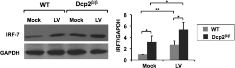
The mammalian Dcp2 mRNA-decapping protein functions primarily on a subset of mRNAs in a transcript-specific manner. Here we show that Dcp2 is an important modulator of genes involved in the type I interferon (IFN) response, which is the initial line of antiviral innate immune response elicited by a viral challenge. Mouse embryonic fibroblast cells with reduced Dcp2 levels (Dcp2(β/β)) contained significantly elevated levels of mRNAs encoding proteins involved in the type I IFN response. In particular, analysis of a key type I IFN transcription factor, IFN regulatory factor 7 (IRF-7), revealed an increase in both IRF-7 mRNA and protein in Dcp2(β/β) cells. Importantly, the increase in IRF-7 mRNA within the background of reduced Dcp2 levels was attributed to a stabilization of the IRF-7 mRNA, suggesting that Dcp2 normally modulates IRF-7 mRNA stability. Moreover, Dcp2 expression was also induced upon viral infection, consistent with a role in attenuating the antiviral response by promoting IRF-7 mRNA degradation. The induction of Dcp2 levels following a viral challenge and the specificity of Dcp2 in targeting the decay of IRF-7 mRNA suggest that Dcp2 may negatively contribute to the innate immune response in a negative feedback mechanism to restore normal homeostasis following viral infection.


Figures

Fig 1
Fig 1
A subset of genes involved in the antiviral immune response are upregulated in Dcp2β/β MEF cells. (A) WT and Dcp2β/β MEF cells were infected with lentivirus for 2 days. Total cellular RNAs were extracted and subjected to microarray analysis. Thirty-five genes involved in the cellular antiviral immune response with signals above the background were selected, and the n-fold change of each gene in Dcp2β/β MEF cells compared to the level in WT MEF cells is plotted. Data represent the average of two independent experiments, with error bars denoting the range of the two experiments. Dark bars indicate genes that were increased more than 1.5-fold in Dcp2β/β MEFs. (B) The levels of the indicated mRNAs were confirmed by real-time PCR analysis. The IRF-7, CXCL10, and OAS2 mRNAs were amplified with gene-specific primers and normalized to the β-actin mRNA level. The mRNA levels in the WT MEF cells were arbitrarily set to 100. The average of two independent experiments is shown with error bars indicating the range of the results in the two experiments.
Fig 2
Fig 2
IFN-mediated antiviral immune response was elevated in Dcp2β/β MEF cells following lentivirus infection. (A) Total RNAs of WT and Dcp2β/β MEF cells were extracted, and mRNA levels of specific genes were determined by qRT-PCR, normalized to β-actin mRNA. mRNA levels in WT cells were arbitrarily set to 1. Data represent the average of three independent experiments, with the standard deviation denoted by the error bar. **, P < 0.01 (Student's t test). (B) WT and Dcp2β/β MEF cells were treated with control lentivirus, and total RNAs were extracted at 0, 12, and 24 h postinfection. mRNA levels of specific genes were determined by qRT-PCR and normalized to β-actin mRNA. IRF-7, OAS2, CXCL10, and IRF-3 mRNA levels in WT cells at time zero were arbitrarily set to 1. IFN-α2 and IFN-β mRNA levels in WT cells at 12 h postinfection were arbitrarily set to 1 due to the very low levels of these two mRNAs before infection. Data represent the average of three independent experiments, with the standard deviation denoted by the error bar. *, P < 0.05; **, P < 0.01; ***, P < 0.001 (Student's t test).
Fig 3
Fig 3
Dcp2 modulates the stability of the IRF-7 mRNAs. (A) WT and Dcp2β/βMEF cells were infected with control lentivirus for 24 h and then treated with actinomycin D (ActD). Total RNAs were harvested at the indicated times posttreatment, and the decay of IRF-7 mRNA was determined by qRT-PCR and normalized to β-actin mRNA. The averages of three independent experiments are plotted, with standard deviations denoted by the error bars. IRF-7 mRNA was considerably stabilized in Dcp2β/β MEF cells. (B) WT and Nudt16KD MEF cells were infected with control lentivirus for 24 h and then treated with actinomycin D, and IRF-7 mRNA levels were determined as described for panel A. (C) Western blotting of Dcp2 expression. Dcp2 levels present in WT MEF cells transduced with a control (Con) retrovirus or Dcp2β/β MEFs stably transduced with either control retrovirus or retrovirus expressing WT human Dcp2 or catalytically inactive mutant Dcp2 (Dcp2 EE/QQ) are shown. Expression of Dcp2 protein was confirmed by Western blot analysis with GAPDH as the loading control. Comparable levels of WT and catalytically inactive Dcp2 are expressed in these cells. (D) MEF cells from panel C were infected with control lentivirus (LV). Total RNAs were extracted before infection and 12 h after infection, and the levels of the mRNAs indicated were determined by qRT-PCR and normalized to β-actin mRNA. The IRF-7 mRNA level in WT cells before infection was arbitrarily set to 1. Data represent the average of three independent experiments, with the standard deviation denoted by the error bar. A key listing the WT or Dcp2β/β cells tested and the particular exogenous protein expressed in the particular cells is shown at the bottom. Overexpression of WT but not the catalytically mutant Dcp2 reversed the increase in IRF-7 in Dcp2β/β MEF cells. *, P < 0.05 (Student's t test). (E) The IRF-7 mRNA 5′ end is selectively targeted by Dcp2. Decapping assays were carried out with 50 ng of bacterially expressed His-Dcp2 using cap-labeled STX7 or a chimeric RNA that harbors 57 nt of the IRF-7 5′ UTR at the 5′ end of STX7 (IRF-STX). The reaction products were resolved by PEI-TLC. The average decapping percentage for each RNA from three independent experiments is shown at the bottom.
Fig 4
Fig 4
IRF-7 protein levels are elevated in Dcp2 knockdown cells. Total cell extract was harvested from WT or Dcp2β/β MEF cells either left uninfected (Mock) or infected with control lentivirus (LV) for 24 h. IRF-7 and GAPDH protein levels were determined from the indicated conditions by Western blotting. Quantitation of IRF-7 protein levels normalized to GAPDH is graphed on the right. The average of three independent experiments was plotted with the standard deviation denoted by the error bar. IRF-7 protein amounts were increased in Dcp2β/β MEF cells relative to WT MEF cells with or without virus infection. *, P < 0.05; **, P < 0.01 (Student's t test).
Fig 5
Fig 5
Enhanced resistance to a viral challenge in Dcp2β/β MEF cells. (A) Diagram of experimental protocol. WT and Dcp2β/β MEFs were treated with NDV or HSV-1 for 24 h, and medium, which would contain secreted cytokines, was collected and transferred to new naive WT MEF cells. Following a 2-h incubation, the cells were infected with HSV-1 for 24 h and cell viability was determined. (B) Cytopathic effect at 24 h after infection. Cell viability was determined by crystal violet staining 24 h after HSV-1 infection. A595 directly correlates with viable cells. Averages of three independent experiments were plotted with error bars denoting the standard deviations. **, P < 0.01. (C) WT and Dcp2β/β MEFs were treated with NDV or HSV-1 for 24 h, and the levels of IRF-7 and IFN-α2 mRNAs were determined by qRT-PCR and normalized to β-actin mRNA. Averages of three independent experiments were plotted, with error bars denoting the standard deviations. *, P < 0.05 (Student's t test).
Fig 6
Fig 6
Dcp2 expression increases upon viral infection and double-stranded RNA treatment. (A) WT MEF cells were treated with lentivirus (LV), poly(I · C) (50 μg/ml), or growth medium containing no virus (Mock) for 24 h. Total RNAs were harvested before and after treatment, and Dcp2 mRNA levels were determined by qRT-PCR and normalized to β-actin mRNA. Dcp2 mRNA levels in untreated MEF cells were arbitrarily set to 1. The average of three independent experiments was plotted with the standard deviation denoted by the error bar. Dcp2 mRNA significantly increased with viral infection or poly(I · C) treatment. (*, P < 0.05; **, P < 0.01). (B) Dcp2 protein was induced upon viral infection and poly(I · C) treatment. WT MEF cells were treated for 24 h with lentivirus (LV), poly(I · C) (50 μg/ml), or growth medium containing no virus (Mock). Total cell extracts were harvested before and after treatment, and Dcp2 protein levels were determined by Western blot analysis with GAPDH as the internal control. Quantitation of Dcp2 protein levels from three independent experiments normalized relative to GAPDH is shown on the right, with the level of Dcp2 in untreated cells set arbitrarily to 1. *, P < 0.05 (Student's t test). (C) WT MEFs were treated with NDV (MOI = 0.5) or HSV-1 (MOI = 1) for 24 h. Dcp2 mRNA levels were analyzed by real-time RT-PCR and normalized to β-actin mRNA levels. *, P < 0.05; **, P < 0.01 (Student's t test).

Similar articles

Cited by

References

    1. Akira S, Uematsu S, Takeuchi O. 2006. Pathogen recognition and innate immunity. Cell 124:783–801. - PubMed
    1. Biggioggero M, Gabbriellini L, Meroni PL. 2010. Type I interferon therapy and its role in autoimmunity. Autoimmunity 43:248–254 - PubMed
    1. Borden EC, et al. 2007. Interferons at age 50: past, current and future impact on biomedicine. Nat. Rev. Drug Discov. 6:975–990 - PMC - PubMed
    1. Carballo E, Lai WS, Blackshear PJ. 2000. Evidence that tristetraprolin is a physiological regulator of granulocyte-macrophage colony-stimulating factor messenger RNA deadenylation and stability. Blood 95:1891–1899 - PubMed
    1. Carballo E, Lai WS, Blackshear PJ. 1998. Feedback inhibition of macrophage tumor necrosis factor-alpha production by tristetraprolin. Science 281:1001–1005 - PubMed

Publication types

MeSH terms