Comparative Study
. 2013 Jun;35(3):549-62.
doi: 10.1007/s11357-011-9378-2. Epub 2012 Jan 15.

Survivin expression increases during aging and enhances the resistance of aged human fibroblasts to genotoxic stress

Affiliations
Comparative Study

Survivin expression increases during aging and enhances the resistance of aged human fibroblasts to genotoxic stress

Huda H Al-Khalaf et al. Age (Dordr). 2013 Jun.

Abstract

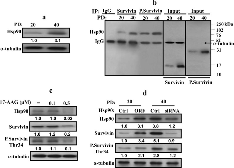
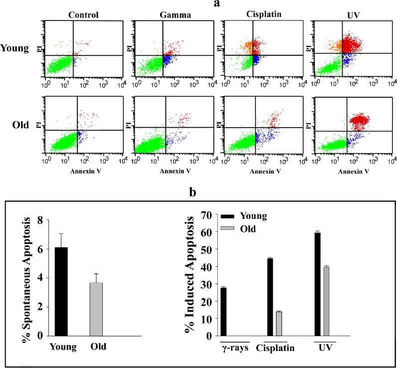
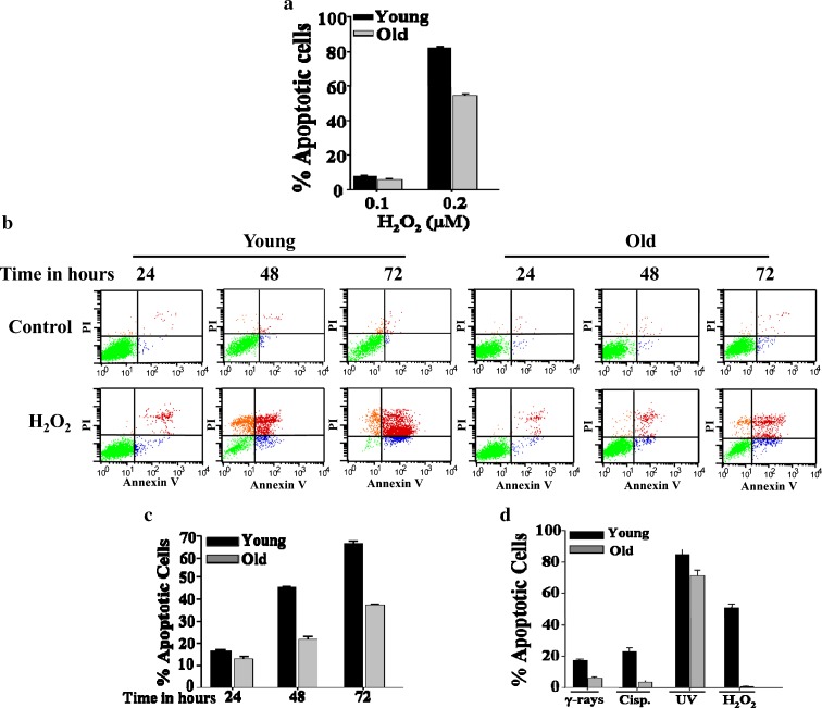
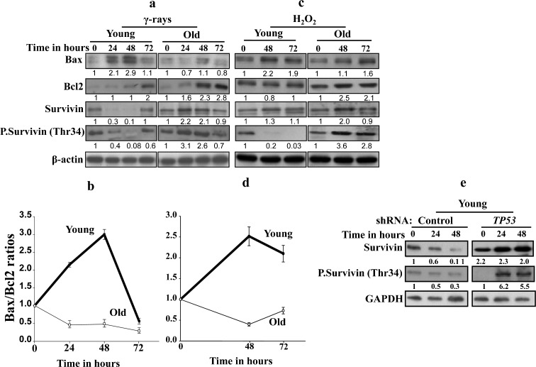
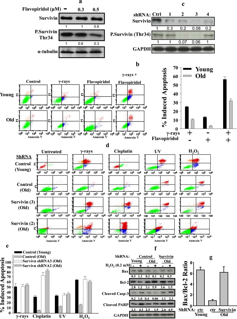
Survivin, an important anti-apoptotic protein, is highly expressed in most cancers, which generally arise in cells of older individuals. We have shown here accumulation of survivin and phospho-survivin in aged normal human skin fibroblasts and mice organs. This age-related accumulation of survivin was due to protein stabilization through association with the molecular chaperone Hsp90 protein, which was also up-regulated during aging. Interestingly, Hsp90 binds preferentially to phospho-survivin, which explains its higher stability. In addition, we provide clear evidence that aged cells exhibit apoptosis resistance when challenged with UV light, cisplatin, γ-rays or H2O2 as compared to their younger counterparts. In response to γ-rays and H2O2, the levels of Bcl-2 and both forms of survivin were up-regulated in old cells, but not in their corresponding young ones. This repression of survivin and phospho-survivin in young cells is p53 dependent. Importantly, survivin inhibition/down-regulation with flavopiridol or specific shRNAs increased the apoptotic response of old fibroblasts to various genotoxic agents, and restored the pro-apoptotic Bax/Bcl2 ratio and the increase in the levels of cleaved caspase-3 and PARP in old cells. These results show the role of survivin in the age-dependent resistance of human fibroblasts, and provide new insights into the molecular mechanisms that underlie the complex relationship between aging, apoptosis, and cancer.


Figures

Fig. 1
Fig. 1
Survivin and phospho-survivin protein levels are higher in old cells. a Cumulative population doublings for HFSN1 cells in culture. b In vitro passaged HFSN1 cells [Population Doubling 20 (PD 20), Population Doubling 40 (PD 40)] were cytospin attached to slides and stained with anti-Ki-67 antibody. Scale bars represent 500 μm. c Proteins were extracted from serially passaged HFSN1 cells as well as from young and old mice tissues, and then used for western blot analysis utilizing the indicated antibodies. The histogram shows the expression level of survivin (PD population doubling, Y young, O old). Error bars represent standard deviation of at least three different experiments. d Cells were harvested and analyzed for DNA content by flow cytometry. The percentage of G0/G1 and G2/M cells are indicated. e Total RNA was extracted from young and old cells, and then RT–PCR was used to assess the level of the survivin mRNA. GAPDH was used as internal control. f Cells were treated with cycloheximide (20 μg/ml), and then were harvested at the indicated periods of time for protein extraction and western blotting. Thirty micrograms of proteins was loaded and the indicated antibodies were used. Signals were quantitated by densitometry and were normalized against GAPDH. The graph shows the proportion of the remaining survivin per time. Error bars indicate standard errors from three different experiments
Fig. 2
Fig. 2
Hsp90 is up-regulated during aging. Whole-cell extracts were prepared from young (PD 20) and old (PD 40) HFSN1 cells. a Western blot analysis using the indicated antibodies. The numbers below the bands indicate the expression level of Hsp90. b Immunoprecipitation with either anti-survivin or anti-phospho-survivin (Thr34) antibodies and mouse IgG was used as control. IP materials were then immunoblotted with anti-Hsp90 antibody. c HFSN1 cells (PD 40) were treated with different concentration of the Hsp90 inhibitor (17-AAG) and then re-incubated for 24 h. Whole-cell extracts were prepared and analyzed by western blot using the indicated antibodies. d Young HFSN1 cells (PD 20) were transfected with a plasmid bearing the HSP90 ORF and a control plasmid, and old HFSN1 cells (PD 40) were transfected with Hsp90-siRNA and control siRNA. Seventy-two hours post-transfection, cells were collected and whole-cell extracts were prepared and analyzed by immunoblotting using the indicated antibodies. The numbers below the bands represent the corresponding fold of variation in the expression levels relative to their respective controls and α-tubulin
Fig. 3
Fig. 3
Aged human skin fibroblasts are resistant to DNA damaging agents. a HFSN1 cells were either mock-treated or challenged with γ-rays (30 Gy), cisplatin (60 μg/ml), or UV light (10 J m−2) and then re-incubated for 72 h. Apoptosis was analyzed by annexin V/PI flow cytometry. b Histograms showing the proportions of spontaneous and induced apoptosis (early + late). The error bars represent standard deviations of at least three different experiments
Fig. 4
Fig. 4
Aged fibroblasts are resistant to H2O2. a HFSN1 cells were either mock-treated or challenged with serial concentrations of H2O2 and then re-incubated for 72 h. Apoptosis was analyzed by annexin V/PI flow cytometry. Histogram showing the proportions of apoptosis in response to the indicated doses of H2O2. b Young and old fibroblasts were either mock-treated or challenged with 0.2 μM H2O2 for the indicated periods of time and then apoptosis was assessed by annexin V/PI flow cytometry. c Histogram showing the proportions of apoptosis after treatment with H2O2 (0.2 μM). The error bars represent standard deviation of at least three different experiments. d Young (PD 6) and old (PD 20) MEF cells were either mock-treated or challenged with the indicated genotoxic agents then re-incubated for 72 h. Apoptosis was analyzed by annexin V/PI flow cytometry. Histogram showing the proportions of induced apoptosis. The error bars represent standard deviation of at least three different experiments
Fig. 5
Fig. 5
The effect of γ-rays and H2O2 on survivin expression is age dependent. a, c Young and old cells were either sham-treated or exposed to γ-rays (30 Gy) or H2O2 (0.2 μM) and then were incubated for the indicated periods of time, before being harvested and proteins extracted. Thirty micrograms of proteins was used for immunoblotting analysis using the indicated antibodies. The numbers below the gels indicate the induction/reduction folds in the expression levels after normalization against β-actin. b, d Graphs showing the age-dependent level of Bax/Bcl-2 ratio after treatment with γ-rays or H2O2, respectively. Error bars represent standard deviation of at least three different experiments. e Western blot analysis using the indicated antibodies in young TP53 shRNA-expressing cells and their control counterparts treated with γ-rays (30 Gy). The numbers below the gels indicate the fold of induction/reduction in the expression levels as compared to the basal level (time 0)
Fig. 6
Fig. 6
Inhibition of survivin enhances apoptotic response of old fibroblasts to DNA damage. a HFSN1 cells (PD 40) were treated with different concentration of flavopiridol and then re-incubated for 24 h. Whole-cell extracts were prepared and analyzed by western blot using the indicated antibodies. b Young and old HFSN1 cells were either mock-treated or challenged with γ-rays (30 Gy), flavopiridol (0.5 μM), or a combination of both and re-incubated for 72 h. Apoptosis was then assessed by annexin V/PI flow cytometry. The histogram shows the proportions of apoptosis induced by the indicated agents. The error bars represent standard deviations of three different experiments. c Western blot showing the expression of the indicated proteins in HFSN1 cells expressing either four shRNA against survivin, independently, or control shRNA (Ctrl). The numbers below the gels indicate the expression level of the indicated proteins as compared to the control. d Young and old HFSN1 fibroblasts expressing either survivin-shRNA or the control shRNA were either mock-treated or challenged with the indicated agents and then cell death was assessed by annexin V/PI flow cytometry. e Histogram showing the proportions of apoptosis induced by the indicated agents. The error bars represent standard deviations of three different experiments. f Young and old HFSN1 fibroblasts expressing the control plasmid and old fibroblasts expressing survivin-shRNA3 were either mock-treated or challenged with H2O2 (0.2 μM) for 72 h. Cells were then collected and cell lysates were prepared to assess the level of the indicated apoptotic proteins. The numbers below the gels indicate the expression levels of the indicated proteins after normalizing against GAPDH. g Graph showing the Bax/Bcl-2 ratio in the indicated cells after treatment with H2O2 (0.2 μM). Error bars represent standard deviation of at least three different experiments

References

    1. Altieri DC. Survivin, versatile modulation of cell division and apoptosis in cancer. Oncogene. 2003;22:8581–8589. doi: 10.1038/sj.onc.1207113. - DOI - PubMed
    1. Altieri DC. Validating survivin as a cancer therapeutic target. Nat Rev Cancer. 2003;3:46–54. doi: 10.1038/nrc968. - DOI - PubMed
    1. Altieri DC. The case for survivin as a regulator of microtubule dynamics and cell-death decisions. Curr Opin Cell Biol. 2006;18:609–615. doi: 10.1016/j.ceb.2006.08.015. - DOI - PubMed
    1. Altieri DC. Survivin and IAP proteins in cell-death mechanisms. Biochem J. 2010;430:199–205. doi: 10.1042/BJ20100814. - DOI - PMC - PubMed
    1. Avula CP, Fernandes G. Inhibition of H2O2-induced apoptosis of lymphocytes by calorie restriction during aging. Microsc Res Tech. 2002;59:282–292. doi: 10.1002/jemt.10206. - DOI - PubMed

Publication types

MeSH terms