. 2012 Jan 15;30(2):165-73.
doi: 10.1038/nbt.2107.

Generation of human vascular smooth muscle subtypes provides insight into embryological origin-dependent disease susceptibility

Affiliations

Generation of human vascular smooth muscle subtypes provides insight into embryological origin-dependent disease susceptibility

Christine Cheung et al. Nat Biotechnol. .

Abstract

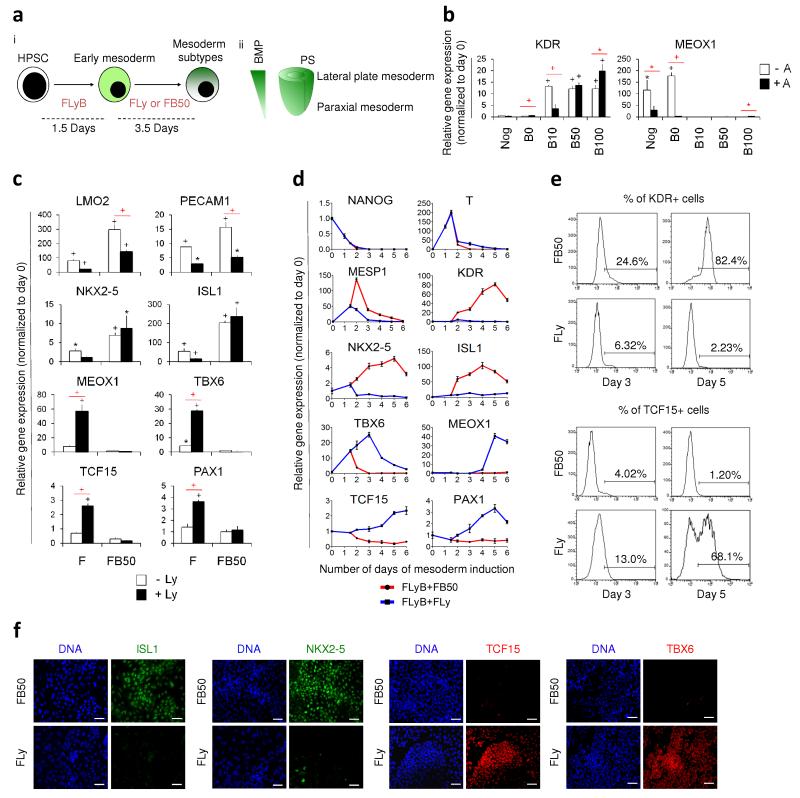
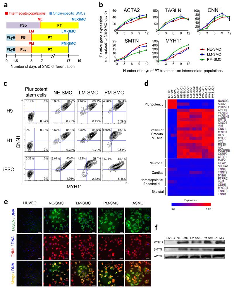
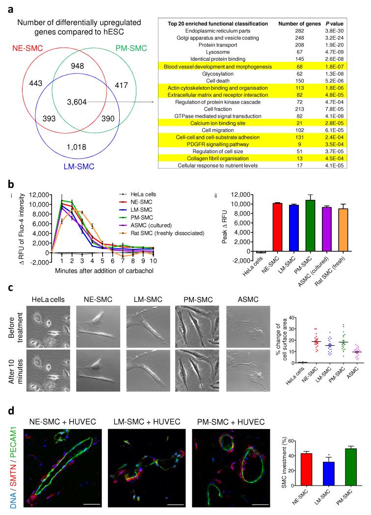
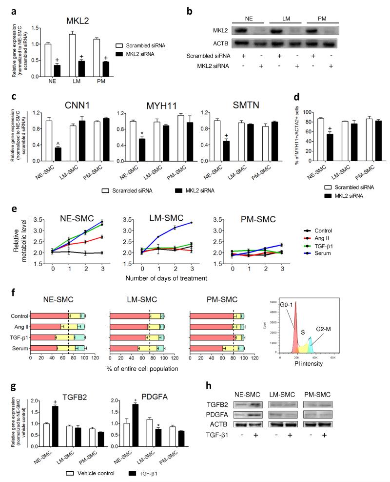
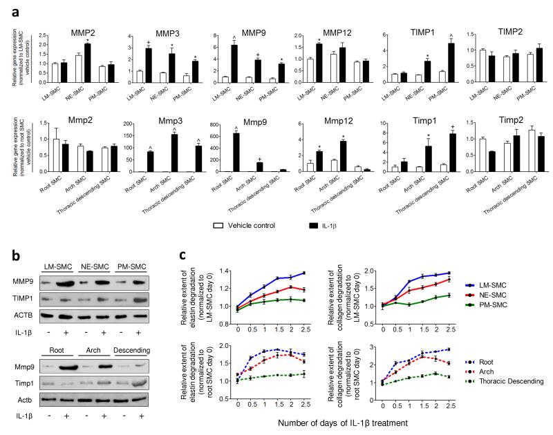
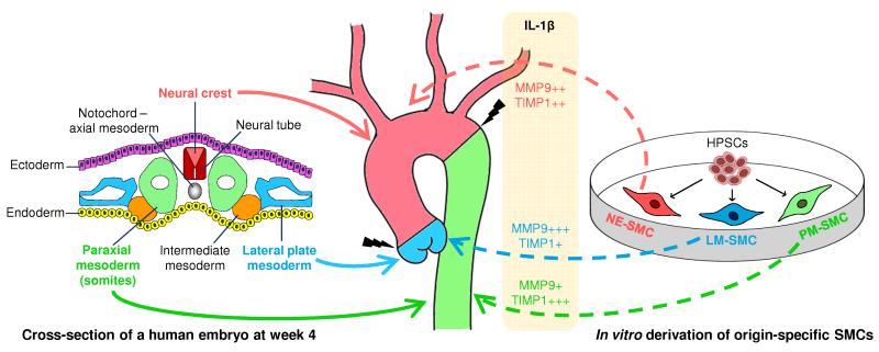
Heterogeneity of embryological origins is a hallmark of vascular smooth muscle cells (SMCs) and may influence the development of vascular disease. Differentiation of human pluripotent stem cells (hPSCs) into developmental origin-specific SMC subtypes remains elusive. Here we describe a chemically defined protocol in which hPSCs were initially induced to form neuroectoderm, lateral plate mesoderm or paraxial mesoderm. These intermediate populations were further differentiated toward SMCs (>80% MYH11(+) and ACTA2(+)), which displayed contractile ability in response to vasoconstrictors and invested perivascular regions in vivo. Derived SMC subtypes recapitulated the unique proliferative and secretory responses to cytokines previously documented in studies using aortic SMCs of distinct origins. Notably, this system predicted increased extracellular matrix degradation by SMCs derived from lateral plate mesoderm, which was confirmed using rat aortic SMCs from corresponding origins. This differentiation approach will have broad applications in modeling origin-dependent disease susceptibility and in developing bioengineered vascular grafts for regenerative medicine.


Figures

Figure 1
Figure 1
Induction of mesoderm subtypes from hPSCs. (a) (i) HPSCs were initially differentiated to early mesoderm using FGF2, LY294002 and BMP4 (depicted as FLyB) for 36 hours. Mesoderm subtype specification required another 3.5 days of FGF2+LY294002 (FLy) or FGF2+BMP4 (FB) for paraxial or lateral plate induction. (ii) BMP concentration gradient exists along the primitive streak (PS). On the basis of fate map studies, mesoderm subtypes emerge along the gradient on the embryo cylinder. (b) KDR and MEOX1 expression levels of hESCs differentiated for 36 hours in FLyB, followed by 3.5 days in FGF2+LY294002 with Noggin (Nog), BMP4 (B) and/or Activin (A) were determined by QRTPCR. BMP4 concentrations ranged from 0 ng/ml (B0) to 100 ng/ml (B100). (c) Lateral plate and paraxial mesoderm marker expression levels in hESCs differentiated for 36 hours in FLyB and then 3.5 additional days in FGF2 (F) or FGF2+BMP4 (FB50, where BMP4 is 50 ng/ml), with (black bars) or without (white bars) LY294002 (Ly) determined by QRTPCR. (d) Temporal QRTPCR to analyze mesoderm specification. Following a common 36-hour treatment of FLyB, lateral and paraxial mesoderms were specified using FB50 (red line) and FLy (blue line) respectively. (e) Percentage of KDR+ and TCF15+ cells at days 3 and 5 determined by flow cytometry. (f) At day 5, FB50- and FLy-treated populations were immunostained for mesoderm subtype-specific markers. Scale bars, 100 μm. Data represent means ± s.e.m. (n = 3). Significant differences compared to day 0 population are indicated in black, while that between two groups are indicated in red (*P < 0.05; +P < 0.01; ^P < 0.001).
Figure 2
Figure 2
Efficient differentiation of intermediate lineages into vascular SMCs. (a) A schematic which outlines the conditions for deriving origin-specific SMCs from hPSCs. FLyB+FB and FLyB+FLy have been described in Figure 1 to generate the lateral plate mesoderm (LM) and paraxial mesoderm (PM) respectively. HPSCs were treated with FGF2+SB431542 (FSb) for 7 days to induce neuroectoderm (NE) differentiation. For further differentiation into vascular SMCs, each intermediate population was subjected to PDGF-BB+TGF-β1 (PT) for 12 additional days. The SMC subtypes, namely the neuroectoderm-derived SMC, lateral mesoderm-derived SMC, paraxial mesoderm-derived SMC are abbreviated as NE-SMC, LM-SMC and PM-SMC respectively. (b) QRTPCR demonstrated increasing expression of SMC markers during the 12 days of PT treatment on the three intermediate populations. Data represent means ± s.e.m. (n = 3). (c) Flow cytometric analysis demonstrated that the SMC differentiation protocols were highly efficient and reproducible in three hPSC lines with 83-92% of the resulting populations double positive for MYH11 and CNN1. (d) Microarray gene expression heat map of control hESCs versus SMC subtypes obtained after 12 days of PT treatment. Red (upregulation) and blue (downregulation) depict differential gene expression from the mean across all samples. (e) The majority of our SMC subtypes immunostained positively for CNN1 (red) and TAGLN (green). Human umbilical vein endothelial cell (HUVEC) was used as a negative control, while ASMC was used as a positive control. Scale bars, 100 μm. (f) Western blot confirmed the presence of the mature SMC proteins, MYH11 and SMTN, in the hPSC-derived SMCs and ASMC but not HUVEC.
Figure 3
Figure 3
Functional characterization of hPSC-derived SMCs. (a) Based on global gene expression profiles, the Venn diagram represent subsets of genes that were differentially upregulated in the SMC subtypes compared to hESC (FDR 0.1%). The commonly upregulated 3,604 genes were analyzed using the functional annotation clustering from DAVID bioinformatics resources. Among the top 20 highly enriched groups, functional characteristics pertaining to vascular SMCs are highlighted in yellow. (b) (i) Change in the relative fluorescence unit (Δ RFU) of Fluo-4 loaded cells was monitored by flow cytometry over 10 minutes after the addition of carbachol, an inducer of contraction. (ii) Peak Δ RFU of Fluo-4 intensity in response to carbachol in the derived SMCs and the SMC controls (n = 3). (c) HPSC-derived SMCs and ASMC displayed contractile ability in response to carbachol treatment. There was a 10-20% change of surface area in contracting cells of all SMCs except HeLa cells (n = 20). (d) Matrigel plugs were harvested 2 weeks after the subcutaneous implantation of hPSC-derived SMCs and HUVECs (1:2). Histological sections were then double immunostained with human-specific SMTN and PECAM1 antibodies. SMC investment was quantified based on the relative length of endothelial luminal structures (green) that had SMC coverage (red) in 10 different optical fields. Statistical test was performed by ANOVA (*P < 0.05). Scale bars, 50 μm.
Figure 4
Figure 4
MKL2 knockdown and cytokine treatments validate the origin-specific characteristics of hPSC-derived SMC subtypes. (a) 60-65% knockdown of the MKL2 expression levels in the intermediate populations (NE, LM and PM) by MKL2 siRNA was verified by QRTPCR. (b) Western blot analysis confirmed the effects of MKL2 siRNA knockdown on the protein levels compared to scrambled siRNA controls. (c) SMC gene expression levels after SMC differentiation of the siRNA-treated intermediate populations were determined by QRTPCR. (d) Percentage of MYH11+/ACTA2+ SMCs obtained from the siRNA-treated intermediate populations was determined by flow cytometry. (e) Proliferation responses of the SMC subtypes were monitored by MTT assay every 24 hours over 3 days of treatment with the cytokines indicated. (f) Cell cycle analysis of the SMC subtypes after 24 hours of cytokine treatments. The percentage of cells in different phases of cell cycle was quantified by the areas under the red peak (G0-G1), the area shaded yellow (S) and under the blue peak (G2-M) (legend on the right). Black dotted lines divide the growth arrested cells in G0-G1 from the proliferating cells in S and G2-M of the control groups. (g) Gene expression levels in control and TGF-β1-treated SMCs were determined by QRTPCR after 10 hours of treatment. (h) Western blot analysis was performed to confirm the distinct secretory responses exhibited by TGF-β1-treated SMC subtypes. Data represent means ± s.e.m. (n = 3). Significant differences compared to the scrambled siRNA or vehicle controls are indicated (*P < 0.05; +P < 0.01, ^P < 0.001).
Figure 5
Figure 5
HPSC-derived SMC subtypes predict MMP and TIMP expression and activity in rat aortic SMCs of corresponding origins. (a) Gene expression levels of MMPs and TIMPs in control (white bars) and IL-1β-treated SMCs (black bars) were determined by QRTPCR after 6 hours of treatment. Differential activation of MMP9 and TIMP1 expression was observed in both the hPSC-derived SMCs (top panel) and rat aortic SMCs (bottom panel). (b) Western blot analysis confirmed the differential amounts of MMP9 and TIMP1 proteins in the IL-1β-treated SMCs of unique origins. (c) Proteolytic abilities of the SMCs were assessed by elastase and collagenase assays over 2.5 days. The origin-specific SMCs (top panel) replicated similar trends of elastin and collagen degradation as the rat aortic SMCs (bottom panel) in response to IL-1β. Data represent means ± s.e.m. (n = 3). Significant differences compared to the vehicle controls (*P < 0.05; +P < 0.01; ^P < 0.001).
Figure 6
Figure 6
The different embryological origins of aortic SMCs may contribute to the site of aortic dissection. Aortic SMCs originate from three distinct developmental lineages. The aortic root is derived from secondary heart field, a lateral plate mesoderm derivative (blue solid arrow), while the ascending aorta and arch are neural crest derived (red solid arrow). The descending aortic SMCs originate from paraxial/somitic mesoderm (green solid arrow). In this study, our in vitro hPSC-derived SMC subtypes predicted the differential MMP and TIMP activation in aortic SMCs of corresponding origins (dotted arrows) in response to an inflammatory mediator, IL-1β. We propose that the origin-specific SMCs display differential proteolytic ability in disease settings, which may result in differential loss of the structural integrity in different regions along the aortic wall. Such a difference in mechanical properties may predispose the sites of aortic dissection to occur preferentially at the boundaries between different SMC lineages (indicated by black jagged bolts).

References

    1. Carmeliet P. Mechanisms of angiogenesis and arteriogenesis. Nature medicine. 2000;6:389–395. - PubMed
    1. Majesky MW. Developmental basis of vascular smooth muscle diversity. Arteriosclerosis, thrombosis, and vascular biology. 2007;27:1248–1258. - PubMed
    1. Waldo KL, et al. Secondary heart field contributes myocardium and smooth muscle to the arterial pole of the developing heart. Developmental biology. 2005;281:78–90. - PubMed
    1. Jiang X, Rowitch DH, Soriano P, McMahon AP, Sucov HM. Fate of the mammalian cardiac neural crest. Development. 2000;127:1607–1616. - PubMed
    1. Wasteson P, et al. Developmental origin of smooth muscle cells in the descending aorta in mice. Development. 2008;135:1823–1832. - PubMed

Publication types

Substances