. 2012 May 9;18(1):549-55.
doi: 10.2119/molmed.2011.00475.

Mice with a D190N mutation in the gene encoding rhodopsin: a model for human autosomal-dominant retinitis pigmentosa

Affiliations

Mice with a D190N mutation in the gene encoding rhodopsin: a model for human autosomal-dominant retinitis pigmentosa

Javier Sancho-Pelluz et al. Mol Med. .

Abstract

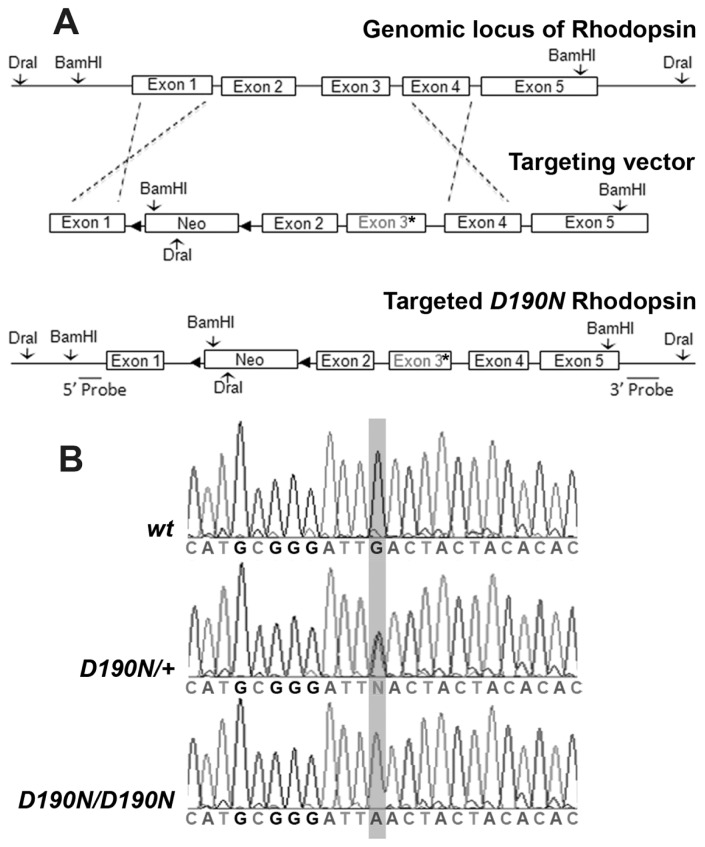
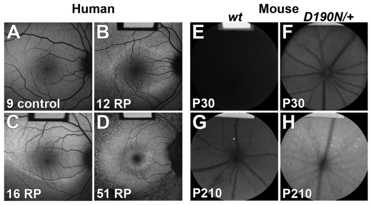
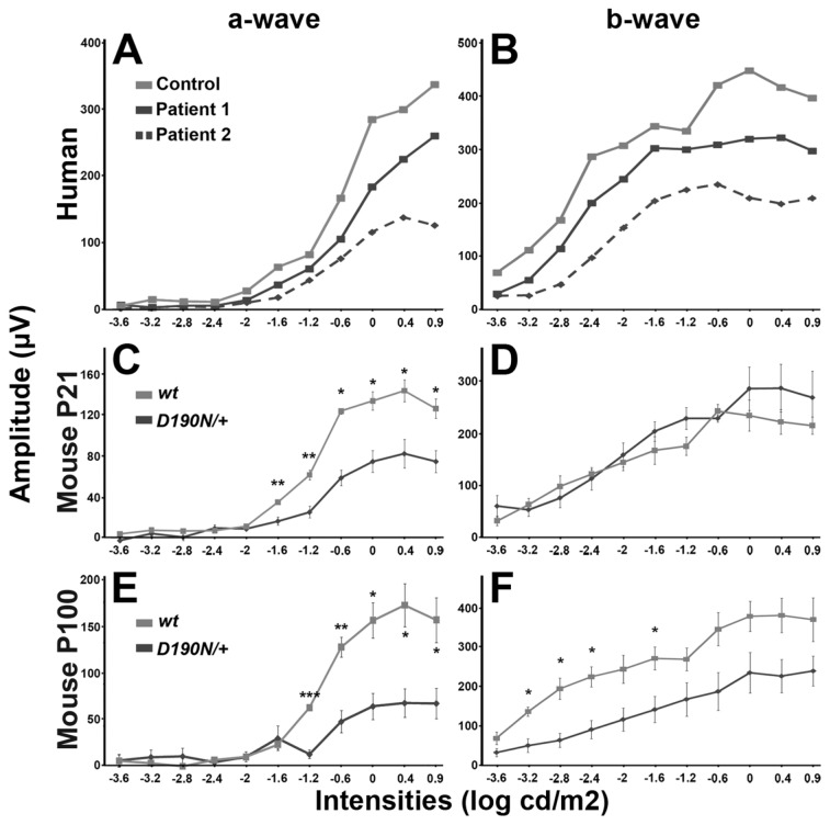
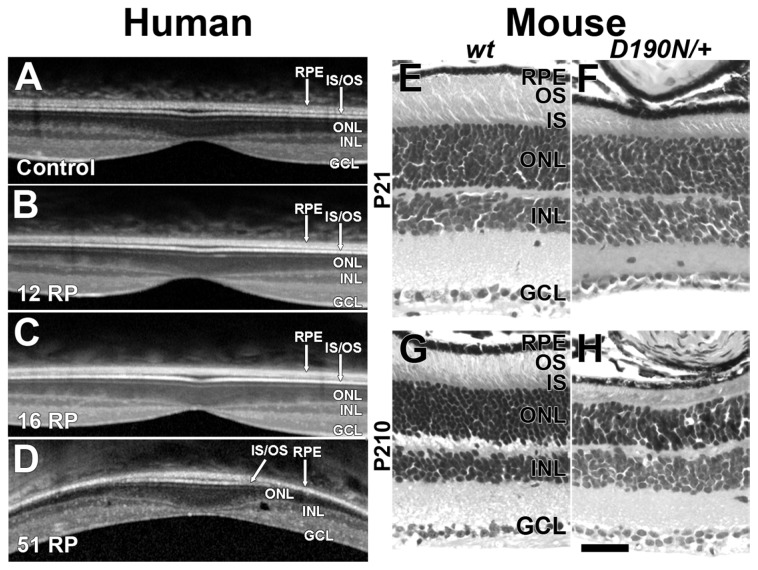
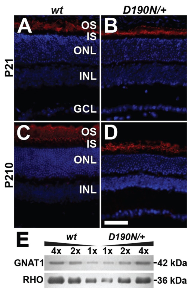
Rhodopsin is the G protein-coupled receptor in charge of initiating signal transduction in rod photoreceptor cells upon the arrival of the photon. D190N (Rho(D190n)), a missense mutation in rhodopsin, causes autosomal-dominant retinitis pigmentosa (adRP) in humans. Affected patients present hyperfluorescent retinal rings and progressive rod photoreceptor degeneration. Studies in humans cannot reveal the molecular processes causing the earliest stages of the condition, thus necessitating the creation of an appropriate animal model. A knock-in mouse model with the D190N mutation was engineered to study the pathogenesis of the disease. Electrophysiological and histological findings in the mouse were similar to those observed in human patients, and the hyperfluorescence pattern was analogous to that seen in humans, confirming that the D190N mouse is an accurate model for the study of adRP.


Figures

Figure 1
Figure 1
Germline transmission of D190N knock-in allele. (A) Genomic locus of rhodopsin (top panel); targeting vector (middle panel) and the resultant targeted D190N allele (bottom panel) are shown. The neomycin cassette was subsequently removed. 5′ and 3′ probes were used to confirm the integrity of the targeted locus. (B) Direct DNA sequencing from D190N/+ mutant mouse DNA showing heterozygous base-substitution at position 190 in D190N/+. Direct sequencing from the D190N/D190N mouse DBA revealed a homozygous base substitution at the 190 codon with a predicted missense mutation of D190N (GAC to AAC): GAC (Asp/D) aspartic acid to AAC (Asn/N) asparagine.
Figure 2
Figure 2
FAF imaging showing generalized increase in lipofuscin accumulation proportional to the severity of the disease. Normal FAF is seen in the right eye of the unaffected 9-year-old sibling (A). FAF imaging demonstrates constriction of a small perifoveal hyper-fluorescent ring in the right eye of a 12-year-old (B), a 16-year-old (C) and a 51-year-old (D) D190N/+ patient. Autofluorescence is elevated inside the ring of the surviving retina and less intense outside of the ring. Compared to the wt mice (E, G), D190N/+ mutant mice show generalized hyperfluorescence and bright dots that increase in intensity and number as the mice age (F, H). Disease severity is highly correlated with increased overall FAF in mice. In (B–H), the reference bar at the top enables intensity comparison.
Figure 3
Figure 3
Reduced photoreceptor scotopic ERG a-wave amplitudes show diminished rod function. Scotopic ERG responses were recorded in two patients and a healthy control. The amplitude of the a-wave (A) was reduced in affected patients at bright intensities, whereas the amplitude of the b-wave (B) was reduced at all light intensities. In D190N/+ mice at both P21 and P100, scotopic a-waves were reduced in comparison to wt, consistent with desensitization of the rod response (C, E). The b-wave in the scotopic test did not significantly differ between D190N/+ and control at P21 (D), although at P100 the difference in the b-wave amplitude was significant (F). Data were composed by measuring the average of different animals (n ≥ 3). Error bars represent standard error of the mean. *P ≤ 0.05; **P ≤ 0.01; ***P ≤ 0.001.
Figure 4
Figure 4
OCT and histology demonstrate slow photoreceptor degeneration in patients and animal models. When compared with a healthy individual (A), young patients (B, C) do not present a difference in ONL thickness on SD-OCT. The oldest patient (D) showed IS/OS loss and diminished ONL as well as cystoid macular edema. H&E-stained paraffin sections of wt and D190N/+ at P21 (E, F) and P210 (G, H) were prepared. Compared to the wt mice at P21 (E), D190N/+ presents slightly reduced ONL thickness and diminished OS length (F). Mutant mice at P210 (H) show a loss of four rows of photoreceptor nuclei compared with age-matched control (G). GCL, ganglion cell layer; INL, inner nuclear layer. Scale bar: 50 μm.
Figure 5
Figure 5
Immunohistochemistry and immunoblotting shows rhodopsin expression in wt and D190N/+ retinas. Rhodopsin correctly localizes in the OS of wt retinas at P21 (A) and P210 (C). In D190N/+ mice, rhodopsin localizes correctly in the OS of the retina at P21 (B) and P210 (D). Compared to the wt controls, D190N/+ mice show shortened OS at both P21 and P210. DAPI was used to counterstain nuclei. GCL, ganglion cell layer; INL, inner nuclear layer. Representative immunoblot of retinal extract prepared from wt and D190N/+ mice demonstrates linearity in rhodopsin levels (E). Each lane represents the amount loaded (mL) per retina (200 mL total sample volume). Scale bar: 50 μm.

References

    1. Boughman JA, Conneally PM, Nance WE. Population genetic studies of retinitis pigmentosa. Am J Hum Genet. 1980;32:223–35. - PMC - PubMed
    1. Berson EL. Retinitis pigmentosa: the Friedenwald Lecture. Invest Ophthalmol Vis Sci. 1993;34:1659–76. - PubMed
    1. Hartong DT, Berson EL, Dryja TP. Retinitis pigmentosa. Lancet. 2006;368:1795–809. - PubMed
    1. Rivolta C, Sharon D, DeAngelis MM, Dryja TP. Retinitis pigmentosa and allied diseases: numerous diseases, genes, and inheritance patterns. Hum Mol Genet. 2002;11:1219–27. - PubMed
    1. Wilson JH, Wensel TG. The nature of dominant mutations of rhodopsin and implications for gene therapy. Mol Neurobiol. 2003;28:149–58. - PubMed

Publication types