. 2012 Apr;56(4):1797-809.
doi: 10.1128/AAC.05708-11. Epub 2012 Jan 17.

SQ109 targets MmpL3, a membrane transporter of trehalose monomycolate involved in mycolic acid donation to the cell wall core of Mycobacterium tuberculosis

Affiliations

SQ109 targets MmpL3, a membrane transporter of trehalose monomycolate involved in mycolic acid donation to the cell wall core of Mycobacterium tuberculosis

Kapil Tahlan et al. Antimicrob Agents Chemother. 2012 Apr.

Abstract

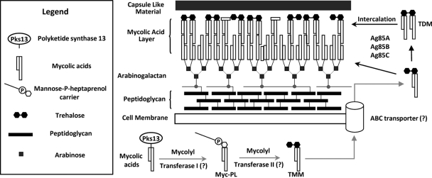
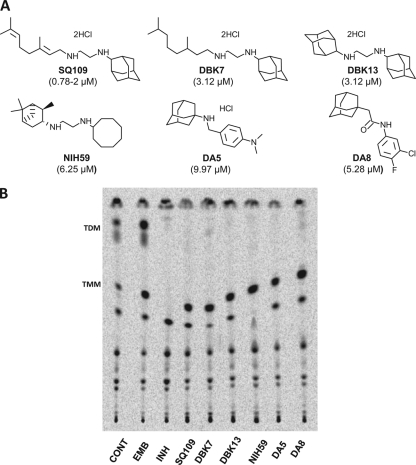
SQ109, a 1,2-diamine related to ethambutol, is currently in clinical trials for the treatment of tuberculosis, but its mode of action remains unclear. Here, we demonstrate that SQ109 disrupts cell wall assembly, as evidenced by macromolecular incorporation assays and ultrastructural analyses. SQ109 interferes with the assembly of mycolic acids into the cell wall core of Mycobacterium tuberculosis, as bacilli exposed to SQ109 show immediate inhibition of trehalose dimycolate (TDM) production and fail to attach mycolates to the cell wall arabinogalactan. These effects were not due to inhibition of mycolate synthesis, since total mycolate levels were unaffected, but instead resulted in the accumulation of trehalose monomycolate (TMM), the precursor of TDM and cell wall mycolates. In vitro assays using purified enzymes showed that this was not due to inhibition of the secreted Ag85 mycolyltransferases. We were unable to achieve spontaneous generation of SQ109-resistant mutants; however, analogs of this compound that resulted in similar shutdown of TDM synthesis with concomitant TMM accumulation were used to spontaneously generate resistant mutants that were also cross-resistant to SQ109. Whole-genome sequencing of these mutants showed that these all had mutations in the essential mmpL3 gene, which encodes a transmembrane transporter. Our results suggest that MmpL3 is the target of SQ109 and that MmpL3 is a transporter of mycobacterial TMM.


Figures

Fig 1
Fig 1
Diagrammatic representation of the mycobacterial cell wall and proposed pathway involved in mycolic acid processing and export (12, 63) (adapted with permission from reference 16a). The cartoon depicts some components of the cell wall while primarily focusing on trehalose monomycolate (TMM) and trehalose dimycolate (TDM). Hypothetical or proposed steps are indicated by gray arrows, and question marks in parentheses represent hypothetical proteins/enzymes not yet identified. The inset legend represents the shapes used to depict the components shown. Note that the diagram is not to scale and does not include detailed structures or all components of the mycobacterial cell wall.
Fig 2
Fig 2
In vitro activity of SQ109. Survival of M. tuberculosis following 10× MIC exposure to EMB, INH, NIH59, and SQ109 under aerobic conditions. Batch cultures were propagated in the presence of the indicated drugs, and samples were withdrawn for determining CFU counts by plating on the days indicated. The increase in CFU counts in the INH treatment is due to the emergence of spontaneous resistance as reported previously (8).
Fig 3
Fig 3
Macromolecular incorporation assays indicate that SQ109 inhibits precursor incorporation during cell wall biosynthesis. (A) Cultures were labeled with [4,5-3H]l-leucine, [1-3H]N-acetyl-d-glucosamine, and [5,6-3H]uracil for 1 h to monitor protein, peptidoglycan, and total nucleic acid biosynthesis, respectively, upon being exposed to different concentrations of SQ109 based on its MIC. (B and C) SQ109 inhibits precursor incorporation into cell wall lipids. Batch cultures were exposed to 10× MIC concentrations of EMB and SQ109 for the indicated times followed by labeling with 14C-acetate for 1 h. Cells were harvested and extracted with CHCl3-CH3OH (2:1), and the solvent extract contained polar extractable lipids, whereas lipids covalently associated with the cell wall remained in the insoluble material. (B) Radioactivity recovered from the solvent-extracted pellet after saponification (representing covalently bound cell wall lipids) showing the rapid decrease in the incorporation of 14C into cell wall-bound lipids on SQ109 treatment. (C) Radioactivity recovered in solvent extract prior to saponification (representing extractable polar lipids).
Fig 4
Fig 4
Ultrastructural changes associated with SQ109 treatment parallel the changes observed with INH and EMB treatment. (A) Scanning electron micrographs of M. tuberculosis treated with 10× MIC of SQ109 (i), EMB (ii), or INH (iii) for 48 h, as opposed to untreated cells (iv), show shortening of cells on drug exposure. Scale bar: 1 μm. (B) Transmission electron micrographs of M. tuberculosis treated with 10× MIC of SQ109 (i, ii), INH (iii, iv), or EMB (v, vi), as opposed to untreated cells (vii, viii), showing longitudinal (i, iii, v, vii) and transverse (ii, iv, vi, viii) sections of representative cells, respectively. Scale bar: 100 nm. Treated cells show cell wall layer thickening with all three agents.
Fig 5
Fig 5
Mycolate lipid profiles of M. tuberculosis on drug exposure. Bacteria in batch cultures were exposed to 10× MIC concentrations of EMB and SQ109 for 1 h and were then labeled with 14C-acetate. Cells were harvested at indicated time points, and the pellets were extracted with CHCl3-CH3OH (2:1). (A) TLC analysis of MAMEs and FAMEs isolated from pellets after free lipids were removed by CHCl3-CH3OH extraction. Solvent system: petroleum ether-diethyl ether (85:15). The figure demonstrates loss of incorporation of de novo-synthesized mycolic acids into the cell wall on SQ109 exposure, contrary to the results for the EMB control. (B) Extractable lipid profiles (CHCl3-CH3OH extract of pellet) showing accumulation of TDM on EMB exposure, as previously reported (49), and loss of TDM production on SQ109 treatment. Solvent system: CHCl3-CH3OH-NH4OH (80:20:2).
Fig 6
Fig 6
Total mycolic acid and fatty acid profiles (intracellular and cell wall associated) from M. tuberculosis cells on SQ109 treatment. Cells were processed as described for Fig. 5 without prior chloroform-methanol extraction to remove free lipids, demonstrating that SQ109 does not reduce mycolic acid biosynthesis. (A) TLC analysis of total MAMEs and FAMEs isolated. (B) Radioactivity recovered from cell pellets after esterification to measure total 14C incorporated into mycolic acids and fatty acids.
Fig 7
Fig 7
SQ109 treatment leads to the loss of TDM biosynthesis and the accumulation of TMM. (A) TLC analysis of extractable polar lipids after EMB, INH, and SQ109 treatment (spots corresponding to TDM and TMM are indicated). Solvent system: CHCl3-CH3OH-H2O (62:25:4). (B) Densitometry analysis of spots corresponding to TDM and TMM from panel A on exposure to the respective drugs, showing the relative levels of the two glycolipids. (C) Two-dimensional TLC analysis and resolution of extractable lipids, showing TDM and TMM migration and accumulation of TMM on SQ109 treatment. Solvent system: first dimension (y axis), CHCl3-CH3OH-H2O (62:25:4), and second dimension (x axis), CHCl3-CH3COOH-CH3OH-H2O (50:60:2.5:3). CONT, control.
Fig 8
Fig 8
SQ109 is not an Ag85 mycolyltransferase inhibitor. Inhibition assays using native Ag85C from M. tuberculosis are shown (36). The known Ag85 inhibitor 6-azido-6-deoxy-α, α′-trehalose (ADT) was used as a positive control, and EMB and NIH59 were used as negative controls. The spots corresponding to TDM and TMM are indicated. Solvent system for TLC analysis: CHCl3-CH3OH-NH4OH (80:20:2).
Fig 9
Fig 9
Identification of adamantyl diamines and related compounds that retain the ability to inhibit TDM production in M. tuberculosis. (A) Identification of the SQ109 analogs and adamantyl amines/amides that inhibit the growth of M. tuberculosis. Structures of the compounds synthesized in the current study (DBK series) and identified in a screen for growth inhibition of M. tuberculosis (DA series) are shown, and their MICs are indicated in parenthesis. (B) Ability of the compounds to inhibit TDM production in M. tuberculosis.

References

    1. Alland D, Steyn AJ, Weisbrod T, Aldrich K, Jacobs WR., Jr 2000. Characterization of the Mycobacterium tuberculosis iniBAC promoter, a promoter that responds to cell wall biosynthesis inhibition. J. Bacteriol. 182:1802–1811 - PMC - PubMed
    1. Backus KM, et al. 2011. Uptake of unnatural trehalose analogs as a reporter for Mycobacterium tuberculosis. Nat. Chem. Biol. 7:228–235 - PMC - PubMed
    1. Barry CE. 2011. Lessons from seven decades of antituberculosis drug discovery. Curr. Top. Med. Chem. 11:1216–1225 - PMC - PubMed
    1. Barry CE, III, Blanchard JS. 2010. The chemical biology of new drugs in the development for tuberculosis. Curr. Opin. Chem. Biol. 14:456–466 - PMC - PubMed
    1. Barry CE, III, et al. 1998. Mycolic acids: structure, biosynthesis and physiological functions. Prog. Lipid Res. 37:143–179 - PubMed

Publication types

MeSH terms

LinkOut - more resources