. 2012 Apr;80(4):1455-66.
doi: 10.1128/IAI.05972-11. Epub 2012 Jan 17.

Hypoxia-mediated impairment of the mitochondrial respiratory chain inhibits the bactericidal activity of macrophages

Affiliations

Hypoxia-mediated impairment of the mitochondrial respiratory chain inhibits the bactericidal activity of macrophages

Melanie Wiese et al. Infect Immun. 2012 Apr.

Abstract

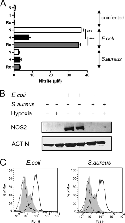
In infected tissues oxygen tensions are low. As innate immune cells have to operate under these conditions, we analyzed the ability of macrophages (Mφ) to kill Escherichia coli or Staphylococcus aureus in a hypoxic microenvironment. Oxygen restriction did not promote intracellular bacterial growth but did impair the bactericidal activity of the host cells against both pathogens. This correlated with a decreased production of reactive oxygen intermediates (ROI) and reactive nitrogen intermediates. Experiments with phagocyte NADPH oxidase (PHOX) and inducible NO synthase (NOS2) double-deficient Mφ revealed that in E. coli- or S. aureus-infected cells the reduced antibacterial activity during hypoxia was either entirely or partially independent of the diminished PHOX and NOS2 activity. Hypoxia impaired the mitochondrial activity of infected Mφ. Inhibition of the mitochondrial respiratory chain activity during normoxia (using rotenone or antimycin A) completely or partially mimicked the defective antibacterial activity observed in hypoxic E. coli- or S. aureus-infected wild-type Mφ, respectively. Accordingly, inhibition of the respiratory chain of S. aureus-infected, normoxic PHOX(-/-) NOS2(-/-) Mφ further raised the bacterial burden of the cells, which reached the level measured in hypoxic PHOX(-/-) NOS2(-/-) Mφ cultures. Our data demonstrate that the reduced killing of S. aureus or E. coli during hypoxia is not simply due to a lack of PHOX and NOS2 activity but partially or completely results from an impaired mitochondrial antibacterial effector function. Since pharmacological inhibition of the respiratory chain raised the generation of ROI but nevertheless phenocopied the effect of hypoxia, ROI can be excluded as the mechanism underlying the antimicrobial activity of mitochondria.


Figures

Fig 1
Fig 1
Impaired ability of murine bone marrow-derived macrophages (Mϕ) to kill intracellular bacteria under hypoxic conditions. (A) Schematic representation of the experimental setup for assaying intracellular survival of E. coli and S. aureus under normoxic and hypoxic conditions. Mϕ were infected with either E. coli or S. aureus at an MOI of 10. One hour after infection extracellular bacteria were removed by a wash with PBS, followed by gentamicin treatment. At 2 h after infection the cells were exposed to either normoxia (N) or hypoxia (H; 0.5% oxygen). For reoxygenation (Re) the cells were first incubated for 8 h under hypoxic conditions and then kept in normoxia. Cell lysates were prepared 2 and 24 h after infection to determine the amount of CFU inside the cells. (B) The graphs shows the percentage of intracellular bacteria in Mϕ under the indicated conditions. Relative survival was calculated by dividing the amount of intracellular bacteria recovered 24 h after infection related to the amount of intracellular bacteria determined 2 h after infection. The data are means + standard errors of the mean (SEM) of five independent experiments. *, P < 0.05; **, P < 0.01; ***, P < 0.001.
Fig 2
Fig 2
Hypoxia does not improve the bacterial growth within Mϕ. (A and B) To study the replication dynamics of E. coli, a strain was used harboring a dual fluorescence reporter plasmid (pDiGc) in which the production of DsRed protein is arabinose inducible, whereas the expression of eGFP is constitutive. The proliferation of bacteria can be monitored by measuring the dilution of DsRed fluorescence after placing the bacterial suspension to arabinose-free conditions. Growing E. coli (pDiGc) strains were analyzed immediately (0 h) after placement into arabinose-free medium (RPMI) and 6 h thereafter. (A) GFP-positive, bacterium-sized particles were identified by flow cytometry and in that population the DsRed fluorescence was analyzed. (B) Mϕ were infected with pDiGc-containing E. coli. After different time points, the cells were lysed, and the DsRed fluorescence of GFP-positive, bacterium-sized particles was measured by flow cytometry. The results of a representative experiment out of at least two similar experiments are displayed. (C and D) The replication of S. aureus was investigated by labeling the bacteria with CFSE. (C) Bacteria were identified after an S. aureus-specific staining. The proliferation of CFSE-labeled S. aureus in medium (RPMI) was accompanied by a reduction in CFSE fluorescence intensity. (D) Mϕ were infected with CFSE-labeled S. aureus. At different time points, the cells were lysed, the bacteria were identified by S. aureus-specific staining, and S. aureus-positive, bacterium-sized particles were analyzed for CFSE fluorescence. The results of a representative experiment out of at least two similar experiments are displayed.
Fig 3
Fig 3
Hypoxia augmented the HIF1A activity of S. aureus-infected Mϕ but does not contribute to the impaired killing of S. aureus under hypoxic conditions. (A) Cellular lysates were prepared of Mϕ infected with E. coli or S. aureus under normoxic or hypoxic conditions for 24 h, and immunoblotting for HIF1A and HIF2A was performed. Equal loading is demonstrated in HIF1A immunoblots by incubating the blots with an actin-specific antibody. To demonstrate equal loading in blots probed for HIF2A, a nonspecific band (n.s.) of the HIF2A antibody is shown. The results of a representative experiment out of at least three similar experiments are displayed. (B) Mϕ were infected with E. coli or S. aureus under normoxic (N) or hypoxic (H) conditions for 24 h, and qRT-PCR was performed with Pgk1 as the target and Hprt1 serving as the internal control. The data are means + the SEM of four experiments. *, P < 0.05; **, P < 0.01; ***, P < 0.001. (C) ns-siRNA (ns) or Hif1a-specific (Hif1a) siRNA was transferred into Mϕ, or the cells were left untreated (untreated). After 24 h, the cells were infected with S. aureus and subjected to normoxic (N) or hypoxic (H) conditions. Cell lysates were prepared 2 and 24 h after infection to determine the CFU count inside the cells. The relative survival was calculated by dividing the amount of intracellular bacteria recovered 24 h after infection related to the amount of intracellular bacteria determined 2 h after infection. The data are means + the SEM of four experiments. *, P < 0.05; **, P < 0.01; ***, P < 0.001.
Fig 4
Fig 4
Hypoxia interferes with the production of oxygen radicals and nitrogen production in response to infection of Mϕ. Mϕ were infected with E. coli or S. aureus at an MOI of 10. One hour after infection, the extracellular bacteria were removed by washing with PBS, followed by gentamicin treatment. At 2 h after infection, the cells were exposed to either normoxia (N) or hypoxia (H; 0.5% oxygen). For reoxygenation (Re), the cells were first incubated for 8 h under hypoxic conditions and then kept in normoxia. (A) After 24 h, the supernatants were collected, and nitrite was measured using the Griess reaction. The data are means + the SEM of five experiments. *, P < 0.05; **, P < 0.01; ***, P < 0.001. (B) Cellular lysates were prepared, and immunoblotting was performed for NOS2 and actin. The results of a representative experiment from at least three similar experiments are shown. (C) Cells were infected as described in panel A. After 24 h, the cells were labeled with CM-H2DCFDA, fixed with 3.5% PFA, and analyzed by flow cytometry. The gray-shaded curve indicates the FL-1 fluorescence of uninfected labeled cells under normoxic conditions. The dotted black curve indicates the FL-1 fluorescence of CM-H2DCFDA-labeled cells under hypoxic conditions, and the solid line curve indicates the FL-1 fluorescence of CM-H2DCFDA-labeled cells under normoxic conditions. The results of a representative experiment out of at least three similar experiments are displayed.
Fig 5
Fig 5
Inhibition of the phagocyte oxidase and NO synthase does not solely explain the impaired killing of S. aureus under hypoxic conditions and is not related with the impaired killing of E. coli in Mϕ. (A) Mϕ from Cybb−/− Nos2−/− or WT littermate controls were infected with S. aureus at an MOI of 10. At 1 h after infection, the extracellular bacteria were removed by washing with PBS, followed by gentamicin treatment. At 2 h after infection, the cells were exposed to either normoxia (N) or hypoxia (H; 0.5% oxygen). For reoxygenation (Re), the cells were first incubated for 8 h under hypoxic conditions and then kept in normoxia. Cell lysates were prepared 2 and 24 h after infection to determine the amount of CFU inside the cells. The relative survival was calculated by dividing the amount of intracellular bacteria recovered 24 h after infection relative to the amount of intracellular bacteria determined 2 h after infection. The data are means + the SEM of at least four experiments. *, P < 0.05; **, P < 0.01; ***, P < 0.001. (B) Mϕ from Cybb−/− Nos2−/− or WT littermate controls were infected with E. coli as described in panel A. The data are means + the SEM of two experiments. *, P < 0.05; **, P < 0.01; ***, P < 0.001.
Fig 6
Fig 6
Hypoxia impairs mitochondrial activity in Mϕ, and inhibition of the mitochondrial respiratory chain completely or partially mimics the effect of hypoxia on the killing of E. coli or S. aureus by Mϕ, respectively. (A) Mϕ were infected with E. coli or S. aureus at an MOI of 10. At 1 h after infection, extracellular bacteria were removed by washing with PBS, followed by gentamicin treatment. At 2 h after infection, the cells were exposed to either normoxia (N) or hypoxia (H; 0.5% oxygen) in the absence or presence of rotenone (100 μM). After at least 24 h, the cells were stained with JC-1. The mitochondrial membrane potential (ΔΨM) was determined and is given in arbitrary units (AU). The data are means + the SD out of three similar experiments performed in triplicate. *, P < 0.05; **, P < 0.01; ***, P < 0.001. (B and C) Cells were infected with E. coli (B) or S. aureus (C) and treated as described in panel A. Cell lysates were prepared 2 and 24 h after infection to determine the number of CFU inside the cells. The relative survival was calculated by dividing the amount of intracellular bacteria recovered 24 h after infection related to the amount of intracellular bacteria determined 2 h after infection. The data are means + the SEM of six experiments. *, P < 0.05; **, P < 0.01; ***, P < 0.001. (D and E) Cells were infected with E. coli (D) or S. aureus (E). At 2 h after infection, the cells were exposed to either normoxia (N) or hypoxia (H; 0.5% oxygen) in the absence or presence of antimycin A (4 μg/ml). Cell lysates were prepared 2 and 24 h after infection to determine the number of CFU inside the cells. The relative survival was calculated by dividing the amount of intracellular bacteria recovered 24 h after infection related to the amount of intracellular bacteria determined 2 h after infection. The data are means + the SEM of at least four experiments. *, P < 0.05; **, P < 0.01; ***, P < 0.001. (F) Mϕ were left untreated or treated with rotenone (100 μM) and subjected to normoxic or hypoxic conditions. After 24 h, the cells were labeled with CM-H2DCFDA, fixed with 3.5% PFA, and analyzed by flow cytometry. The gray-shaded curve indicates the FL-1 fluorescence of labeled cells under normoxic conditions. The black dotted curve indicates the FL-1 fluorescence of CM-H2DCFDA-labeled cells after stimulation with rotenone and incubation under hypoxic conditions, and the black solid curve demonstrates the FL-1 fluorescence of CM-H2DCFDA-labeled cells after stimulation with rotenone and incubation under normoxic conditions.
Fig 7
Fig 7
Inhibition of the mitochondrial respiratory chain is the PHOX- and NOS2-independent component of the impaired killing of S. aureus by Mϕ during hypoxia. Mϕ from Cybb−/− Nos2−/− or WT littermate controls were infected with S. aureus. At 1 h after infection, extracellular bacteria were removed by washing with PBS, followed by gentamicin treatment. At 2 h after infection, the cells were exposed to either normoxia (N) or hypoxia (H; 0.5% oxygen) in the absence or presence of rotenone (100 μM). Cell lysates were prepared 2 and 24 h after infection to determine the amount of CFU inside the cells. The relative survival was calculated by dividing the amount of intracellular bacteria recovered 24 h after infection relative to the amount of intracellular bacteria determined 2 h after infection. The data are means + the SEM of at least four experiments. *, P < 0.05; **, P < 0.01; ***, P < 0.001.

Similar articles

Cited by

References

    1. Ackermann M, et al. 2008. Self-destructive cooperation mediated by phenotypic noise. Nature 454:987–990 - PubMed
    1. Albina JE, Henry WL, Jr, Mastrofrancesco B, Martin BA, Reichner JS. 1995. Macrophage activation by culture in an anoxic environment. J. Immunol. 155:4391–4396 - PubMed
    1. Anand RJ, et al. 2007. Hypoxia causes an increase in phagocytosis by macrophages in a HIF-1α-dependent manner. J. Leukoc. Biol. 82:1257–1265 - PubMed
    1. Arsenijevic D, et al. 2000. Disruption of the uncoupling protein-2 gene in mice reveals a role in immunity and reactive oxygen species production. Nat. Genet. 26:435–439 - PubMed
    1. Atkuri KR, Herzenberg LA, Niemi AK, Cowan T, Herzenberg LA. 2007. Importance of culturing primary lymphocytes at physiological oxygen levels. Proc. Natl. Acad. Sci. U. S. A. 104:4547–4552 - PMC - PubMed

Publication types

MeSH terms

LinkOut - more resources