. 2012 Jan 17;5(207):ra6.
doi: 10.1126/scisignal.2002636.

Plexins are GTPase-activating proteins for Rap and are activated by induced dimerization

Affiliations

Plexins are GTPase-activating proteins for Rap and are activated by induced dimerization

Yuxiao Wang et al. Sci Signal. .

Erratum in

  • Sci Signal. 2012 Mar 13;5(215):er2

Abstract

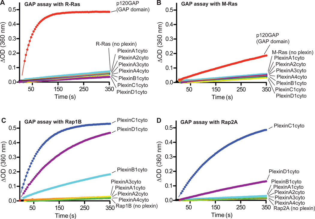
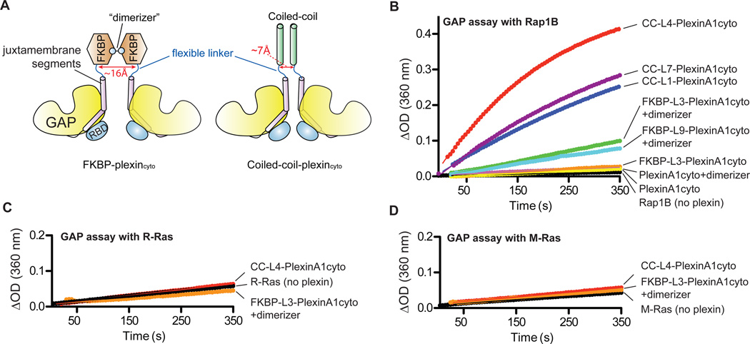
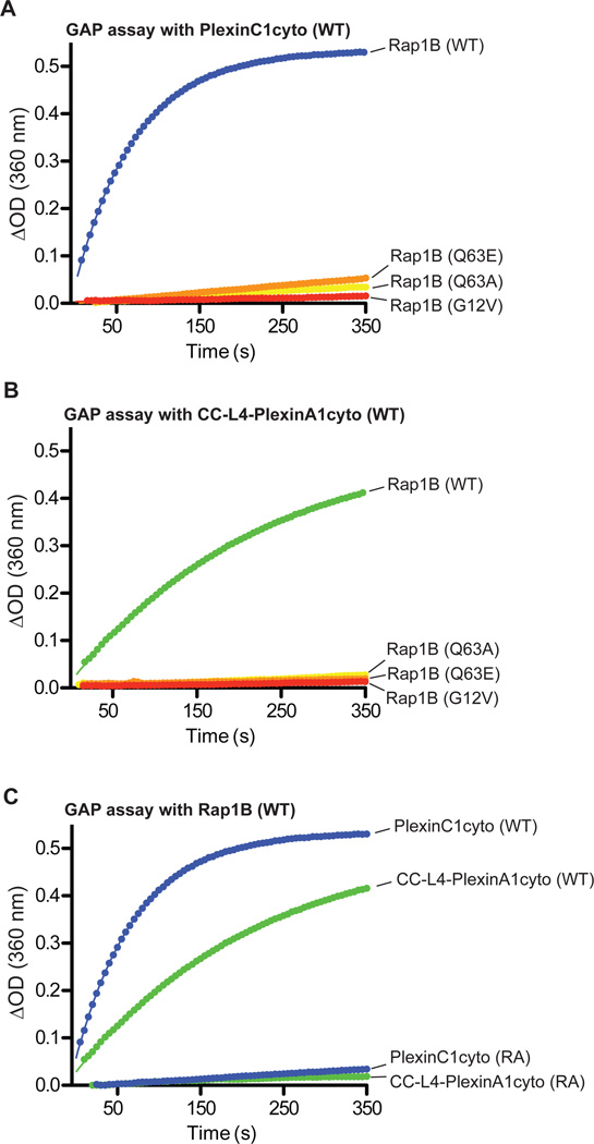
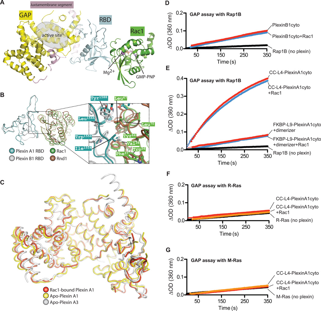
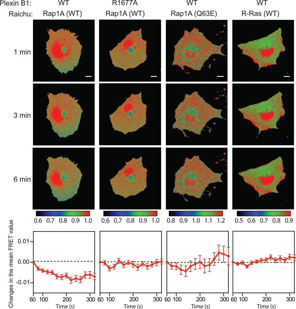
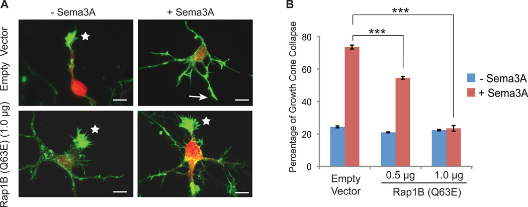
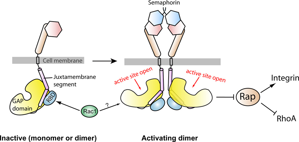
Plexins are cell surface receptors that bind to semaphorins and transduce signals that regulate neuronal development, immune responses, and other processes. Signaling through plexins has been proposed to rely on specific guanosine triphosphatase (GTPase)-activating protein (GAP) activity for R-Ras and M-Ras. Activation of this GAP activity of plexins appears to require simultaneous binding of semaphorin to the plexin extracellular domain and of the Rho GTPases Rac1 or Rnd1 to the cytoplasmic region. However, GAP activity of plexins has eluded detection in several recent studies. We show that the purified cytoplasmic region of plexin uses a noncanonical catalytic mechanism to act as a GAP for Rap, but not for R-Ras or M-Ras. The RapGAP activity of plexins was autoinhibited and was activated by induced dimerization. Biochemical and crystallographic analyses demonstrated that binding of Rho GTPases did not directly contribute to activation of plexin RapGAP activity. Semaphorin stimulated the RapGAP activity of full-length plexin in cells, which was required for plexin-mediated neuronal growth cone collapse. Together, these findings define a pathway for plexin signaling and provide insights into the mechanism for semaphorin-induced activation of plexins.


Figures

Fig. 1
Fig. 1. Purified plexinscyto display GAP activity for Rap but not for R-Ras or M-Ras
(A to D) None of the tested plexinscyto accelerate GTP hydrolysis for R-Ras (A) or M-Ras (B). Plexinscyto show GAP activity towards both Rap1B (C) and Rap2A (D). Data shown are representative of at least 3 independent experiments.
Fig. 2
Fig. 2. Effect of dimerization on the GAP activity of plexinscyto
(A) Schematic diagrams of the FKBP or coiled-coil plexincyto fusions to enable induced or constitutive dimerization. (B) The RapGAP activity of PlexinA1cyto is stimulated by dimerization. “+dimerizer” indicates pre-incubation of the FKBP fusions with the dimerizer. (C to D) CC-L4-PlexinA1cyto and dimerized FKBP-PlexinA1cyto do not show GAP activity for R-Ras (C) or M-Ras (D). Data shown are representative of at least 3 independent experiments.
Fig. 3
Fig. 3. Mutational analysis of the RapGAP activity of plexinscyto
(A to B) The G12V, Q63A or Q63E mutation in Rap1B inhibits GTP hydrolysis catalyzed by PlexinC1cyto (A) or CC-L4-PlexinA1cyto (B). (C) Mutation of the arginine finger (RA) abolishes the RapGAP activity of both PlexinC1cyto and CC-L4-PlexinA1cyto. Data shown are representative of at least 3 independent experiments.
Fig. 4
Fig. 4. Binding of Rac1 does not affect the structure or the GAP activity of plexinscyto
(A) Overall structure of the PlexinA1cyto-Rac1(Q61L) complex. The second plexin molecule in the asymmetric unit, which is not bound to Rac1, is not shown. (B) Comparison of the PlexinA1cyto-Rac1 complex and the PlexinB1 RBD-Rnd1 complex (PDB ID: 2REX). The superimposition is based on the RBDs in the two structures. The juxtamembrane segment and the GAP domain of PlexinA1 are omitted for clarity. (C) Superimposition of Apo-PlexinA3 (PDB ID: 3IG3), Apo-PlexinA1 and Rac1-bound PlexinA1. (D) Active Rac1 does not affect the RapGAP activity of PlexinB1cyto. For GAP assays with Rac1, plexin proteins were incubated with GTP-loaded Rac1(Q61L) before the assays. (E) Active Rac1 does not affect the RapGAP activity of dimeric PlexinA1cyto. (F to G) CC-L4-PlexinA1cyto in the presence of Rac1 remains inactive towards R-Ras (F) and M-Ras (G). Data shown are representative of at least 3 independent experiments.
Fig. 5
Fig. 5. Semaphorin activates the RapGAP activity of full-length PlexinB1 in cells
FRET images were recorded after semaphorin treatment of the cells transfected with the combinations of plasmids as indicated at the top panel. Each column contains three timelapse images of one representative cell for each transfection combination. Scale bars: 10 µm. The images are pseudo-colored based on the FRET value of each pixel. The calibration bars for the coloring schemes are shown below each column. In the bottom panels, the changes in the mean FRET value of all the imaged cells in each group are plotted as a function of time. The plots are combined data from at least 3 independent experiments (n = 8~12 cells). Error bars represent standard error of the mean.
Fig. 6
Fig. 6. The RapGAP activity of plexin is required for semaphorin induced neuronal growth cone collapse
(A) Representative images of semaphorin-induced growth cone collapse of rat cortical neurons. Growth cone morphology was visualized by staining of F-actin with Alexa-554 conjugated phalloidin (green). Transfected neurons were identified based on the mCherry fluorescence signal (red). Uncollapsed and collapsed growth cones are indicated by stars and arrows, respectively. Scale bars: 10 µm. (B) Quantification of growth cone collapse of the neurons that are transfected with empty vector or various amounts of Rap1B Q63E mutant. Error bars represent standard error of the mean. Asterisks (***) indicate significant differences (p<0.001) determined by analysis of variance (ANOVA) followed by Bonferroni post-tests.
Fig. 7
Fig. 7. A schematic model for the regulation of the RapGAP activity of plexin
Prior to semaphorin binding, plexin is an inactive monomer or dimer, in which the RapGAP activity is autoinhibited. Semaphorin-induced dimerization of the plexin extracellular region promotes formation of the activating dimer of the cytoplasmic region, which converts the GAP domain to the active state through an allosteric mechanism. Rac1 or other activator RhoGTPases are not involved in this allosteric activation process, and their roles in plexin activation are not clear. Plexin inactivates Rap to promote neuronal growth cone collapse and other changes in cell morphology, which may rely on the ability of Rap to regulate cell adhesion and cytoskeleton dynamics through integrin and RhoA, respectively.

Similar articles

Cited by

References

    1. Tran TS, Kolodkin AL, Bharadwaj R. Semaphorin regulation of cellular morphology. Annual review of cell and developmental biology. 2007;23:263–292. - PubMed
    1. Kruger RP, Aurandt J, Guan KL. Semaphorins command cells to move. Nature reviews. 2005;6:789–800. - PubMed
    1. Capparuccia L, Tamagnone L. Semaphorin signaling in cancer cells and in cells of the tumor microenvironment--two sides of a coin. Journal of cell science. 2009;122:1723–1736. - PubMed
    1. Rujescu D, Meisenzahl EM, Krejcova S, Giegling I, Zetzsche T, Reiser M, Born CM, Moller HJ, Veske A, Gal A, Finckh U. Plexin B3 is genetically associated with verbal performance and white matter volume in human brain. Mol Psychiatry. 2007;12:190–194. 115. - PubMed
    1. Antipenko A, Himanen JP, van Leyen K, Nardi-Dei V, Lesniak J, Barton WA, Rajashankar KR, Lu M, Hoemme C, Puschel AW, Nikolov DB. Structure of the semaphoring-3A receptor binding module. Neuron. 2003;39:589–598. - PubMed

Publication types

Associated data