

Imaging features of paediatric haemophilic pseudotumour of the maxillary bone: report of three cases and review of the literature
- PMID: 22253339
- PMCID: PMC3587104
- DOI: 10.1259/bjr/12938443
Imaging features of paediatric haemophilic pseudotumour of the maxillary bone: report of three cases and review of the literature
Abstract
Objectives: Haemophilic pseudotumour (HP) is an extremely rare lesion. The purpose of this study was to describe the CT and MRI features of maxillary bone HPs and introduce the key points to differentiate HP from the mimicking entities in the region.
Methods: We retrospectively reviewed three paediatric patients with histology-proven HPs arising from the maxillary bone. All three patients underwent CT and/or MRI. Combined with six previously reported cases in the literature, the imaging features were comprehensively analysed.
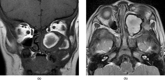
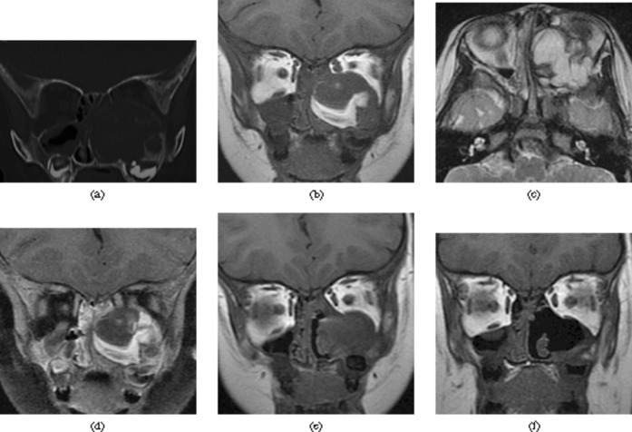
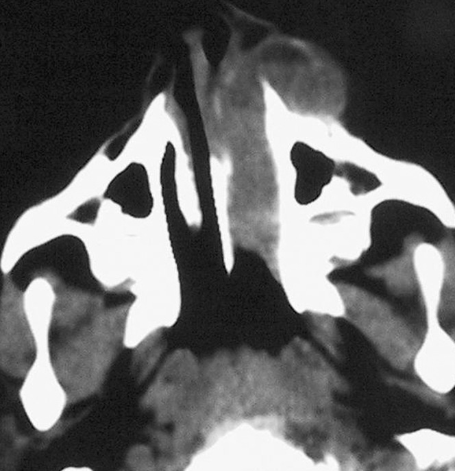
Results: All HPs showed a well-demarcated, multilobulated expansile osteolytic lesion in the maxillary bone. On non-enhanced CT, HPs appeared of mixed density relative to grey matter. The lesions appeared to have markedly heterogeneous signal intensity on both T(1) and T(2) weighted images, with septa-like enhancement following the administration of contrast material, which corresponded to blood products in various stages of evolution. The lesions caused cortical thinning and even focal disappearance and multiple bone septa were identified within the involved maxillary bone. Some HPs were associated with radiated periosteal proliferation, which can easily be misdiagnosed as a malignant bone tumour.
Conclusion: A high index of suspicion for HP and a familiarity with imaging findings may help to accurately diagnose this rare entity.
Figures
References
-
- de Sousa SO, de Piratininga J, Pinto Júnior DS, de Araújo N. Hemophilic pseudotumor of the jaws: report of two cases. Oral Surg Oral Med Oral Pathol Oral Radiol Endod 1995;79:216–19 - PubMed
-
- Zheng K, Zheng P. Hemophilic pseudotumor involving maxilla and tibia. Chin Med J (Engl) 1997;110:233–5 - PubMed
-
- Stafford JM, James TT, Allen AM, Dixon LR. Hemophilic pseudotumor: radiologic-pathologic correlation. Radiographics 2003;23:852–6 - PubMed
-
- Park JS, Ryu KN. Hemophilic pseudotumor involving the musculoskeletal system: spectrum of radiologic findings. AJR Am J Roentgenol 2004;183:55–61 - PubMed
-
- Rodriguez-Merchan EC. Haemophilic cysts(pseudotumours). Haemophilia 2002;8:393–401 - PubMed
Publication types
MeSH terms
LinkOut - more resources
Full Text Sources
Medical
Research Materials
Miscellaneous
