. 2012 Mar 9;287(11):8495-506.
doi: 10.1074/jbc.M111.323915. Epub 2012 Jan 17.

Calcium/calmodulin-dependent protein kinase II (CaMKII) inhibition induces neurotoxicity via dysregulation of glutamate/calcium signaling and hyperexcitability

Affiliations

Calcium/calmodulin-dependent protein kinase II (CaMKII) inhibition induces neurotoxicity via dysregulation of glutamate/calcium signaling and hyperexcitability

Nicole M Ashpole et al. J Biol Chem. .

Abstract

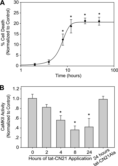
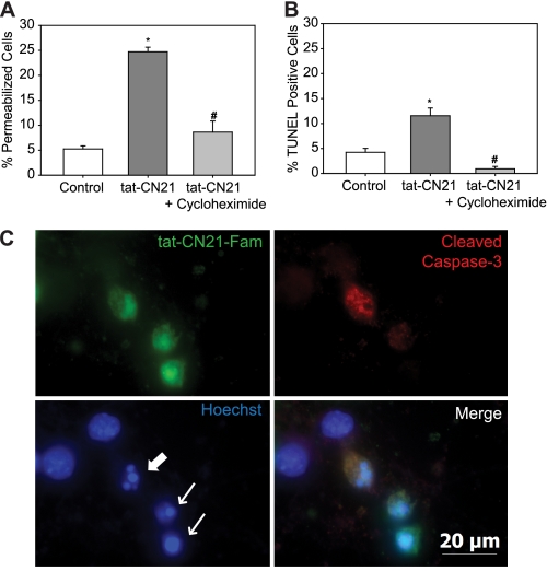
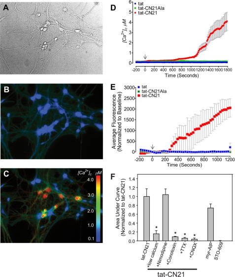
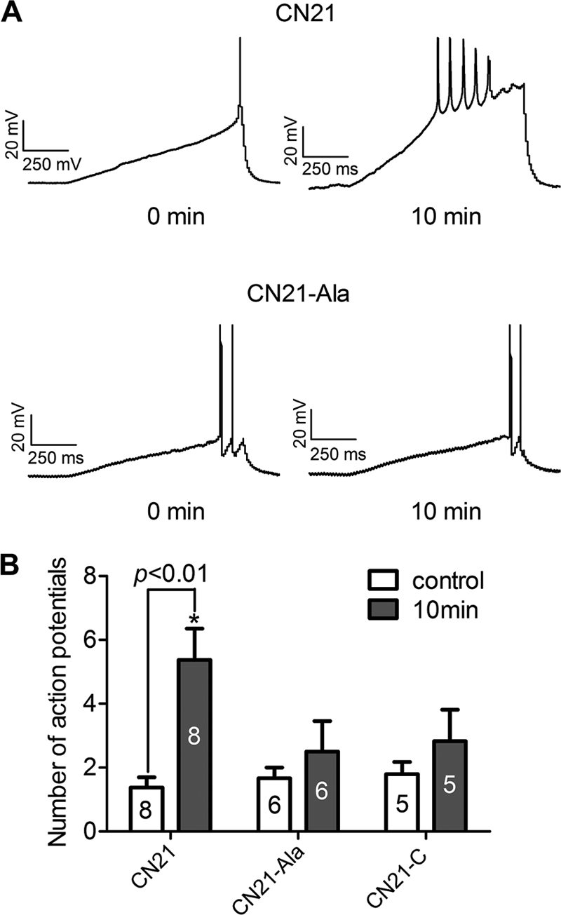
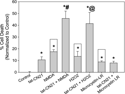
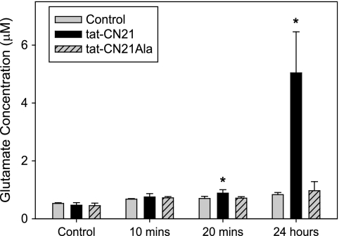
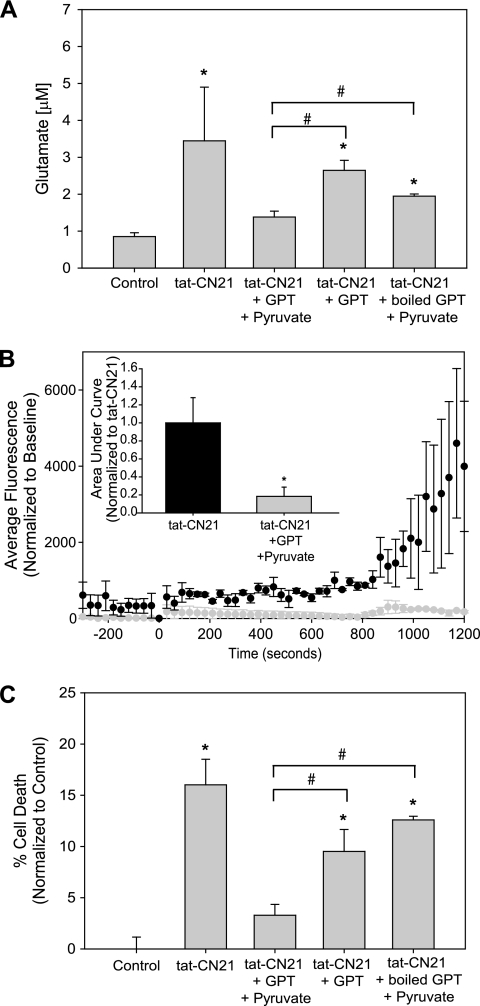
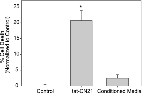
Aberrant glutamate and calcium signalings are neurotoxic to specific neuronal populations. Calcium/calmodulin-dependent kinase II (CaMKII), a multifunctional serine/threonine protein kinase in neurons, is believed to regulate neurotransmission and synaptic plasticity in response to calcium signaling produced by neuronal activity. Importantly, several CaMKII substrates control neuronal structure, excitability, and plasticity. Here, we demonstrate that CaMKII inhibition for >4 h using small molecule and peptide inhibitors induces apoptosis in cultured cortical neurons. The neuronal death produced by prolonged CaMKII inhibition is associated with an increase in TUNEL staining and caspase-3 cleavage and is blocked with the translation inhibitor cycloheximide. Thus, this neurotoxicity is consistent with apoptotic mechanisms, a conclusion that is further supported by dysregulated calcium signaling with CaMKII inhibition. CaMKII inhibitory peptides also enhance the number of action potentials generated by a ramp depolarization, suggesting increased neuronal excitability with a loss of CaMKII activity. Extracellular glutamate concentrations are augmented with prolonged inhibition of CaMKII. Enzymatic buffering of extracellular glutamate and antagonism of the NMDA subtype of glutamate receptors prevent the calcium dysregulation and neurotoxicity associated with prolonged CaMKII inhibition. However, in the absence of CaMKII inhibition, elevated glutamate levels do not induce neurotoxicity, suggesting that a combination of CaMKII inhibition and elevated extracellular glutamate levels results in neuronal death. In sum, the loss of CaMKII observed with multiple pathological states in the central nervous system, including epilepsy, brain trauma, and ischemia, likely exacerbates programmed cell death by sensitizing vulnerable neuronal populations to excitotoxic glutamate signaling and inducing an excitotoxic insult itself.


Figures

FIGURE 1.
FIGURE 1.
Neurotoxicity with CaMKII inhibition. A, neuronal death (mean ± S.E., n = 3–15) normalized to control when CaMKII inhibitors (10 μm peptide inhibitors and 1 μm small molecule inhibitors) were applied to neuronal cultures (8 DIV) for 1 h (gray bars) or 24 h (black bars). *, p < 0.05 compared with control (one-way ANOVA, post hoc Dunnett's test). B, neuronal death (mean ± S.E., n = 4–17) normalized to control when 10 μm tat-CN21 or tat-CN21Ala was applied to neuronal cultures for 24 h at 8, 14, and 21 DIV. *, p < 0.05 compared with control at that time point; #, p < 0.05 compared with 8 DIV tat-CN21 treatment (one-way ANOVA, post hoc Dunnett's test). @, p < 0.05 for 14 DIV tat-CN21 treatment versus 21 DIV tat-CN21 treatment (t test). C, neuronal death (mean ± S.E., n = 4) normalized to control when 10 μm tat-CN21 or tat-CN21Ala was applied to co-cultures for 24 h. *, p < 0.05 compared with control (one-way ANOVA, post hoc Dunnett's test).
FIGURE 2.
FIGURE 2.
Time dependence of neurotoxicity and CaMKII inactivation with CaMKII inhibition. A, neuronal death (mean ± S.E., n = 7–12) normalized to control when 10 μm tat-CN21 was applied to neuronal cultures for varying lengths of time. *, p < 0.05 compared with control (one-way ANOVA, post hoc Dunnett's test). B, kinase activity (mean ± S.D., n = 3–4) in neuronal lysates subjected to an in vitro CaMKII assay in the presence of calcium/calmodulin after 10 μm tat-CN21 (or tat-CN21Ala) was applied to cortical neurons for varying lengths of time. *, p < 0.05 compared with control (one-way ANOVA, post hoc Dunnett's test).
FIGURE 3.
FIGURE 3.
Neuronal apoptosis with CaMKII inhibition. Neuronal death (mean ± S.E., n = 5–10) following treatment with tat-CN21 with or without 0.5 mg/ml cycloheximide as measured by ethidium homodimer membrane permeability dye (A) or TUNEL staining (B). *, p < 0.05 compared with control; #, p < 0.05 compared with tat-CN21 alone (one-way ANOVA, post hoc Dunnett's test). C, representative image of a field of neurons treated with 10 μm tat-CN21-Fam for 24 h (top left) immunostained for cleaved caspase-3 (top right), nuclear marker Hoechst (bottom left), and a merge of all three channels (bottom right). Arrows indicate fragmented or pyknotic nuclei.
FIGURE 4.
FIGURE 4.
Calcium dysregulation with CaMKII inhibition in hippocampal neurons. A, representative bright field; B and C, fluorescent images of Fura-2FF-loaded hippocampal neurons. B, before treatment; C, after treatment with 10 μm tat-CN21. D, cytoplasmic calcium levels ([Ca2+]c) (mean ± S.E.) before and after application of 10 μm tat-CN21, tat-CN21Ala, or tat. E, neuronal intracellular calcium levels (mean ± S.E.) before (−300 to 0 s) and after (0 to 1200 s) application of 10 μm tat-CN21, tat-CN21Ala, or tat, as measured by Fluo-4 (n = 4). F, average integral of fluorescent intensity from 0 to 1200 s in E (mean ± S.E., n = 3–6) with application of CaMKII inhibitors with and without pharmacological blockers of neuronal excitability. The integral was normalized to the calcium influx observed with application of 10 μm tat-CN21. *, p < 0.05 compared with tat-CN21 alone (one-way ANOVA, post hoc Dunnett's test).
FIGURE 5.
FIGURE 5.
CaMKII inhibition augments neuronal excitability. A, representative traces from cortical neurons at time 0 and 10 min following diffusion of either 1 μm CN21 or 1 μm CN21Ala. Neurons were held at their resting membrane potentials and injected with 1-s depolarizing current ramps to evoke action potentials. B, number of action potentials (mean ± S.D.) evoked at time 0 or 10 min after whole cell configuration in the presence of CN21 or control CN21Ala or CN21C. *, p < 0.01 between the number of action potentials between time 0 and 10 min (one-way ANOVA, post hoc Bonferroni).
FIGURE 6.
FIGURE 6.
Prolonged CaMKII inhibition sensitizes neurons to excitotoxicity-related insults. Neuronal death (mean ± S.E., n = 3–7) following treatment with various combinations of tat-CN21, NMDA, H2O2, or microcystin-LR. All cell death measurements were made 48 h from the start of treatments. Cultures were treated with NMDA, H2O2, or microcystin-LR independently or in combination with a 24-h pretreatment of 10 μm tat-CN21. The clear boxes highlight the potential levels of cytotoxicity if the average death induced by tat-CN21 treatment alone would be additive. *, p < 0.05 compared with control (one-way ANOVA, post hoc Dunnett's test). #, p < 0.05 compared with NMDA treatment alone (t test). @, p < 0.05 compared with H2O2 alone (t test).
FIGURE 7.
FIGURE 7.
CaMKII inhibition results in increased glutamate in conditioned neuronal media. Glutamate concentration (mean ± S.D., n = 3–6) in neuronal media following incubation with 10 μm tat-CN21 or tat-CN21Ala for varying lengths of time, as measured by a glutamate oxidase assay. *, p < 0.05 compared with vehicle control (DMSO) (one-way ANOVA, post hoc Dunnett's test).
FIGURE 8.
FIGURE 8.
Enzymatic catalysis of glutamate prevents acute and prolonged effects of CaMKII inhibition. A, glutamate concentration (mean ± S.D., n = 4–8) in neuronal media following incubation with 10 μm tat-CN21 for 24 h with and without co-application of GPT/pyruvate, GPT alone, or boiled GPT/pyruvate. *, p < 0.05 compared with control; #, p < 0.05 compared with tat-CN21/GPT/pyruvate treatment (one-way ANOVA, post hoc Dunnett's test). B, neuronal intracellular calcium levels (mean ± S.E., n = 3) following application of tat-CN21 in the presence or absence of GPT/pyruvate. Bar graph inset indicates the average integral from 0 to 1200 s (mean ± S.E., n = 3) for these treatment groups. C, neuronal death (mean ± S.E., n = 3–6) after 24 h of treatment with 10 μm tat-CN21 alone or co-application with GPT/pyruvate, GPT alone, or boiled GPT/pyruvate. *, p < 0.05 compared with control; #, p < 0.05 compared with tat-CN21/GPT/pyruvate treatment (one-way ANOVA, post hoc Dunnett's test).
FIGURE 9.
FIGURE 9.
Pharmacological antagonism of the NMDA receptor prevents acute and prolonged effects of CaMKII inhibition. A, average integral of fluorescent intensity from 0 to 1200 s (mean ± S.E., n = 3–6) reflecting calcium influx in control neurons, or neurons subjected to treatment with tat-CN21 alone or in combination with 20 μm MK-801 or in combination with a prior synaptic NMDA-R blockade. To block synaptic NMDA-Rs before tat-CN21 treatment, 10 μm bicuculline was applied to allow synaptic activity, followed by the addition of 20 μm MK-801 to inhibit the synaptic NMDA-Rs opened as a result of this synaptic activity. *, p < 0.05 compared with control; #, p < 0.05 compared with tat-CN21 (one-way ANOVA, post hoc Dunnett's test). B, neuronal death (mean ± S.E., n = 4–24) after 24 h of treatment with 10 μm tat-CN21 alone or in the presence of 20 μm MK-801, 10 μm ifenprodil, 1 μm memantine, or 200 nm tetrodotoxin (TTX). *, p < 0.05 compared with control; #, p < 0.05 compared with tat-CN21 (one-way ANOVA, post hoc Dunnett's test).
FIGURE 10.
FIGURE 10.
Conditioned media from neurons treated with CaMKII inhibitors does not induce neurotoxicity. Neuronal death (mean ± S.E., n = 3–9) in neurons treated with 10 μm tat-CN21 for 24 h or naive neurons treated for 24 h with media removed from tat-CN21-treated neurons. *, p < 0.05 compared with control (one-way ANOVA, post hoc Dunnett's test).

Similar articles

Cited by

References

    1. Coultrap S. J., Vest R. S., Ashpole N. M., Hudmon A., Bayer K. U. (2011) CaMKII in cerebral ischemia. Acta Pharmacol. Sin. 32, 861–872 - PMC - PubMed
    1. Hudmon A., Schulman H. (2002) Neuronal Ca2+/calmodulin-dependent protein kinase II. The role of structure and autoregulation in cellular function. Annu. Rev. Biochem. 71, 473–510 - PubMed
    1. Colbran R. J. (2004) Targeting of calcium/calmodulin-dependent protein kinase II. Biochem. J. 378, 1–16 - PMC - PubMed
    1. Perlin J. B., Churn S. B., Lothman E. W., DeLorenzo R. J. (1992) Loss of type II calcium/calmodulin-dependent kinase activity correlates with stages of development of electrographic seizures in status epilepticus in rat. Epilepsy Res. 11, 111–118 - PubMed
    1. Zalewska T., Domanska-Janik K. (1996) Brain ischemia transiently activates Ca2+/calmodulin-independent protein kinase II. Neuroreport 7, 637–641 - PubMed

Publication types

MeSH terms