. 2012 May;26(5):1946-59.
doi: 10.1096/fj.11-199851. Epub 2012 Jan 17.

Identification of oligomers at early stages of tau aggregation in Alzheimer's disease

Affiliations

Identification of oligomers at early stages of tau aggregation in Alzheimer's disease

Cristian A Lasagna-Reeves et al. FASEB J. 2012 May.

Abstract

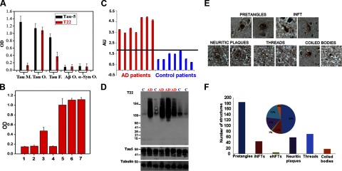
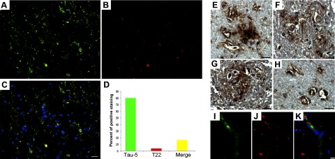
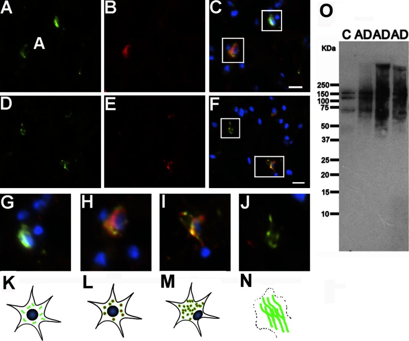
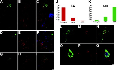
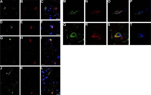
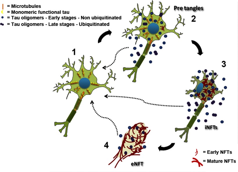
Neurofibrillary tangles (NFTs) are a pathological hallmark of Alzheimer's disease (AD); however, the relationship between NFTs and disease progression remains controversial. Analyses of tau animal models suggest that phenotypes coincide with accumulation of soluble aggregated tau species but not the accumulation of NFTs. The pathological role of prefilamentous tau aggregates, e.g., tau oligomeric intermediates, is poorly understood, in part because of methodological challenges. Here, we engineered a novel tau oligomer-specific antibody, T22, and used it to elucidate the temporal course and biochemical features of oligomers during NFT development in AD brain. We found that tau oligomers in human AD brain samples were 4-fold higher than those in the controls. We also revealed the role of oligomeric tau conformers in pretangles, neuritic plaques, and neuropil threads in the frontal cortex tissue from AD brains; this analysis uncovers a consistent code that governs tau oligomerization with regard to degree of neuronal cytopathology. These data are the first to characterize the role of tau oligomers in the natural history of NFTs, and they highlight the suitability of tau oligomers as therapeutic targets in AD and related tauopathies.


Figures

Figure 1.
Figure 1.
Specificity of T22 antibody for tau oligomers. A) ELISA analysis shows that T22 does not show any significant reactivity for monomeric tau, but some signal is observed in tau fibrils, possibly due to the cross-reactivity of the antibody or the presence of oligomers in the preparation. T22 does not recognize oligomers from other amyloidogenic proteins, such as Aβ or α-synuclein; Tau-5, which recognizes all forms of tau, was used as a positive control for the tau samples. B) T22 neutralizes tau oligomer toxicity when preincubated at an equimolar ratio, as assessed using the MTT toxicity assay and SY5Y human neuroblastoma cells. Tau oligomers (bar 1) and Aβ oligomers (bar 2) are highly toxic to neuroblastoma cells. T22 can neutralize the toxicity caused by tau oligomers (bar 3) but not Aβ oligomers (bar 4); tau fibrils were not toxic (bar 5). The same is true for monomeric tau (bar 6), as well as for the PBS control (bar 7). C–F) Antibody T22 preferentially recognizes tau oligomers in AD brain. C) ELISA analysis of PBS soluble fraction from AD and age-matched control brains. In all patients with AD, T22 signal corresponding to tau oligomers (red) was higher than that in age-matched controls (blue). Values were normalized with tubulin. AU, arbitrary units. D) Western blot of PBS-soluble fraction from AD and control frontal cortex. Top blot, T22; middle blot, Tau-5; bottom blot, tubulin loading controls. High-molecular-mass bands corresponding to tau oligomers detected by T22 are elevated in AD brain compared with control brain, in which the major T22 immunoreactive band probably corresponds to a dimer. T22 does not recognize monomeric tau; however, Tau-5-immunoreactive material corresponding to tau monomer is abundant in both AD and control samples (middle blot). E) Tau oligomer localization during the evolution of NFTs as assessed using T22. Photomicrographs of T22 staining developed using avidin-biotin complex with hematoxylin-counterstained paraffin sections. T22 labels pre-NFTs, iNFTs, neuritic coronae, threads, and coiled bodies. F) Quantitation of T22 immunoreactivity from a total of 377 structures; 184 of the lesions corresponded to pretangles, 71 to neuropil threads, and a lower quantity to other strutures: 44 iNFTs, 58 neuritic plaques, 17 coiled bodies, and 3 eNFTs. Inset: pie graph indicating percentage distribution of total T22 immunoreactivity among these various structures.
Figure 2.
Figure 2.
Tau oligomers constitute a small portion of total tau in AD frontal cortex. A–C) Representative immunofluorescent images of AD frontal cortex showing Tau-5 (A, green), T22 (B, red), and merge (C; also including DAPI, blue) confirm the presence of tau oligomers in situ. D) Histogram summarizing quantitation of 16 AD cases; only 20% of total aggregates corresponded to oligomers. Extracellular tau oligomers were also detected. E–H) Photomicrographs demonstrating diffuse extracellular tau oligomers (as detected by T22), including those in association with arterioles. Double immunofluorescence with von Willebrand factor antibody (I, green), T22 (J, red), and merge (K; also with DAPI, blue) suggested the presences of extracellular tau oligomers. Sections were stained with DAPI. Scale bar = 20 μm (A–C); 15 μm (E–K).
Figure 3.
Figure 3.
Phosphorylation of tau at Thr231 precedes the formation of tau oligomers. A–F) Double staining for anti-phospho Thr231 (A, D; green), T22 (B, E; red), and merge (C, F, plus DAPI in blue) demonstrate that tau oligomers are present at early and intermediate stages of aggregation and are preceded by phosphorylation at Thr231. G) Phospho-tau (Thr231) appears throughout the soma before its aggregation (stage 0). H) At early stages, the cells are positive for T22 and Thr231; nuclei and neuronal outlines appear normal (stage 1). I) In a more advanced stage, intraneuronal oligomers and phospho-tau aggregates colocalize, but the nucleus is eccentric (stage 2). J) Finally, at late stages, phospho-tau Thr231 signal remains, whereas T22 staining and nuclei are gone. K–N) Schematic diagram of tau oligomer formation and cell death, leading to the formation of a ghost tangle. Functional monomeric tau is phosphorylated in healthy neurons (K), then tau oligomerization occurs in the cytoplasm (L). Later, tau oligomers cause cell dysfunction and displacement of the nucleus to the side of the cell (M); these events finally lead to cell death and the formation of NFTs (N). O) Western blot analysis, using anti-phospho Thr231, indicated that tau is phosphorylated at this epitope in age-matched controls and detected high-molecular-mass bands in AD corresponding to tau filaments. Scale bars = 10 μm.
Figure 4.
Figure 4.
Tau oligomerization occurs before phosphorylation at Ser202/Thr205. A–I) Photomicrographs showing immunofluorescence signals for AT8 (A, D; green), T22 (B, E; red), and merge (C, F; also including DAPI, blue) localize tau oligomers in pretangles (F) and iNFTs (C), but not in eNFTs that are labeled with AT8 (C, F). In some instances, extracellular phosphorylated tau at Ser202/Thr205 (G, green) and extracellular oligomers (H, red) do not correlate (I, merge + DAPI). J, K) Distribution of oligomeric tau and phospho-Ser202/Thr205 tau among NFT species. J) Oligomeric species are distributed 79% in pretangles, 19% in iNFTs, and 2% in eNFTs. K) AT8 signal corresponded 72.4% to eNFTs, 22.3% to iNFTs, and only 5.21% to pretangles. L–Q) It is also possible to see some of AT8 and T22 signals in iNFTs, but in these cases the colocalization between AT8 (L, O; green) and T22 (M, P; red) is negligible (N, Q; merge + DAPI). Scale bars = 10 μm (A–I); 5 μm (L–N); 2 μm (O–Q).
Figure 5.
Figure 5.
Ubiquitination of tau occurs at intermediate stages during NFT evolution, after appearance of oligomers. A–C) Immunofluorescence images of pretangles fail to show colocalization of anti-ubiquitin staining (A, green), T22 (B, red), and merge (C; with DAPI, blue). D–I) Tau oligomers (E, H; red) are highly ubiquitinated (D, G; green) at intermediate stages. F, I) Merged images plus DAPI. J–L) Structures labeled with anti-ubiquitin (J, green; arrow) that resemble eNFTs are not detected with T22 (K, red), suggesting that ubiquitination continues after the transition of oligomer to fibril. L) Merged image + DAPI. M–T) Ubiquitinated tau oligomers positive for FRET are present in iNFTs. Laser scanning confocal images depicting anti-ubiquitin staining (M, Q; green), T22 signal (N, R; red), and merged images plus DAPI (O, S); colocalization of anti-ubiquitin and tau oligomers is seen only in iNFTs (O, S). Increased donor fluorescence (ubiquitin, Alexa Fluor 488) is observed only in iNFTs, showing the presence of FRET exclusively in these structures (P, T). Scale bars = 10 μm.
Figure 6.
Figure 6.
Schematic diagram of NFT life cycle and the role of oligomers in NFT formation and disease progression. 1) Under normal conditions, tau participates in the association and dissociation of microtubules, conferring dynamics to the system. 2) Post-translational modifications of tau, e.g., phosphorylation, can dissociate tau from microtubules. This dissociation produces an increase in the cytosolic concentration of the protein that exceeds the minimal tau concentration necessary to support conformational changes and leads to an electrostatic modification in the molecule that enables it to form a side chain-side chain interaction, culminating in the formation of a tau-tau dimer. Once these dimers are formed and adopt a stable structure, they can begin a process of nucleation, forming oligomers. 3) Subsequently, tau continues the aggregation process and undergoes additional post-translational modifications, such as phosphorylation and ubiquitination. These oligomers (in 2 and 3) are highly toxic and can induce memory deficits in mice and seed the aggregation of monomeric unmodified tau. Then tau oligomers form tau filaments, termed PHFs; these tau structures undergo new modifications and form NFTs. 4) Finally, after cell death, NFTs maintain their flame shape and form “ghost tangles” or eNFTs. Dashed arrows indicate that extracellular tau oligomers can induce the aggregation of monomeric tau from healthy neurons.

References

    1. Schultz C., Ghebremedhin E., Del Tredici K., Rub U., Braak H. (2004) High prevalence of thorn-shaped astrocytes in the aged human medial temporal lobe. Neurobiol. Aging 25, 397–405 - PubMed
    1. Umahara T., Tsuchiya K., Ikeda K., Kanaya K., Iwamoto T., Takasaki M., Mukai K., Shibata N., Kato S. (2002) Demonstration and distribution of tau-positive glial coiled body-like structures in white matter and white matter threads in early onset Alzheimer's disease. Neuropathology 22, 9–12 - PubMed
    1. Braak H., Braak E. (1988) Neuropil threads occur in dendrites of tangle-bearing nerve cells. Neuropathol. Appl. Neurobiol. 14, 39–44 - PubMed
    1. Adalbert R., Nogradi A., Babetto E., Janeckova L., Walker S. A., Kerschensteiner M., Misgeld T., Coleman M. P. (2009) Severely dystrophic axons at amyloid plaques remain continuous and connected to viable cell bodies. Brain 132, 402–416 - PubMed
    1. Augustinack J. C., Schneider A., Mandelkow E. M., Hyman B. T. (2002) Specific tau phosphorylation sites correlate with severity of neuronal cytopathology in Alzheimer's disease. Acta Neuropathol. 103, 26–35 - PubMed

Publication types