. 2012;7(1):e29561.
doi: 10.1371/journal.pone.0029561. Epub 2012 Jan 12.

Capsules from pathogenic and non-pathogenic Cryptococcus spp. manifest significant differences in structure and ability to protect against phagocytic cells

Affiliations

Capsules from pathogenic and non-pathogenic Cryptococcus spp. manifest significant differences in structure and ability to protect against phagocytic cells

Glauber de S Araujo et al. PLoS One. 2012.

Abstract

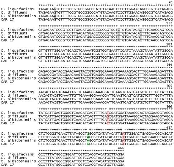
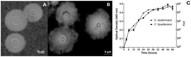
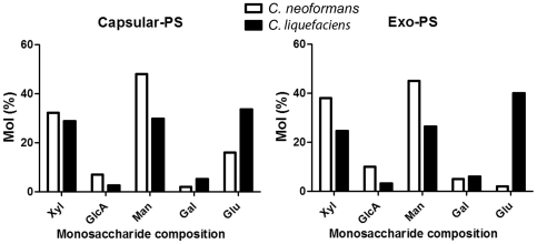
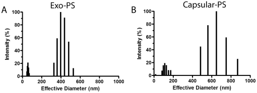
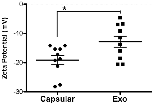
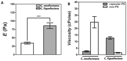
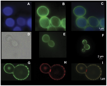
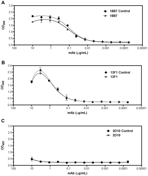
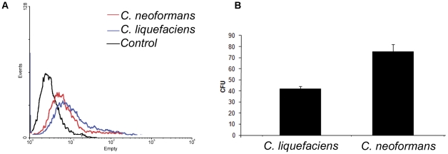
Capsule production is common among bacterial species, but relatively rare in eukaryotic microorganisms. Members of the fungal Cryptococcus genus are known to produce capsules, which are major determinants of virulence in the highly pathogenic species Cryptococcus neoformans and Cryptococcus gattii. Although the lack of virulence of many species of the Cryptococcus genus can be explained solely by the lack of mammalian thermotolerance, it is uncertain whether the capsules from these organisms are comparable to those of the pathogenic cryptococci. In this study, we compared the characteristic of the capsule from the non-pathogenic environmental yeast Cryptococcus liquefaciens with that of C. neoformans. Microscopic observations revealed that C. liquefaciens has a capsule visible in India ink preparations that was also efficiently labeled by three antibodies generated to specific C. neoformans capsular antigens. Capsular polysaccharides of C. liquefaciens were incorporated onto the cell surface of acapsular C. neoformans mutant cells. Polysaccharide composition determinations in combination with confocal microscopy revealed that C. liquefaciens capsule consisted of mannose, xylose, glucose, glucuronic acid, galactose and N-acetylglucosamine. Physical chemical analysis of the C. liquefaciens polysaccharides in comparison with C. neoformans samples revealed significant differences in viscosity, elastic properties and macromolecular structure parameters of polysaccharide solutions such as rigidity, effective diameter, zeta potential and molecular mass, which nevertheless appeared to be characteristics of linear polysaccharides that also comprise capsular polysaccharide of C. neoformans. The environmental yeast, however, showed enhanced susceptibility to the antimicrobial activity of the environmental phagocytes, suggesting that the C. liquefaciens capsular components are insufficient in protecting yeast cells against killing by amoeba. These results suggest that capsular structures in pathogenic Cryptococcus species and environmental species share similar features, but also manifest significant difference that could influence their potential to virulence.


Conflict of interest statement

Competing Interests: The authors have declared that no competing interests exist.

Figures

Figure 1
Figure 1. Alignment of ribosomal region D1/D2, in 28 S rDNA, between C. liquefaciens strain Car17 and related species.
Figure 2
Figure 2. Microscopic visualization of C. liquefaciens and C. neoformans capsules and growth profile.
Panel A) India ink stain of C. neoformans cells. Panel B) Counterstaining of C. liquefaciens with India ink. Panel C) Growth curves of C. neoformans and C. liquefaciens at 30°C in minimal media.
Figure 3
Figure 3. Monosaccharide composition of capsular and exo-PS of C. liquefaciens and C. neoformans.
Xyl (Xylose), GlcA (Glucuronic acid), Man (Mannose), Gal (Galactose), Glu (Glucose) and GlcNAc (N-acetylglucosamine).
Figure 4
Figure 4. Physical properties of capsular and exo-PS from C. liquefaciens obtained by DLS.
Panel A and B show size distribution of PS fibers capsular and exo-PS samples, respectively.
Figure 5
Figure 5. Zeta Potential of exo- and capsular-PS from C. liquefaciens.
The charges of the different C. liquefaciens PS forms were significantly different (P Value<0.01 calculated by Student's T test (two-tailed)).
Figure 6
Figure 6. Elastic and viscosity properties of C. liquefaciens polysaccharide.
Panel A) Young modulus (E) of C. neoformans and C. liquefaciens capsular-PS demonstrates significant differences in their elastic properties (*** P value<0.01 calculated by Student's T test (two-tailed)). Panel B) Viscosity values for C. neoformans and C. liquefaciens capsular and exo-PS. The differences between exo- and capsular-PS in C. neoformans and C. liquefaciens were statistically significant (P value<0.01 calculated by Student's T test (two-tailed)). Also, the differences between PS types for both yeast species were statistically significant (P value<0.01 calculated by Student's T test (two-tailed)).
Figure 7
Figure 7. Surface architecture of C. liquefaciens.
Panel A) Uvitex staining (blue) of C. liquefaciens strain showing chitin distribution at the cell wall. Panel B) Staining of C. liquefaciens cell with mAb 18B7 showing that the antibody raised to C. neoformans GXM cross reacts with C. liquefaciens components. Panel C) Merge of panels A and B. Panel D) Light microscopy of acapsular C. neoformans cap67 cells. Panel E) Exo-PS from C. liquefaciens attaching to an acapsular C. neoformans mutant with the PS labeled with mAb 18B7-FITC. Panel F) Incorporation of C. neoformans (control) exo-PS by acapsular C. neoformans cells with labeling of PS by mAb 18B7-FITC. Panel G) C. liquefaciens labeled with 1 mAb 8B7-FITC Panel H) C. liquefaciens stained with WGA-rhodamine. Panel I). Merge of G and H with C. neoformans labeled with mAb 18B7 (green fluorescence) and WGA (red fluorescence).
Figure 8
Figure 8. Binding of monoclonal antibodies to C. liquefaciens polysaccharide.
Serological reactivity of C. liquefaciens PS with antibodies 18B7 (panel A; IgG1), 13F1 (panel B; IgM) and 2D10 (panel C; IgM) are shown. C. neoformans PS was used in control systems.
Figure 9
Figure 9. Phagocytosis assays of fungal cells by Acanthamoeba castellanii.
Panel A) Profiles of interaction of FITC-labeled C. liquefaciens at 30°C with Acanthamoeba castellanii as determined by flow cytometry. In red, fluorescence population of C. neoformans cells in amoebae. In blue, fluorescence population of C. liquefaciens cells in amoebae. In black, non-fluorescence control amoebae. Panel B) Fungal survival (CFU counts) of yeast cells after interaction with amoebae is shown for C. liquefaciens and C. neoformans at 30°C. All experiments were done in triplicates and Student's T test (two-tailed) was carried for statistical studies.

References

    1. Khawcharoenporn T, Apisarnthanarak A, Mundy LM. Non-neoformans cryptococcal infections: a systematic review. Infection. 2007;35:51–8. - PMC - PubMed
    1. Petter R, Kang BS, Boekhout T, Davis BJ, Kwon-Chung KJ. A survey of heterobasidiomycetous yeasts for the presence of the genes homologous to virulence factors of Filobasidiella neoformans, CNLAC1 and CAP59. Microbiology. 2001;147:2029–36. - PubMed
    1. Casadevall A, Perfect JR. Cryptococcus neoformans, vol. 1998. ASM Press.
    1. Burnik C, Altintas ND, Ozkaya G, Serter T, Selcuk ZT, et al. Acute respiratory distress syndrome due to Cryptococcus albidus pneumonia: case report and review of the literature. Med Mycol. 2007;45:469–73. - PubMed
    1. De Castro LE, Sarraf OA, Lally JM, Sandoval HP, Solomon KP, et al. Cryptococcus albidus keratitis after corneal transplantation. Cornea. 2005;24:882–3. - PubMed

Publication types

MeSH terms