. 2012;7(1):e29769.
doi: 10.1371/journal.pone.0029769. Epub 2012 Jan 13.

Transcription factor STOX1A promotes mitotic entry by binding to the CCNB1 promotor

Affiliations

Transcription factor STOX1A promotes mitotic entry by binding to the CCNB1 promotor

Daan van Abel et al. PLoS One. 2012.

Abstract

Background: In this study we investigated the involvement of the transcription factor STOX1A in the regulation of the cell cycle.

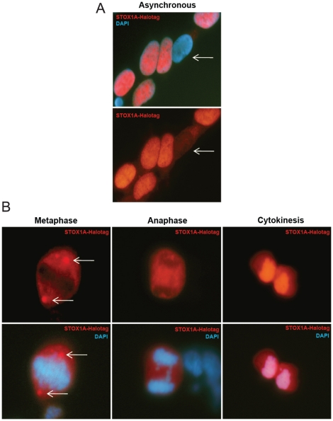
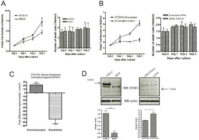
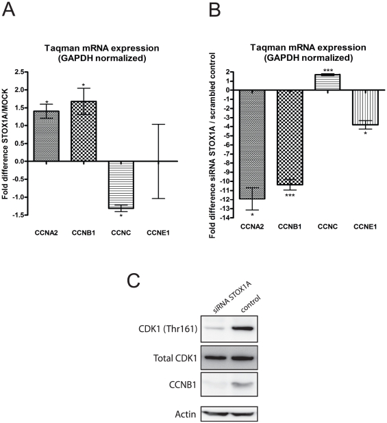
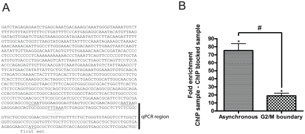
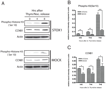
Methodology/principal findings: We found that several major cell cycle regulatory genes were differentially expressed upon STOX1A stimulation and knockdown in the neuroblastoma cell line SH-SY5Y. This includes STOX1A dependent differential regulation of cyclin B1 expression, a cyclin which is known to regulate mitotic entry during the cell cycle. The differential regulation of cyclin B1 expression by STOX1A is direct as shown with chromatin immunoprecipitation. Results furthermore suggest that mitotic entry is enhanced through the direct upregulation of cyclin B1 expression effectuated by STOX1A.

Conclusions: In conclusion we hereby show that STOX1A is directly involved in the regulation of the cell cycle.


Conflict of interest statement

Competing Interests: The authors have declared that no competing interests exist.

Figures

Figure 1
Figure 1. Expression analysis of STOX1A in stably transfected SH-SY5Y cells.
(A) Immunofluorescence shows exclusively nuclear or cytoplasmic (white arrows) staining for STOX1A-Halotag protein in STOX1A stably transfected SH-SY5Y cells. (B) Cells undergoing mitosis showing a non-overlapping STOX1A/DAPI immunofluorescence pattern during metaphase and anaphase until cytokinesis when STOX1A immunofluoresence overlaps with DAPI (DNA) immunofluorescence.
Figure 2
Figure 2. The effect of STOX1A on cell proliferation in the neuroblastoma cell-line SH-SY5Y.
(A) The proliferation curve shows significantly decreased cell proliferation for STOX1A siRNA transfected SH-SY5Y cells compared to scrambled controls after 1, 2, and 3 days in culture. (A. left bar) In parallel we did not found significant differences in cell death between STOX1A siRNA and scrambled siRNA transfected cells at each of the indicated time points. (B) The proliferation curve shows significantly increased cell proliferation for STOX1A compared to MOCK stably transfected SH-SY5Y cells after 1, 2, and 3 days in culture. (B. left bar) In parallel we did not found significant differences in cell death between STOX1A and MOCK transfected cells at each of the indicated time points. Bars are mean ± SEM. P-values for each timepoint were calculated using two-tailed unpaired t-test. (C) Overexpression and knockdown of STOX1A was determined with quantitative RT-PCR showing a mean 21 fold (mean ΔΔCt 4.4) increase in mRNA expression for STOX1A in stably transfected SH-SH5Y cells compared to the controls (Left bar) and a mean 71 fold (mean ΔΔCt 6.15) decreased mRNA expression for STOX1A siRNA transfected SH-SY5Y cells compared to the scrambled controls (right bar). Bars are mean ± SEM. *** indicate P<0.001 (one sample t-test with theoretical mean 0). N = 4, each sample was measured in triplicate. (D, Bottom left graph) Quantification of STOX1A-Halotag protein was performed using densitometry. The ratio number of obtained band intensities for STOX1A (at the expected band size of 150 Kd) divided by actin was compared to the ratio number of obtained band intensities for MOCK divided by actin for 3 independent experiments. A significant increase for the STOX1A ratio number was found compared to the MOCK ratio number. (D, Bottom right graph) Quantification of endogenous STOX1A protein was performed using densitometry. The ratio number of obtained band intensities for STOX1A siRNA (at the expected band size of 110 Kd) divided by actin was compared to the ratio number of obtained band intensities for scrambled siRNA divided by actin for 3 independent experiments. A significant increase for the STOX1A siRNA ratio number was found compared to the scrambled siRNA ratio number. P-values were calculated using two-tailed unpaired t-test, error bars represent ± SEM, * indicate P<0.05, *** indicate P<0.001. Westernblot images are a representative of at least 3 independent experiments.
Figure 3
Figure 3. STOX1A induces changes in several major cell cycle regulatory genes.
(A) The effect of stable STOX1A overexpression in the SH-SY5Y neuroblastoma cell line on four major mammalian cell cycle regulatory genes was determined with quantitative RT-PCR showing a mean 1,4 fold (mean ΔΔCt is −0,49) and mean 1,72 fold (mean ΔΔCt is −0,78) increased mRNA expression for CCNA2 and CCNB1, respectively, and a mean 1,22 fold (mean ΔΔCt is −0,29) decreased mRNA expression for CCNC in STOX1A stably transfected cell compared to their negative controls. (B) The effect of STOX1A knockdown in the SH-SY5Y neuroblastoma cell line on four major mammalian cell cycle regulatory genes was determined with quantitative RT-PCR showing a mean 11,8 fold (mean ΔΔCt is −3,56), mean 10.3 fold (mean ΔΔCt is −3,37) and mean 3,8 fold (mean ΔΔCt is −1,92) increased mRNA expression for CCNA2, CCNB1 and CCNE1 respectively, and a mean 1,7 fold (mean ΔΔCt is −0,77) increased mRNA expression for CCNC in STOX1A siRNA treated cells compared to their negative controls. (A,B) Bars are mean ± SEM. * indicate P<0.05, ** indicate P<0.01, *** indicate P<0.001 (one sample t-test with theoretical mean 0). N = 4, each sample was measured in triplicate. (C) Expression of endogenous CCNB1 protein and the active form of the CDK1 protein was determined by western blot using total cell protein extracts obtained from STOX1A siRNA and control treated SH-SY5Y cells. CCNB1 proteins were detected by a specific antibody recognizing total CCNB1 protein. CDK1 proteins were detected using an antibody detecting total CDK1 protein levels and a specific antibody recognizing the active form of CDK1 phosphorylated at threonine 161. An antibody specific for actin was used as a loading control. Westernblot image is a representative of at least 3 independent experiments.
Figure 4
Figure 4. STOX1A binds to the 5′ upstream regulatory region of the CCNB1 gene.
(A) The DNA sequence previously characterized as the 5′ upstream regulatory region of the CCNB1 gene , indicating the fragment used for quantitative PCR. (B) Chromatin immunoprecipitation was performed with asynchronized STOX1A stably transfected cells in parallel with STOX1A stably transfected cells synchronized at the G2/M phase boundary. Significantly higher enrichment for the 5′ upstream regulatory region of the CCNB1 gene was found in asynchronized cells compared to cells synchronized at the G2/M-phase boundary (left vs right bar). Bars are mean ± SEM, P-values were calculated using two-tailed unpaired t-test. # indicates P<0.001. (B, left bar). Quantitative PCR results show a mean 75 fold (mean ΔΔCt is −6.23) enrichment for the 5′ upstream regulatory region of the CCNB1 gene in STOX1A stimulated ChIP DNA, compared to their negative controls (ChIP sample vs ChIP negative sample) obtained from asynchronized STOX1A stably transfected SH-SY5Y cells. (B, right bar) Results show a mean fold 18 (mean ΔΔCt is −4,18), enrichment for the 5′ upstream regulatory region of the CCNB1 gene in STOX1A stimulated ChIP DNA, compared to their negative controls (ChIP sample vs ChIP negative sample) obtained from STOX1A stably transfected SH-SY5Y cells synchronized at the G2/M-phase boundary. Bars are mean ± SEM. Asterisks indicate P<0.05 (one sample t-test with theoretical mean 0).
Figure 5
Figure 5. Direct upregulation of CCNB1 effectuates enhanced mitotic entry.
(A) Stably transfected STOX1A and MOCK SH-SY5Y cells were synchronized in the S-phase and total cell extracts were prepared from 0, 4 and 8 h after release from the 2× thymidine block. Western blotting was performed using antibodies to Phospho-Histone H3 (Ser 10) and CCNB1. An antibody specific for actin was used as a loading control. Westernblot image is a representative of at least 3 independent experiments. (B) Westernblot (see 5A) quantification of Phospho-H3 (ser 10) was performed using densitometry. The ratio number of obtained band intensities for STOX1A divided by actin was compared to the ratio number of obtained band intensities for MOCK divided by actin for 3 independent experiments. A significant increase for the STOX1A ratio number was found at all time points compared to the MOCK ratio number. (C) Westernblot (see 5A) quantification of CCNB1 was performed using densitometry. The ratio number of obtained band intensities for STOX1A divided by actin was compared to the ratio number of obtained band intensities for MOCK divided by actin for 3 independent experiments. A significant increase for the STOX1A ratio number was found at all time points compared to the MOCK ratio number. P-values were calculated using two-tailed unpaired t-test, error bars represent ± SEM, * indicate P<0.05, ** indicate P<0.01, *** indicate P<0.001.

Similar articles

Cited by

References

    1. Bloom J, Cross FR. Multiple levels of cyclin specificity in cell-cycle control. Nat Rev Mol Cell Biol. 2007;8:149–60. - PubMed
    1. Woo RA, Poon RY. Cyclin-dependent kinases and S phase control in mammalian cells. Cell cycle. 2003;2:316–24. - PubMed
    1. Porter LA, Donoghue DJ. Cyclin B1 and CDK1: nuclear localization and upstream regulators. Prog Cell Cycle Res. 2003;5:335–47. - PubMed
    1. Murray AW. Recycling the cell cycle: cyclins revisited. Cell. 2004;116:221–34. - PubMed
    1. Pines J. Mitosis: a matter of getting rid of the right protein at the right time. Trends Cell Biol. 2006;16:55–63. - PubMed

MeSH terms