. 2012;7(1):e29914.
doi: 10.1371/journal.pone.0029914. Epub 2012 Jan 11.

Persistence of Borrelia burgdorferi in rhesus macaques following antibiotic treatment of disseminated infection

Affiliations

Persistence of Borrelia burgdorferi in rhesus macaques following antibiotic treatment of disseminated infection

Monica E Embers et al. PLoS One. 2012.

Erratum in

  • PLoS One. 2012;7(4):10.1371/annotation/4cafed66-fb84-4589-a001-131d9c50aea6
  • PLoS One. 2013;8(9). doi:10.1371/annotation/f84663e3-0a2c-4243-8f97-3a58133c1b0f
  • Correction: Persistence of Borrelia burgdorferi in Rhesus Macaques following Antibiotic Treatment of Disseminated Infection.
    Embers ME, Barthold SW, Borda JT, Bowers L, Doyle L, Hodzic E, Jacobs MB, Hasenkampf NR, Martin DS, Narasimhan S, Phillippi-Falkenstein KM, Purcell JE, Ratterree MS, Philipp MT. Embers ME, et al. PLoS One. 2013 Sep 24;8(9):10.1371/annotation/f84663e3-0a2c-4243-8f97-3a58133c1b0f. doi: 10.1371/annotation/f84663e3-0a2c-4243-8f97-3a58133c1b0f. eCollection 2013. PLoS One. 2013. PMID: 24116243 Free PMC article.

Abstract

The persistence of symptoms in Lyme disease patients following antibiotic therapy, and their causes, continue to be a matter of intense controversy. The studies presented here explore antibiotic efficacy using nonhuman primates. Rhesus macaques were infected with B. burgdorferi and a portion received aggressive antibiotic therapy 4-6 months later. Multiple methods were utilized for detection of residual organisms, including the feeding of lab-reared ticks on monkeys (xenodiagnosis), culture, immunofluorescence and PCR. Antibody responses to the B. burgdorferi-specific C6 diagnostic peptide were measured longitudinally and declined in all treated animals. B. burgdorferi antigen, DNA and RNA were detected in the tissues of treated animals. Finally, small numbers of intact spirochetes were recovered by xenodiagnosis from treated monkeys. These results demonstrate that B. burgdorferi can withstand antibiotic treatment, administered post-dissemination, in a primate host. Though B. burgdorferi is not known to possess resistance mechanisms and is susceptible to the standard antibiotics (doxycycline, ceftriaxone) in vitro, it appears to become tolerant post-dissemination in the primate host. This finding raises important questions about the pathogenicity of antibiotic-tolerant persisters and whether or not they can contribute to symptoms post-treatment.


Conflict of interest statement

Competing Interests: The authors have declared that no competing interests exist.

Figures

Figure 1
Figure 1. Experimental design for assessment of treatment efficacy in the late, disseminated phase of infection (Experiment 1).
A) diagram of animal groups, showing inoculation (B. burgdorferi or sham) and treatment groups (treated with antibiotics or untreated); and B) the time line of the study.
Figure 2
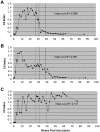
Figure 2. Patterns of antibody response to the C6 peptide in infected animals.
The C6 index measured as a function of time PI with B. burgdorferi followed three distinct patterns in Experiment 1. Each graph is the longitudinal antibody response to C6 from one representative animal for each pattern. In treated (n = 12) animals (A) the response declined sharply, almost to background levels, within the antibiotic administration periods (solid vertical lines, weeks 27–31 for ceftriaxone, weeks 31–39 for doxycycline). In sham-treated animals (B, C) the response either declined, but independently from the sham treatment intervals, to eventually reach background level (B, n = 7), or oscillated throughout the study period without ever declining steadily (C, n = 5). The pattern in (C) corresponds to the C6-positive animals, indicated in Table 1. The dotted lines in B and C indicate the sham-treatment intervals. The index cut-off value was calculated as described in Material and Methods.
Figure 3
Figure 3. Images of tissue inflammation and B. burgdorferi antigen in tissues from animals treated in the late, disseminated phase (Experiment 1).
For antigen detection, samples of tissue were stained for fluorescent detection (IFA) with anti-B. burgdorferi monoclonal (see Materials and Methods) antibody. (A) Hematoxylin &Eosin stain showing monocytic and lymphocytic infiltrate in a heart section (20×) of a treated animal (AM38). (B) Image of positive IFA staining from the heart tissue of the same animal.
Figure 4
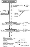
Figure 4. Experimental outline and sample collection for assessment of treatment efficacy in the disseminated phase of infection (Experiment 2).
Figure 5
Figure 5. Spirochetes recovered by xenodiagnosis from animals treated in the disseminated phase of infection.
Images from direct fluorescent staining of B. burgdorferi spirochetes found in xenodiagnostic tick midgut culture (A) or tick midgut preparation (B) from treated animals GA59 and GB56, respectively.
Figure 6
Figure 6. Nucleic acid detection of B. burgdorferi (Experiment 2).
Detection by PCR or RT-PCR using primers for the indicated genes from: A) skin biopsy samples; B) xenodiagnostic ticks; C) organ tissue culture pellets; and D) directly from tissues known to harbor the spirochetes. Animal numbers in bold are of those that were treated. Asterisks indicate clear positive amplimers.sk = skin; h t = heart; bl = bladder; spl = pleen. Labels include: M = marker (100 bp ladder); (+) = B.burgdorferi, strain B31 DNA/RNA; ssc = spirochete strain negative control (Bb strain JD1 DNA); ntc = no template control; tnc = tick negative control; uim = uninfected monkey DNA/RNA.

References

    1. Steere AC, Coburn J, Glickstein L. The emergence of Lyme disease. Journal of Clinical Investigation. 2004;113:1093–1101. - PMC - PubMed
    1. Wormser Gary P, Dattwyler Raymond J, Shapiro Eugene D, Halperin John J, Steere A, et al. The Clinical Assessment, Treatment, and Prevention of Lyme Disease, Human Granulocytic Anaplasmosis, and Babesiosis: Clinical Practice Guidelines by the Infectious Diseases Society of America. Clinical Infectious Diseases. 2006;43:1089–1134. - PubMed
    1. Asch ES, Bujak DI, Weiss M, Peterson MG, Weinstein A. Lyme disease: an infectious and postinfectious syndrome. Journal of Rheumatology. 1994;21:454–461. - PubMed
    1. Shadick NA, Phillips CB, Logigian EL, Steere AC, Kaplan RF, et al. The long-term clinical outcomes of Lyme disease. A population-based retrospective cohort study. Annals of Internal Medicine. 1994;121:560–567. - PubMed
    1. Steere AC, Levin RE, Molloy PJ, Kalish RA, Abraham JH, 3rd, et al. Treatment of Lyme arthritis. Arthritis & Rheumatism. 1994;37:878–888. - PubMed

Publication types

MeSH terms