. 2012;7(1):e29933.
doi: 10.1371/journal.pone.0029933. Epub 2012 Jan 10.

Epigallocatechin-3-gallate as a potential therapeutic drug for TTR-related amyloidosis: "in vivo" evidence from FAP mice models

Affiliations

Epigallocatechin-3-gallate as a potential therapeutic drug for TTR-related amyloidosis: "in vivo" evidence from FAP mice models

Nelson Ferreira et al. PLoS One. 2012.

Abstract

Background: Familial amyloidotic polyneuropathy (FAP) is a neurodegenerative disease caused by the extracellular deposition of mutant transthyretin (TTR), with special involvement of the peripheral nervous system (PNS). Currently, hepatic transplantation is considered the most efficient therapy to halt the progression of clinical symptoms in FAP since more than 95% of TTR is produced by the liver. However, less invasive and more reliable therapeutic approaches have been proposed for FAP therapy, namely based on drugs acting as inhibitors of amyloid formation or as amyloid disruptors. We have recently reported that epigallocatechin-3-gallate (EGCG), the most abundant catechin in green tea, is able to inhibit TTR aggregation and fibril formation, "in vitro" and in a cellular system, and is also able to disrupt pre-formed amyloid fibrils "in vitro".

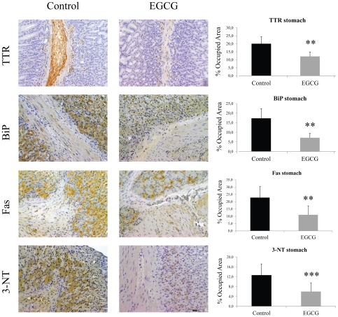
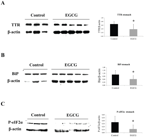
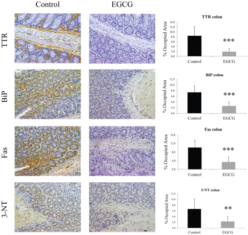
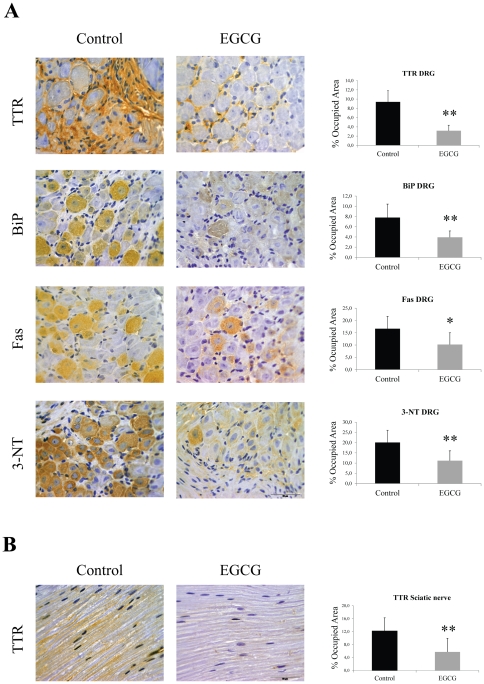
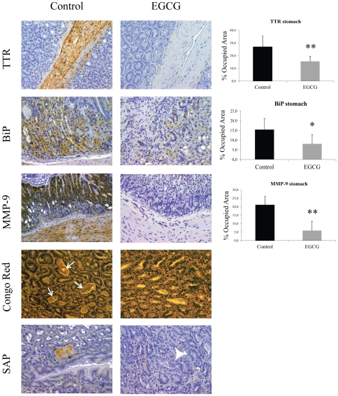
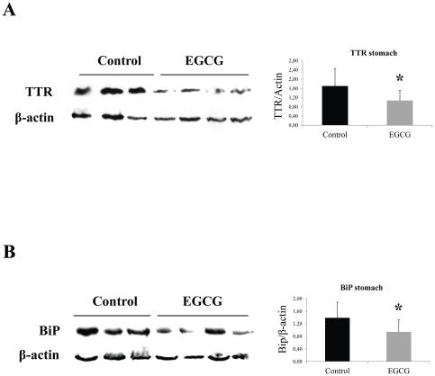
Methodology and principal findings: In the present study, we assessed the effect of EGCG subchronic administration on TTR amyloidogenesis "in vivo", using well characterized animal models for FAP. Semiquantitative immunohistochemistry (SQ-IHC) and Western blot analysis of mice tissues after treatment demonstrated that EGCG inhibits TTR toxic aggregates deposition in about 50% along the gastrointestinal tract (GI) and peripheral nervous system (PNS). Moreover EGCG treatment considerably lowered levels of several biomarkers associated with non-fibrillar TTR deposition, namely endoplasmic reticulum (ER)-stress, protein oxidation and apoptosis markers. Treatment of old FAP mice with EGCG resulted not only in the decrease of non-fibrillar TTR deposition but also in disaggregation of amyloid deposits. Consistently, matrix metalloproteinase (MMP)-9 and serum amyloid P component (SAP), both markers of amyloid deposition, were also found reduced in treated old FAP mice.

Conclusions and significance: The dual effect of EGCG both as TTR aggregation inhibitor and amyloid fibril disruptor together with the high tolerability and low toxicity of EGCG in humans, point towards the potential use of this compound, or optimized derivatives, in the treatment of TTR-related amyloidoses.


Conflict of interest statement

Competing Interests: The authors have declared that no competing interests exist.

Figures

Figure 1
Figure 1. EGCG treatment decreases TTR deposition and associated biomarkers in stomach of hTTR V30M mice.
Representative immunohistochemistry of TTR, BiP, Fas and 3-nitrotyrosine in stomach of hTTR V30M mice treated with EGCG (right panels; n = 10) and age-matched controls (left panels; n = 6). Scale bar 100 µm. Histogram: quantification of immunohistochemical images is represented as percentage of occupied area ± SD (**P<0.01; ***P<0.005).
Figure 2
Figure 2. EGCG treatment decreases ER-stress markers associated with TTR deposition in stomach of hTTR V30M mice.
Representative (A) anti-TTR, (B) anti-BiP and (C) anti-P-eIF2α Western blots of stomachs from TTR V30M mice treated with EGCG and non-treated mice. Histogram: normalized TTR/β-actin, BiP/β-actin and anti-P-eIF2α/β-actin density quantifications ±SD (*P<0.05).
Figure 3
Figure 3. EGCG treatment decreases TTR deposition and associated biomarkers in colon of hTTR V30M mice.
Representative immunohistochemistry of TTR, BiP, Fas and 3-nitrotyrosine in colon of hTTR V30M mice treated with EGCG (right panels; n = 10) and age-matched non-treated animals (left panels; n = 6). Scale bar 100 µm. Histogram: quantification of immunohistochemical images is represented as percentage of occupied area ± SD (**P<0.01; ***P<0.005).
Figure 4
Figure 4. EGCG treatment decreases TTR deposition and associated biomarkers in the peripheral nervous system (PNS) of hTTR V30M/HSF mice.
(A) Representative immunohistochemistry of TTR, BiP, Fas and 3-nitrotyrosine in dorsal root ganglia of hTTR V30M/HSF treated with EGCG for 6 weeks (right panels; n = 8) and age-matched controls (left panels; n = 5). Scale bar 50 µm. (B) Immunohistochemistry of TTR in sciatic nerve of hTTR V30M treated mice (right panels; n = 8) and age-matched non-treated animals (left panels; n = 5). Scale bar 50 µm. Histograms: quantification of the levels of the referred markers expressed as percentage of occupied area ± SD (*P<0.05; **P<0.01).
Figure 5
Figure 5. EGCG treatment decreases TTR deposition and associated biomarkers in the gastrointestinal tract of old hTTR V30M mice.
Immunohistochemistry and Congo Red analysis of stomachs from aged hTTR V30M mice treated with EGCG (right panels; n = 11) and aged-matched controls (left panels; n = 8). The top 3 panels represent TTR, BiP and MMP-9 immunohistochemistry. The bottom two panels are representative Congo Red staining (white arrows pointing at CR birefringence in a non-treated mouse) and mouse SAP immunohistochemistry. Scale bar 100 µm. Histograms: quantification of the levels of the referred markers expressed as percentage of occupied area ± SD (*P<0.05; **P<0.01).
Figure 6
Figure 6. EGCG treatment decreases TTR amyloid deposition in the gastrointestinal tract of old hTTR V30M mice.
Representative (A) anti-TTR and (B) anti-BiP Western blots of stomachs from aged hTTR V30M mice treated with EGCG and non-treated mice. Histogram: normalized TTR/β-actin and BiP/β-actin density quantifications ±SD (*P<0.05).

References

    1. Saraiva MJ, Birken S, Costa PP, Goodman DS. Amyloid fibril protein in familial amyloidotic polyneuropathy, Portuguese type. Definition of molecular abnormality in transthyretin (prealbumin). J Clin Invest. 1984;74:104–119. - PMC - PubMed
    1. Benson MD, Kincaid JC. The molecular biology and clinical features of amyloid neuropathy. Muscle Nerve. 2007;36(4):411–23. - PubMed
    1. Quintas A, Vaz DC, Cardoso I, Saraiva MJ, Brito RM. Tetramer dissociation and monomer partial unfolding precedes protofibril formation in amyloidogenic transthyretin variants. J Biol Chem. 2001;276(29):27207–13. - PubMed
    1. Cardoso I, Goldsbury CS, Müller SA, Olivieri V, Wirtz S, et al. Transthyretin fibrillogenesis entails the assembly of monomers: a molecular model for in vitro assembled transthyretin amyloid-like fibrils. J Mol Biol. 2002;317(5):683–95. - PubMed
    1. Sousa MM, Cardoso I, Fernandes R, Guimaraes A, Saraiva MJ. Deposition of transthyretin in early stages of familial amyloidotic polyneuropathy: evidence for toxicity of nonfibrillar aggregates. Am J Pathol. 2001;159:1993–2000. - PMC - PubMed

Publication types

MeSH terms