. 2012;7(1):e30063.
doi: 10.1371/journal.pone.0030063. Epub 2012 Jan 11.

Rescue of dystrophic skeletal muscle by PGC-1α involves a fast to slow fiber type shift in the mdx mouse

Affiliations

Rescue of dystrophic skeletal muscle by PGC-1α involves a fast to slow fiber type shift in the mdx mouse

Joshua T Selsby et al. PLoS One. 2012.

Abstract

Increased utrophin expression is known to reduce pathology in dystrophin-deficient skeletal muscles. Transgenic over-expression of PGC-1α has been shown to increase levels of utrophin mRNA and improve the histology of mdx muscles. Other reports have shown that PGC-1α signaling can lead to increased oxidative capacity and a fast to slow fiber type shift. Given that it has been shown that slow fibers produce and maintain more utrophin than fast skeletal muscle fibers, we hypothesized that over-expression of PGC-1α in post-natal mdx mice would increase utrophin levels via a fiber type shift, resulting in more slow, oxidative fibers that are also more resistant to contraction-induced damage. To test this hypothesis, neonatal mdx mice were injected with recombinant adeno-associated virus (AAV) driving expression of PGC-1α. PGC-1α over-expression resulted in increased utrophin and type I myosin heavy chain expression as well as elevated mitochondrial protein expression. Muscles were shown to be more resistant to contraction-induced damage and more fatigue resistant. Sirt-1 was increased while p38 activation and NRF-1 were reduced in PGC-1α over-expressing muscle when compared to control. We also evaluated if the use a pharmacological PGC-1α pathway activator, resveratrol, could drive the same physiological changes. Resveratrol administration (100 mg/kg/day) resulted in improved fatigue resistance, but did not achieve significant increases in utrophin expression. These data suggest that the PGC-1α pathway is a potential target for therapeutic intervention in dystrophic skeletal muscle.


Conflict of interest statement

Competing Interests: The authors have declared that no competing interests exist.

Figures

Figure 1
Figure 1. Virally-mediated gene transfer.
Six weeks following PGC-1α gene delivery total (PGC-1α) and viral (V-PGC-1α) expression was increased in treated limbs compared to untreated limbs (n = 6/group). * indicates p<0.05.
Figure 2
Figure 2. Relative force reduction in 6 wk old EDL's following lengthening contractions.
PGC-1α over-expressing limbs were better able to maintain force during lengthening contractions when compared to control limbs (n = 13/group). Data from age matched C57 animals is included for reference purposes. * indicates p<0.05. Con – contraction.
Figure 3
Figure 3. PGC-1α protects against muscle fatigue.
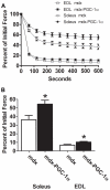
Muscle fatigue curves in the soleus (n = 6/group) and EDL (n = 7/group) (A) during 10 minutes of a fatigue protocol where muscles are contracted every second for 10 minutes. Force of the final contraction (B) was significantly higher in the soleus and EDL muscles over-expressing PGC-1α when compared to control muscle. * indicates p<0.05.
Figure 4
Figure 4. PGC-1α over-expression reduces disease-related muscle injury.
10× micrographs of six week old soleus muscles following H and E staining (A and B). (C) The total areas of necrotic, H&E negative, or regenerating cells were quantified and is expressed as a percent of the total soleus area. In addition, laminin was detected with a fluorescently labeled antibody (not shown) in order to determine central nucleation. (D) PGC-1α caused a reduction in central nucleation. N = 5/group; * indicates p<0.05.
Figure 5
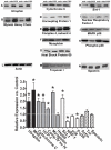
Figure 5. Protein expression following 6 weeks of PGC-1α over-expression.
Slow genes are shown in black (utrophin – A, slow myosin heavy chain – B) oxidative genes are shown in gray (cytochrome C – C, uncoupling protein-1 – D, complex IV subunit IV – E, myoglobin – F, Hsp 60 – G), pathway genes are shown in white (Sirt-1 – H, nuclear respiratory factor -1 – I, p38 – J, phospho-p38 – K), and controls have diagonal lines (Actin – L, Troponin – M, Spectrin – N). Relative change compared to control limbs (n = 6/group) (O). * indicates p<0.05 compared to control limbs.
Figure 6
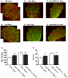
Figure 6. Myosin fiber type distribution in treated and control soleus muscles.
Histological sections of soleus (10×) were exposed to an antibody against the slow isoform of myosin heavy chain (A). A C57 (healthy) section is included as reference (Left). The control limb (middle) shows moderate type I content. The corresponding injected limb (Right) clearly has an elevation in type I content. In serial sections, we also evaluated type II myosin heavy chain content (B). A C57 (healthy) section is included for reference (Left). The control limb (middle) shows high levels of type II expression. PGC-1α caused a reduced expression of type II fibers (Right). The absolute fiber numbers from each section expressing type I and II fibers were counted and recorded (C). PGC-1α caused a shift toward type I fibers and away from type II fibers. These were then made relative to total fiber number (D). Once again, a type I shift is observed. N = 9/group; * indicates p<0.05.
Figure 7
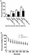
Figure 7. PGC-1α partially maintains diaphragmatic function six months following gene transfer.
Neonatal mdx mice were injected in the sub-xyphiod region in order to cause diaphragmatic infection and sacrificed 6 mo later. Diaphragms over-expressing PGC-1α were more resistant to contraction induced injury (n = 4 Con; n = 7 PGC-1α) (A; Contraction 4 – p = 0.08), however, fatigue resistance was similar between groups (n = 7/group) (B). * indicates p<0.05.
Figure 8
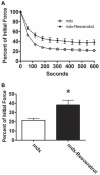
Figure 8. Resveratrol supplementation increased fatigue resistance in dystrophic skeletal muscle.
One month old mdx mice were fed a diet containing 100 mg/kg/day resveratrol or control diet for eight weeks. Resveratrol feeding increased fatigue resistance in the soleus (A) and force generated during the final contraction (B ) was higher in treated animals when compared to control. n = 8 Con; n = 6 Res; * indicates p<0.05.
Figure 9
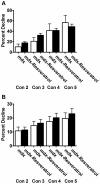
Figure 9. Resveratrol supplementation did not improve resistance to contraction induced injury.
Feeding a diet containing 100 mg/kg/day resveratrol for eight weeks did not improve resistance to contraction induced injury in (A) the EDL (n = 8 Con; n = 6 Res) or (B) the soleus (n = 8/group) when compared to control.

References

    1. Nowak KJ, Davies KE. Duchenne muscular dystrophy and dystrophin: pathogenesis and opportunities for treatment. EMBO Rep. 2004;5:872–876. - PMC - PubMed
    1. Petrof BJ, Shrager JB, Stedman HH, Kelly AM, Sweeney HL. Dystrophin protects the sarcolemma from stresses developed during muscle contraction. Proc Natl Acad Sci U S A. 1993;90:3710–3714. - PMC - PubMed
    1. Hopf FW, Turner PR, Steinhardt RA. Calcium misregulation and the pathogenesis of muscular dystrophy. Subcell Biochem. 2007;45:429–464. - PubMed
    1. Whitehead NP, Yeung EW, Allen DG. Muscle damage in mdx (dystrophic) mice: role of calcium and reactive oxygen species. Clin Exp Pharmacol Physiol. 2006;33:657–662. - PubMed
    1. Spencer MJ, Croall DE, Tidball JG. Calpains are activated in necrotic fibers from mdx dystrophic mice. J Biol Chem. 1995;270:10909–10914. - PubMed

Publication types

MeSH terms

LinkOut - more resources