. 2012;7(1):e30122.
doi: 10.1371/journal.pone.0030122. Epub 2012 Jan 11.

Region specific and worldwide distribution of collagen-binding M proteins with PARF motifs among human pathogenic streptococcal isolates

Affiliations

Region specific and worldwide distribution of collagen-binding M proteins with PARF motifs among human pathogenic streptococcal isolates

Silvana Reissmann et al. PLoS One. 2012.

Abstract

Some of the variety of Streptococcus pyogenes and Streptococcus dysgalactiae ssp. equisimilis (SDSE) M proteins act as collagen-binding adhesins that facilitate acute infection. Moreover, their potential to trigger collagen autoimmunity has been implicated in the pathogenesis of acute rheumatic fever and attributed to a collagen-binding motif called PARF (peptide associated with rheumatic fever). For the first time we determine the rate of clinical isolates with collagen-binding M proteins that use a PARF motif (A/T/E)XYLXX(L/F)N in a defined geographic region, Vellore in South India. In this region both, incidence of streptococcal infections and prevalence of acute rheumatic fever are high. M proteins with PARF motif conferred collagen-binding activity to 3.9% of 153 S. pyogenes and 10.6% of 255 SDSE clinical isolates from Vellore. The PARF motif occurred in three S. pyogenes and 22 SDSE M protein types. In one of the S. pyogenes and five of the SDSE M proteins that contained the motif, collagen-binding was impaired, due to influences of other parts of the M protein molecule. The accumulated data on the collagen binding activity of certain M protein types allowed a reanalysis of published worldwide emm-typing data with the aim to estimate the rates of isolates that bind collagen via PARF. The results indicate that M proteins, which bind collagen via a PARF motif, are epidemiologically relevant in human infections, not only in Vellore. It is imperative to include the most relevant collagen-binding M types in vaccines. But when designing M protein based vaccines it should be considered that collagen binding motifs within the vaccine antigen remain potential risk factors.


Conflict of interest statement

Competing Interests: The authors have declared that no competing interests exist.

Figures

Figure 1
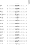
Figure 1. Known S. pyogenes and SDSE collagen-binding M proteins harboring PARF.
M protein designations are given on the left. The PARF motif is shown in black letters and eleven flanking amino acids on both the N-terminal and C-terminal side in grey letters. Collagen-binding activity is indicated on the right with ‘+’ when positive or with ‘−’ when negative. Numbers flanking the sequence indicate its position within the mature protein. Amino acids conserved or typical for PARF are shown in bold. Identical amino acids (*), conserved substitutions (:), and semi-conserved substitutions (.) are indicated as determined by multiple alignment. Amino acids with basic side chains in positions 2, 5 or 6 are highlighted in grey.
Figure 2
Figure 2. Collagen binding to M3, M31 and M55.
(A) S. pyogenes strains that expressed the M protein M3.22 (white bars) or M3.23 (black bars) or (B) S. pyogenes strains that expressed M protein M55 (white bars) or M31 (black bars) and a SDSE strains that expressed M31.5 (grey bar) were compared for binding of collagen IV. Strain designations are indicated in the abscissa. The binding capacity was expressed as the bound percentage of added radiolabelled collagen IV. The average of three experiments is depicted with standard deviation. In (A) PARF sequences of the M proteins are shown above. (C and D) SPR sensograms of the interaction between immobilized human collagen IV (400 RU) and M protein M31.5 (C) or M55 (D) as analyte in the liquid phase were recorded upon injection of the analyte in concentrations that are indicated on the right in nM. Injections started at t = 0 s and ended at t = 120 s.
Figure 3
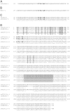
Figure 3. M proteins with predicted PARF motifs that did not bind collagen.
Protein sequences of four types or groups of M proteins that harbored a prototypical PARF motif but had members that did not bind collagen (A–D). M protein designations are given on the left together with the results of collagen IV binding experiments that are given in brackets with ‘+’ for binding and ‘−’ for non-binding proteins. Numbers that flank the sequence indicate its position within the mature M protein without signal peptide. In C and D identical amino acids (*), conserved substitutions (:), and semi-conserved substitutions (.) in two or three compared sequences are marked below. Amino acids characteristic for PARF are printed in bold. In D residues that distinguish the collagen-IV binding stG120.1 from the non-binding stG120.0 and stGM220 are highlighted in grey.
Figure 4
Figure 4. Prediction of N-terminal coiled-coil structures in PARF-positive M proteins.
(A) Coiled-coil structure prediction for the N-terminal sequences of the PARF-positive M proteins is given as P-scores vs. the amino acid position relative from the PARF motif with the first amino acid of the motif being position 1. The figure shows the superposition of all curves without indicating protein designations, however, collagen-binding M proteins are shown in blue and non-binding M proteins are shown in red. Single curves for all M proteins are provided as a supplementary figure (Figure S1). The position of the PARF motif is highlighted in light blue. A black dashed line indicates the threshold value for coiled-coil prediction. Values below 0.025 indicate a coiled-coil structure. The prediction separates the M proteins into two classes; one class with (B) and one without (C) an N-terminal coiled-coil domain. They are shown in schematic representations that are not drawn to scale. B and C are not based on structural data other than coiled-coil prediction. The PARF motif is highlighted in red. The C-terminal end is labelled (-COOH).
Figure 5
Figure 5. Prediction of coiled-coil structures in PARF-positive M proteins.
Coiled-coil structure prediction for the N-terminal sequences of the PARF-positive M proteins stG120.0 (red dotted line), stG120.1 (light blue solid line) and stGM220 (black dotted line) is given as P-scores vs. the amino acid position relative from the PARF motif with the first amino acid of the motif being position 1. Isolates with M protein stG120.1 bind collagen IV, while strains that carry one of the other two M proteins do not. The position of the PARF motif is highlighted in light blue. A black dashed line indicates the threshold value for coiled-coil prediction. Values below 0.025 indicate a coiled-coil structure.

References

    1. Cunningham MW. Pathogenesis of group A streptococcal infections. Clin Microbiol Rev. 2000;13:470–511. - PMC - PubMed
    1. Efstratiou A. Pyogenic streptococci of Lancefield groups C and G as pathogens in man. Soc Appl Bacteriol Symp Ser. 1997;26:72S–79S. - PubMed
    1. Baracco GJ, Bisno AL. Group C and group G streptococcal infections: epidemiologic and clinical aspects. In: Fischetti VA, editor. Gram-positive pathogens. 2nd ed. Washington, D.C.: ASM Press; 2006. pp. 223–229.
    1. Haidan A, Talay SR, Rohde M, Sriprakash KS, Currie BJ, et al. Pharyngeal carriage of group C and group G streptococci and acute rheumatic fever in an Aboriginal population. Lancet. 2000;356:1167–1169. - PubMed
    1. Wannamaker LW. The chain that links the heart to the throat. Circulation. 1973;48:9–18. - PubMed

Publication types