. 2012 Jan;6(1):e1466.
doi: 10.1371/journal.pntd.0001466. Epub 2012 Jan 10.

Orientia tsutsugamushi in human scrub typhus eschars shows tropism for dendritic cells and monocytes rather than endothelium

Affiliations

Orientia tsutsugamushi in human scrub typhus eschars shows tropism for dendritic cells and monocytes rather than endothelium

Daniel H Paris et al. PLoS Negl Trop Dis. 2012 Jan.

Abstract

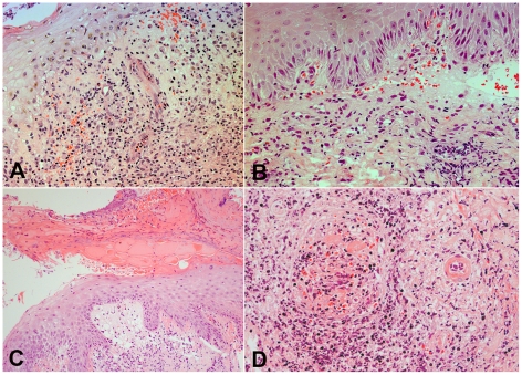
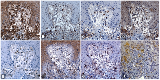
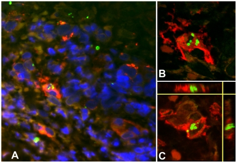
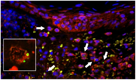
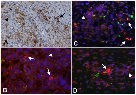
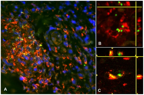
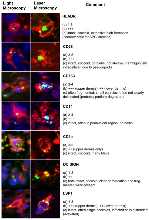
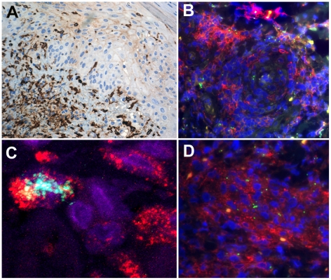
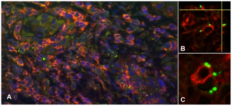
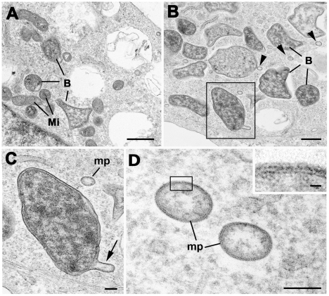
Scrub typhus is a common and underdiagnosed cause of febrile illness in Southeast Asia, caused by infection with Orientia tsutsugamushi. Inoculation of the organism at a cutaneous mite bite site commonly results in formation of a localized pathological skin reaction termed an eschar. The site of development of the obligate intracellular bacteria within the eschar and the mechanisms of dissemination to cause systemic infection are unclear. Previous postmortem and in vitro reports demonstrated infection of endothelial cells, but recent pathophysiological investigations of typhus patients using surrogate markers of endothelial cell and leucocyte activation indicated a more prevalent host leucocyte than endothelial cell response in vivo. We therefore examined eschar skin biopsies from patients with scrub typhus to determine and characterize the phenotypes of host cells in vivo with intracellular infection by O. tsutsugamushi, using histology, immunohistochemistry, double immunofluorescence confocal laser scanning microscopy and electron microscopy. Immunophenotyping of host leucocytes infected with O. tsutsugamushi showed a tropism for host monocytes and dendritic cells, which were spatially related to different histological zones of the eschar. Infected leucocyte subsets were characterized by expression of HLADR+, with an "inflammatory" monocyte phenotype of CD14/LSP-1/CD68 positive or dendritic cell phenotype of CD1a/DCSIGN/S100/FXIIIa and CD163 positive staining, or occasional CD3 positive T-cells. Endothelial cell infection was rare, and histology did not indicate a widespread inflammatory vasculitis as the cause of the eschar. Infection of dendritic cells and activated inflammatory monocytes offers a potential route for dissemination of O. tsutsugamushi from the initial eschar site. This newly described cellular tropism for O. tsutsugamushi may influence its interaction with local host immune responses.


Conflict of interest statement

The authors have declared that no competing interests exist.

Figures

Figure 1
Figure 1. Histological features of eschars: Intra- and subepidermal changes.
Panels A and B. Intraepidermal splitting and sub-epidermal blistering with basal vacuolar changes, inflammatory exocytosis and focal red blood cell extravasation in the superficial dermis. Patient TM2644 (Panel A) and TM2646 (Panel B), magnification ×400. Panel C. Acute inflammatory sloughing and artefactual separation of the epidermis. A demarcated area of ulceration and necrosis of the overlying epidermis with subepidermal vacuolation, necrosis and mononuclear infiltration, patient TM2663, magnification ×250. Panel D. Perivascular infiltrates and leucocytoclastic vasculitis. On the right hand side the cross-section of an arteriole, on the left, a venous vessel with intravascular obliteration and secondary thrombus. There is prominent perivascular cuffing with mononuclear cells and fibrinoid necrosis of the arteriolar wall. Patient TM2193, magnification ×250. All panels Haematoxylin & Eosin (H&E) staining.
Figure 2
Figure 2. Immunohistochemical phenotyping of cellular infiltrates at the dermal-epidermal border.
Serial sections at the dermal-epidermal border, demonstrate that APCs are the dominant cell type adjacent to the central necrotic zone of the eschar. Immunophenotyping by MHC class II receptor for APCs (Panel A: HLADR), macrophages (B: CD68) and DCs (C: DC-SIGN, D: FXIIIa, G: S100 and H: LSECtin), neutrophils (E: CD15) and lymphocytes (F: CD4). The proportion of dendritic cells was higher in the dermal-epidermal border zone, than in the deeper dermis. Subepidermal vacuolization is prominent. Patient TM2663, magnification ×400, counterstained with haematoxylin, peroxidase immunostaining in brown.
Figure 3
Figure 3. Multiple intracellular O. tsutsugamushi within antigen presenting cells (APCs) in the superficial dermis.
APCs are characterised by MHC class II receptor (HLADR) positivity and were associated with intracellular O. tsutsugamushi in admission samples of an eschar from a patient with acute scrub typhus. Panel A: HLADR-positive cells in red, O. tsutsugamushi in green. Panels B and C: Laser Scanning Micrographs. Panel B depicts the same infected cell as in Panel A as a 0.3 µm thin section. Panel C shows a 3D stack projection (of 0.3 µm thin sections) of intracytosolic O. tsutsugamushi (in green), with small, high-density granules with high refractive index, staining positively for O. tsutsugamushi antigen. Patient TM2193, magnification ×400, LSM inserts ×1000, double-immunolabeling: HLADR in red, O. tsutsugamushi in green and DAPI nuclear counterstain in blue.
Figure 4
Figure 4. Multiple intracellular O. tsutsugamushi within CD1a positive Langerhans cells in the superficial dermis.
Local proliferation of Langerhans cells in the epidermis, with a distinct CD1a+ nodule formation and O. tsutsugamushi infected cells located in the dermis (white arrows) of an eschar from a patient with acute scrub typhus. Erythrocytes appear yellow due to autofluorescence. Insert: Laser Scanning Micrograph (LSM), 0.3 µm thin section visualizing the intracytosolic location of O. tsutsugamushi, accompanied by small intracellular microparticles with high refractile index. Patient TM2193, magnification ×400, insert ×1000. Labeling: CD1a in red, O. tsutsugamushi in green and DAPI nuclear counterstain in blue.
Figure 5
Figure 5. Dermal dendritic cells in the dermal-epidermal zone of the eschar.
Panel A: High numbers of DCSIGN+ cells in the dermal-epidermal zone of the eschar. The variance of staining-intensity of individual positive cells ranged from strong (long black arrow) to weak (short black arrow). Patient TM2644, magnification ×400, peroxidase immunostaining in brown. Panels B, C and D: Immuno-double-labelling with HLADR (Panel B), DC-SIGN (Panel C) and LSP-1 (Panel D) stained red. The signal intensity of these markers in infected cells with O. tsutsugamushi (in green) appeared more pronounced (long white arrows) than in uninfected cells (short white arrows). Patient TM2193 (Panel B), patient TM2508 (Panel C and D). Magnification ×400, blue nuclear counterstaining with DAPI.
Figure 6
Figure 6. O. tsutsugamushi co-localize with CD68 positive macrophages in perivascular infiltrates.
Panel A: Perivascular cuff formation and clusters of predominantly CD68+ macrophages, co-localizing closely with O. tsutsugamushi. Patient TM2193, magnification ×400. Magnification ×400, insert ×1000, double-immunolabeling: CD1a in red, O. tsutsugamushi in green and DAPI nuclear counterstain in blue. Panels B and C: Laser Scanning Micrographs with 3D stack projections of multiple 0.3 µm thin sections, depicting intact O. tsutsugamushi associated with CD68+ macrophages (panel B). Unambigous intracellular location of O. tsutsugamushi in a CD68+ cell (panel C). Patient TM2508, magnification ×1000, CD68 labeled in red, O. tsutsugamushi in green.
Figure 7
Figure 7. Overview of host-cell phenotypes associated with O. tsutsugamushi infection in vivo in human eschars.
Characteristics were defined as follows: (a) Number of O. tsutsugamushi per cell per section. (b) Proportion of cells in infiltrate. (c) Morphological aspects of intracellular O. tsutsugamushi. Infiltrates were estimated in a semi-quantitative manner: The extent of immunopositivity in the inflammatory cells for specific cell markers was graded in the following manner: (−) in 0%, (±) in <1%, (+) in 1%–25%, (++) in 26%–50%, (+++) in >50% and (++++) in >75% of infiltrated cells.
Figure 8
Figure 8. O. tsutsugamushi-infected monocytes/macrophages in dense perivascular infiltrates express CD14 and scavenger receptor CD163.
Panel A: High density of CD163+ monocye/macrophages at the dermal-epidermal border and after intraepidermal translocation adjacent to the inoculation of O. tsutsugamushi. Patient TM2391 (panel A), magnification ×200, peroxidase immunostaining in brown. Panels B and D: Perivascular CD14+ monocyte/macrophages conglomerates containing O. tsutsugamushi. Cross-section of perivascular cuff formation (panel B) and longitudinal section (panel D). Patient TM2644, magnification ×400, double-immunolabeling: CD14 in red, O. tsutsugamushi in green and DAPI nuclear counterstain in blue. Panel C: Laser Scanning Micrograph, 0.3 µm thin section confirms the intracellular co-localization of O. tsutsugamushi in a CD163+ monocyte/macrophage. A large proportion of O. tsutsugamushi in these cells appeared in a fragmented state. Patient TM2508, magnification ×1000, double-immunolabeling: CD163 in red, O. tsutsugamushi in green and DAPI nuclear counterstain in blue.
Figure 9
Figure 9. The co-localisation pattern of O. tsutsugamushi with lymphocytes.
Panel A: Perivascular cuff formation of predominantly mononuclear cells containing large numbers of CD3+ cells co-localising with O. tsutsugamushi. Panels B and C: Laser Scanning Micrographs. The association of T cells with O. tsutsugamushi was commonly by external attachment rather than definite intracellular location. Panel B depicts a 3-D stack projection showing external O. tsutsugamushi attachment to the T lymphocyte, in the lateral vertical sections. Panel B is a 0.6 µm thin section. Patient TM2193, magnification in panel A ×400, in panels B and C ×1000. Double-immunolabeling: CD3 in red, O. tsutsugamushi in green and DAPI nuclear counterstain in blue.
Figure 10
Figure 10. Ultrastructural examination of O. tsutsugamushi in U937 cells reveals microparticle formation.
Panel A: Bizarre morphology of O. tsutsugamushi (B) in the U937 cell line, with corners and tent-like stretching within the cytoplasm with adjacent mitochondria (Mi). Bar length corresponds to 1 µm. Panel B: A number of O. tsutsugamushi (B) with associated microparticles (arrowheads) within the host cell cytoplasm. Bar represents 500 nm. Panel C: Enlargement of the enclosed bacterium from Panel B showing blebbing of the outer membrane (arrow). ‘mp’ - detached microparticle. Bar length corresponds to 100 nm. Panel D: Detail of two microparticles (mp) formed by the outer, but not inner leaflet of O. tsutsugamushi. These microparticles carry the surface antigens of the ‘mother’ Orientia, and their function remains unclear. Bar length corresponds to 100 nm. Insert - Enlargement of the enclosed area in D showing the structure of the limiting membrane of the microparticle. Bar length corresponds to 10 nm.

References

    1. Phongmany S, Rolain JM, Phetsouvanh R, Blacksell SD, Soukkhaseum V, et al. Rickettsial infections and fever, Vientiane, Laos. Emerg Infect Dis. 2006;12:256–262. - PMC - PubMed
    1. Rosenberg R. Drug-resistant scrub typhus: Paradigm and paradox. Parasitol Today. 1997;13:131–132. - PubMed
    1. Paris DH, Jenjaroen K, Blacksell SD, Phetsouvanh R, Wuthiekanun V, et al. Differential patterns of endothelial and leucocyte activation in ‘typhus-like’ illnesses in Laos and Thailand. Clin Exp Immunol. 2008;153:63–67. - PMC - PubMed
    1. Blacksell SD, Bryant NJ, Paris DH, Doust JA, Sakoda Y, et al. Scrub typhus serologic testing with the indirect immunofluorescence method as a diagnostic gold standard: a lack of consensus leads to a lot of confusion. Clin Infect Dis. 2007;44:391–401. - PubMed
    1. Lee YS, Wang PH, Tseng SJ, Ko CF, Teng HJ. Epidemiology of scrub typhus in eastern Taiwan, 2000–2004. Jpn J Infect Dis. 2006;59:235–238. - PubMed

Publication types

MeSH terms