Case Reports
. 2011 Nov;26(6):447-50.
doi: 10.5001/omj.2011.114.

Uterus didelphys with obstructed right hemivagina, ipsilateral renal agenesis and right pyocolpos: a case report

Case Reports

Uterus didelphys with obstructed right hemivagina, ipsilateral renal agenesis and right pyocolpos: a case report

Hansa Dhar et al. Oman Med J. 2011 Nov.

Abstract

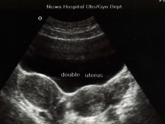
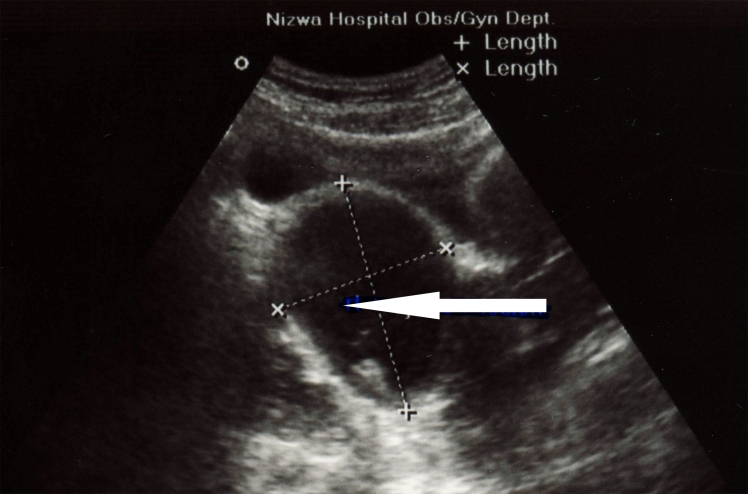
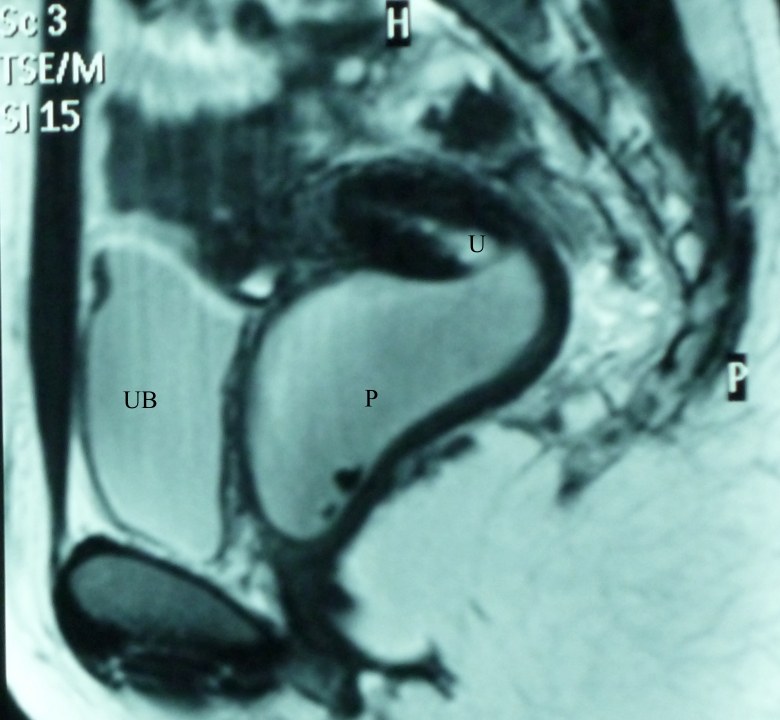
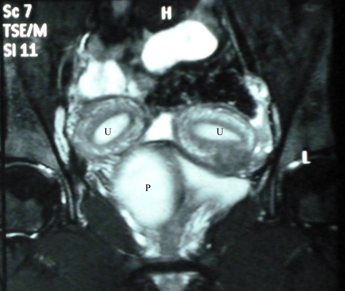
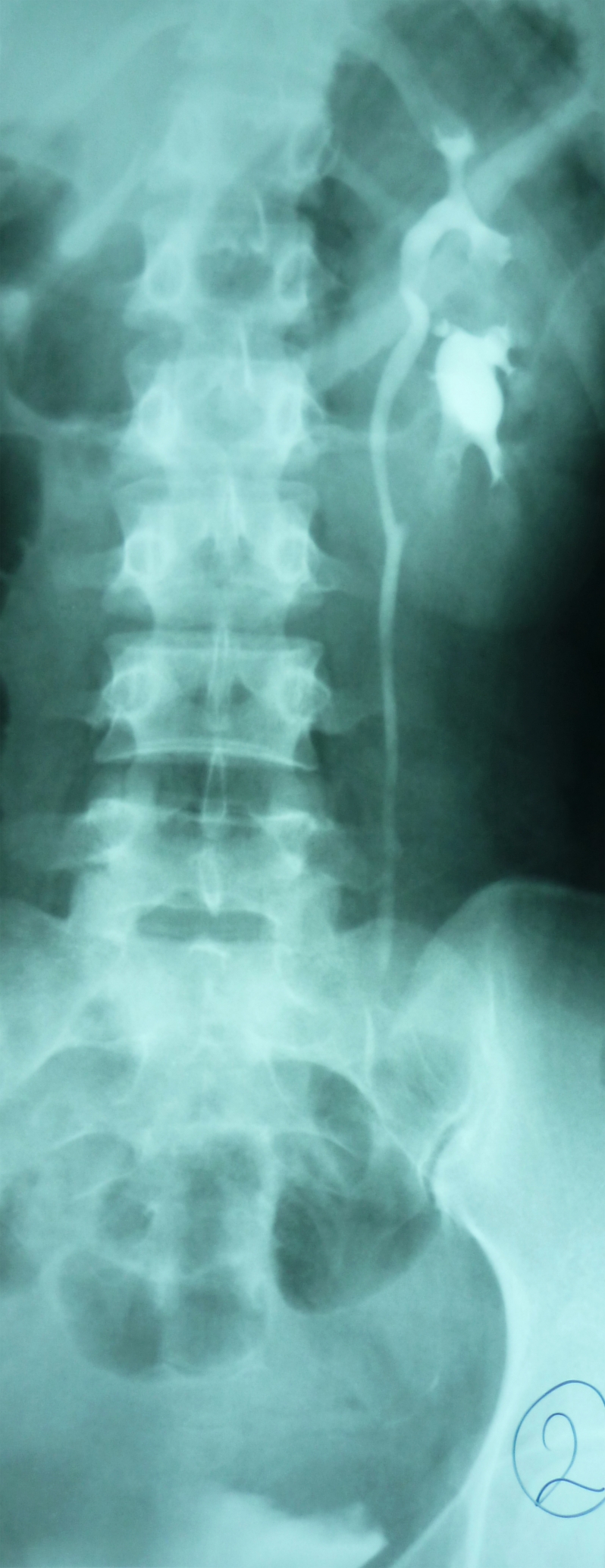
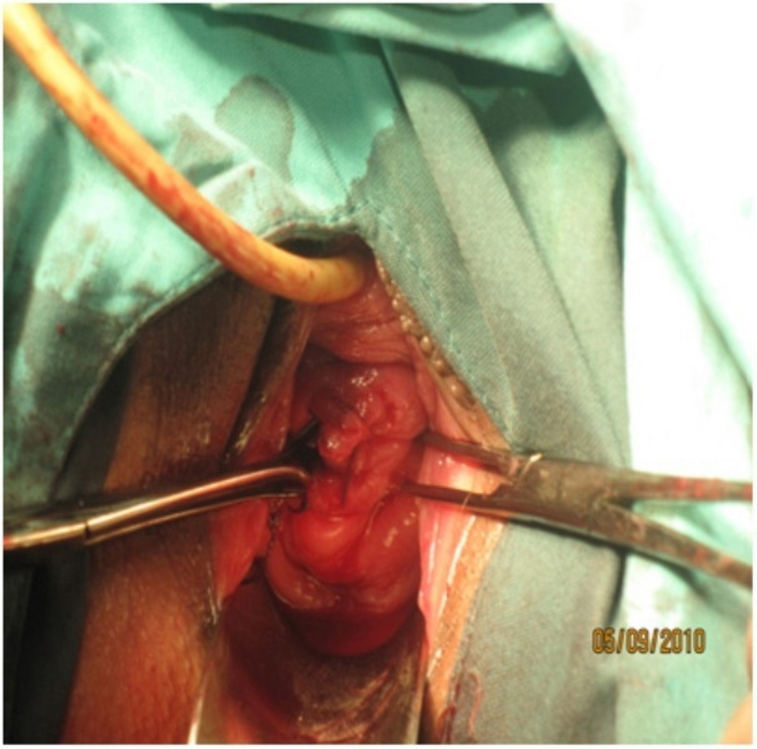
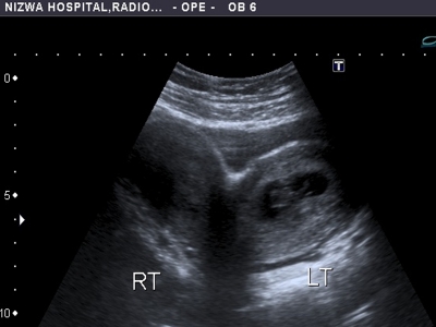
Uterus didelphys with obstructed hemivagina and ipsilateral renal agenesis (OHVIRA Syndrome) is a rare congenital anomaly of the female genital tract. Uterus didelphys occurs when the midline fusion of the mullerian ducts is arrested, either completely or incompletely. Women with didelphic uterus may be asymptomatic and unaware of having a double uterus. They may present with complaints of dysmenorrhoea and dyspareunia. A 25 year old woman attending the infertility clinic at Nizwa regional referral hospital presented with history of dysmenorrhoea and foul vaginal discharge with right cystic pelvic mass. She was diagnosed as a case of double uterus with obstructed right hemivagina and right pyocolpos with ipsilateral renal agenesis after routine ultrasonography in the clinic followed by MRI. Excision of the right vaginal septum with drainage of 200 ml of purulent discharge was performed. She was relieved of her symptoms and conceived promptly after the surgical excision of the partial vaginal septum.

Keywords: Dysmenorrhea; Mullerian duct; Pyocolpos; Renal agenesis; Uterus didelphys.


Figures

Figure 1
Figure 1
Transverse transabdominal sonographic image through the pelvis demonstrates the two uteri.
Figure 2
Figure 2
Right pyocolpos: Longitudinal transabdominal sonographic image through the pelvis demonstrates cystic structure with echogenic material inside, representing distended right upper hemivagina.
Figure 3
Figure 3
Axial T2-weighted MR image demonstrates dilated right hemivagina with high T2-signal-intensity fluid (pyocolpos, P) compressing the left hemivagina (V). Normal appearance both uteri (U) with normal endometrium plate.
Figure 4
Figure 4
Sagittal T2-weighted MR image demonstrates dilated right hemivagina with high T2-signal-intensity fluid (pyocolpos, P) extending into endocervical canal of right uterus (U), Urinary bladder (UB).
Figure 5
Figure 5
Coronal T2-weighted MR image with fat suppression, demonstrates dilated right hemivagina with high T2-signal-intensity fluid (pyocolpos, P). Normal-appearing both uteri (U) with normal endometrium plate.
Figure 6
Figure 6
Intravenous urogram (IVU) demonstrates incomplete duplex of left kidney with ureters of upper and lower moieties seen united at level of L3 transverse process level.
Figure 7
Figure 7
Double cervices in volsellum after right side septotomy.
Figure 8
Figure 8
Transverse transabdominal sonographic image through the pelvis demonstrates the two uteri with gestational sac and fetal pole in the left uterus.

References

    1. Madureira AJ, Mariz CM, Bernardes JC, Ramos IM. Case 94: Uterus didelphys with obstructing hemivaginal septum and ipsilateral renal agenesis. Radiology 2006. May;239(2):602-606 10.1148/radiol.2392031187 - DOI - PubMed
    1. Kim TE, Lee GH, Choi YM, Jee BC, Ku SY, Suh CS, et al. Hysteroscopic resection of the vaginal septum in uterus didelphys with obstructed hemivagina: a case report. J Korean Med Sci 2007. Aug;22(4):766-769 10.3346/jkms.2007.22.4.766 - DOI - PMC - PubMed
    1. Nahum GG. Uterine anomalies. How common are they, and what is their distribution among subtypes? J Reprod Med 1998. Oct;43(10):877-887 - PubMed
    1. Golan A, Langer R, Bukovsky I, Caspi E. Congenital anomalies of the müllerian system. Fertil Steril 1989. May;51(5):747-755 - PubMed
    1. Smith NA, Laufer MR. Obstructed hemivagina and ipsilateral renal anomaly (OHVIRA) syndrome: management and follow-up. Fertil Steril 2007. Apr;87(4):918-922 10.1016/j.fertnstert.2006.11.015 - DOI - PubMed

Publication types

LinkOut - more resources